

Abstract
Odontogenic myxoma (OM) is a rare benign painless, slow-growing lesion with local aggressive behavior. Pain and sensory disturbance and fibro-osseous appearance in histopathology have been rarely reported in OM. The authors reported a 52-year-old male case presented with a large gingival mass around a mobile mandibular right first molar extended to the distal aspect of the third molar. Microscopic examination of the incisional and excisional biopsy revealed an OM with numerous newly formed bone or cementum-like material present throughout the specimen like those seen in fibro-osseous lesions. For avoiding to recurrence, a segmental mandibulectomy was performed and a metal plate was inserted to the right mandible defect under general anesthesia. Rehabilitation was completed with the placement of implants. We review and discuss about this variety.
Keywords: Bone disease, case report, odontogenic tumor
INTRODUCTION
Rudolf Virchow in 1871 reported first case of myxoma.[1] Myxomas of head and neck region are rare.[2,3] Myxoma classified into two types: odontogenic myxoma and those originated from soft tissue like perioral soft tissue, parotid gland, ear or larynx.[4] Thoma and Goldman first introduce odontogenic myxoma (OM) in 1974 when they observed the histologic appearance resemble to fibromyxoid tissue.[5] OM compromised 3-11 % of all odontogenic tumors.[6] OM is originated from mesenchymal stem cell of odontogenic mesenchyme originated from a developing tooth bud or embryonic mesenchymal cell of the dental follicle, dental papilla or periodontal ligament.[7] Formerly, investigators made a distinction between odontogenic myxomas (derived from odontogenic mesenchyme) and osteogenic myxomas (presumably derived from primitive bone tissue). However, most orthopedic pathologist do not accept that myxomas occur in the extra-gnathic skeleton, and all myxomas of the jaws are currently considered to be of odontogenic origin.[8,9,10,11] OMs comprising 3-20 % of all odontogenic tumors, with incidence of 0.07 new case per million per year.[1,12] The age distribution range between 5 and 70 years, reported a peak at second and third decade of life.[4] Approximately more than 60 percent are located in mandible and female predilection reported.[13]
CASE REPORT
A 52-year-old male patient referred to the Oral Medicine Clinic of Shiraz University of Medical Sciences complained of swelling and diffused pain in the right side mandibular teeth and recently paresthesia and dysesthesia of the right side of the lower lip. He reported the mass and expansion of his jaw occurred 1 year ago without any symptom, but diffused pain occurred 3 months ago. His medical history was significant for deafness and blood hypertension for which he was under treatment with a beta-blocker (atenolol).
In clinical examination, a mild diffused, painful and tender, bony hard swelling in the right side of the mandible that extended from the middle part of the mandible to the angle of mandible was observed. The surface color is normal and no sign of lymphadenopathy observed.
In intraoral evaluation, a tender swelling exhibited smooth surface and indistinct border soft mass extended at the right mandibular side from the first premolar to the second molar, caused displacement and mobility of first and second molars. Pus formation around the first molar occurred. Palpation of mass clarified expansion and thinning of buccal and lingual cortexes [Figure 1]. Multiple decayed teeth, calculus and stain were observed on this side. A small amount of blood was obtained in aspiration from the anterior border of the lesion, but in aspiration from the posterior part of the lesion, nothing was found.
Figure 1.
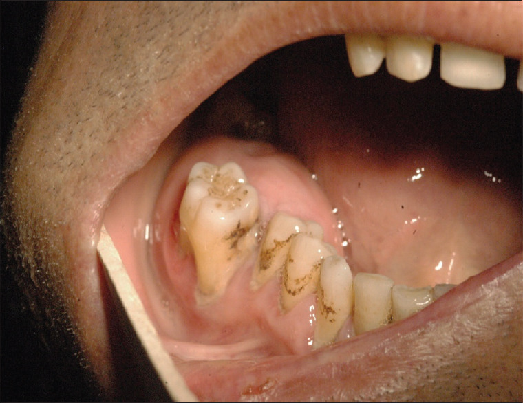
Intraoral presentation of soft solid mass involving the right molar area
A plain panoramic radiography revealed an approximately 7 cm × 3.5 cm, ill-defined, multinodular radiolucent lesion extending from the apical and distal root of the third molar to the mesial side of the apex of the canine tooth in the right lingual side, with mild root resorption. The anterior aspect of the lesion showed soap bubble appearance. The inferior alveolar canal was eroded and discontinued [Figure 2a]. Conventional occlusal view showed that buccal cortical perforation occurred [Figure 2b]. Further investigation with a cone-beam computed tomography revealed radiolucency in the right mandibular area resorbing the buccal cortex, alveolar bone and the root of lateral root 6–8 representing an invasive process. A minimal and fine intralesional calcification was reported. A differential diagnosis consisted of uncommon odontogenic myxoma (OM), ameloblastoma and central giant cell granuloma was made by clinicopathologic correlation.
Figure 2.

(a) Radiograph showing a multilocular radiolucency cause tooth displacement (arrow show borders of lesion). (b) The conventional occlusal radiographic view show expansion and buccal perforation
An incisional biopsy was performed under local anesthesia with a partial flap. The semi-hard gelatinous tissue was removed and excised. The histopathologic evaluation presented tissue consisted of a fragment of fibro collagenous connective tissue with many myxoid areas and infective area and pus accumulation observed. Residual bony trabeculae were seen and very small-sized trabeculae of mineralized tissue were occupied the mainly myxoid areas [Figure 3].
Figure 3.

(a) Photomicrograph showing myxoid stroma containing spindle shape and stellate cell and little amount of collagen and multiple foci of calcification (H&E staining, ×40 magnification) (b) Another view showing myxoid stroma and fewer calcified particles derived from other side of the lesion (c) Calcified products or trabecular particles (H&E staining, ×200 magnification) (d) Photomicrograph showing a polarized light comparison between residual lamellar bone and osteo-cementum particle
A surgical management involved a segmental resection under general anesthesia and a metal plate was inset to the defected area. The inferior alveolar nerve was preserved. The patient has been under regular follow-up and rehabilitation. A surgical defect has been healing gradually and no evidence of recurrence has been observed on the latest radiograph obtained 12 months after surgery.
DISCUSSION
In our presented case, the lesion showed uncommon clinical and histopathologic properties. The age of the patient was higher than the common presentation age. The pain and lip paresthesia reported in this case in contrast to common occurrences. The histopathology of this case showed haphazardly myxoid stroma with multiple cementum-like particles.
Clinically, smaller OM commonly is painless, asymptomatic and slow growing, and soft tissue surrounding the lesion is normal but advanced and large cases have cortical expansion and facial asymmetry.[14] The most frequent location is the tooth-bearing areas and in rare cases reported in association with missing or unerupted teeth.[15] The majority of OM occurred in the mandible at the molar region[16] In contrast to our case, in many case reports of OM, pain and paresthesia and sensory disturbances are uncommon findings but loosening and displacement of the teeth are usual events.[17,18]
OM appears as a suddenly detected multilocular or unilocular radiolucent bony lesion with irregular, scalloped and ill-defined or well-defined borders.[19] The lesion usually is well defined, and it may have a corticated margin but most often is poorly defined, especially in the maxilla.[20] The radiolucent defect may contain thin, wispy trabeculae of residual bone, which are often arranged at right angles to one another; therefore, the “soap bubble “or” tennis racquet strings” appearances are common radiologic findings.[21] Perforation of the cortical plate and radiopaque line extended from the periosteum, resembling “sunray” appearance – a rare feature of OM.[22,23]
Since OMs most often have a multilocular pattern, the differential diagnosis should include other multilocular lesions, such as ameloblastoma, central giant cell granulomas and central hemangiomas. The finding of characteristic thin straight septa with less than expected bone expansion is very useful in the differential diagnosis. In cases that fine calcification is observed in radiography, fibro-osseous lesions and central ossifying fibroma can be included in the differential diagnosis. OM can be distinguished by histopathologic report, according to encapsulation and density of stroma.[18,24]
Histopathologically, the tumor is composed of random oriented stellate, spindle-shaped cells in a plentiful, loose myxoid stroma that contains only a few collagen fibrils.[25] The matrix substance is composed of glycosaminoglycan, chiefly hyaluronic acid and chondroitin sulfate. In immunohistochemistry, the cells present diffuse immunoreactivity with antibodies directed against vimentin, with focal reactivity for muscle-specific actin.[26] OM very rarely exhibits boney or cementum-like calcifications.[10,18,27] This variant of OM includes the multiple case reports with similar histopathologic features already reported. Rennie et al. in 1985 reported the first case of actively forming cementum-like calcified material in OM.[28] According to Table 1, we found few published case reports about OM containing osteocement-like tissue. Effiom et al.[29] reported the clinical and histological findings in 63 diagnosed OM cases. They reported that there is a tendency for calcification to occur in OM in certain ethnic groups. Mamabolo et al.[30] in South Africa reported intralesional calcifications detected radiographically in nine cases of OM. The World Health Organization classification of odontogenic recognized that OM can contain odontogenic epithelium like as seen in central odontogenic fibroma. The local aggressive behavior can distinguish between these tumors.[31]
Table 1.
List of case reports of odontogenic myxoma with osteocementum like calcification
| Authors | Location | Age/gender | Year |
|---|---|---|---|
| Rennie et al. (28) | Mandible | 5 y/o , Male | 1985 |
| Oygür et al. (34) | Maxilla | 19 y/o , Female | 2001 |
| Lin Y & Basile (27) | Mandible | 29 y/o , Female | 2010 |
| Lahey et al. (18) | Mandible | 69 y/o, Female | 2013 |
| Rallis et al. (10) | Maxilla | 32 y/o, Male | 2014 |
| Hirai et al. (35) | Mandible | 54 y/o, Male | 2014 |
| Hammad et al. (31) | Mandible | 45 y/o , Female | 2016 |
| Rezaee et al. | Mandible | 52 y/o , Male | 2020 |
The initial and most important treatment for OM is surgery. There are currently no clear surgical management guidelines for OM, and a variety of approaches may be used.[12,32] Small lesions are treated by curettage, but careful periodic reevaluation is necessary for at least 5 years. For larger lesions, more aggressive approaches such as segmental or block resection and hemimandibulectomy with reconstruction surgery may be required.[33] Oygür et al.[34] and Hirai et al.[35] reported unusual histopathological features of odontogenic myxoma was the presence of calcified spherules that removed as segmental resection. High recurrence rates (10%–30%) after conservative surgical treatment were reported.[26] Gelatinous nature and infiltrative properties and the absence of capsule are the most important description for recurrence.[20]
Finally, since there have been a few reports of this clinical and histopathological finding, we suggest that it should be acknowledged among the features of OM to avoid misdiagnosis and any resultant effects on managements.
Patient consent
Written informed consent was obtained from the patient for publication of this Case Report and any accompanying images, photograph and radiographic view.
Declaration of patient consent
The authors certify that they have obtained all appropriate patient consent forms. In the form, the patient has given his consent for his images and other clinical information to be reported in the journal. The patient understands that name and initials will not be published and due efforts will be made to conceal identity, but anonymity cannot be guaranteed.
Financial support and sponsorship
This study was financially supported by Oral Medicine department of Shiraz University of Medical Science.
Conflicts of interest
There are no conflicts of interest.
Acknowledgment
The authors thank the Oral Medicine department and Vice-Chancellery of Research Shiraz University of Medical Science for supporting this research. We are thankful to Dr. Ali Dehghani Najvani for histopathological considerations.
REFERENCES
- 1.Carvalho de Melo AU, de Farias Martorelli SB, Cavalcanti PH, Gueiros LA, Martorelli Fde O. Maxillary odontogenic myxoma involving the maxillary sinus: Case report. Braz J Otorhinolaryngol. 2008;74:472–5. doi: 10.1016/S1808-8694(15)30586-3. [DOI] [PMC free article] [PubMed] [Google Scholar]
- 2.Shah A, Lone P, Latoo S, Ahmed I, Malik A, Hassan S, et al. Odontogenic myxoma of the maxilla: A report of a rare case and review on histogenetic and diagnostic concepts. Natl J Maxillofac Surg. 2011;2:189–95. doi: 10.4103/0975-5950.94480. [DOI] [PMC free article] [PubMed] [Google Scholar]
- 3.Ansari A, Thomas A, Bohra R, Vare R, Rathod J, Dongardive SJOCR. Recurrent myxoma of the maxilla: A rare case. J Otolaryngology Case Reports. 2019;10:1–4. [Google Scholar]
- 4.Thabusum DA, Rajesh N, Reddy RS, Ravikanth M, Raju US. Odontogenic myxoma of maxilla–A rare case report. WJPMR. 2017;3:282–5. [Google Scholar]
- 5.Thoma KH, Goldman HM. Central myxoma of the jaw. Oral Surg Oral Med Oral Pathol. 1947;33:B532–40. doi: 10.1016/0096-6347(47)90315-3. [DOI] [PubMed] [Google Scholar]
- 6.Regezi JA, Kerr DA, Courtney RM. Odontogenic tumors: Analysis of 706 cases. J Oral Surg. 1978;36:771–8. [PubMed] [Google Scholar]
- 7.Dotta JH, Miotto LN, Spin-Neto R, Ferrisse TM. Odontogenic myxoma: Systematic review and bias analysis. Eur J Clin Invest. 2020;50:e13214. doi: 10.1111/eci.13214. [DOI] [PubMed] [Google Scholar]
- 8.Reddy GS, Kumar BS, Muppa R, Regonda SK, Tvs HK. Odontogenic fibromyxoma of maxilla: A rare case report. Case Rep Dent. 2013;2013:345479. doi: 10.1155/2013/345479. https://doi.org/10.1155/2013/345479. [DOI] [PMC free article] [PubMed] [Google Scholar]
- 9.Noffke CE, Raubenheimer EJ, Chabikuli NJ, Bouckaert MM. Odontogenic myxoma: Review of the literature and report of 30 cases from South Africa. Oral Surg Oral Med Oral Pathol Oral Radiol Endod. 2007;104:101–9. doi: 10.1016/j.tripleo.2007.01.026. [DOI] [PubMed] [Google Scholar]
- 10.Rallis G, Dais P, Kostakis G, Stathopoulos P. Osteo-cementum producing odontogenic myxomas. A literature review of a distinctive variant. J Maxillofac Oral Surg. 2015;14:176–81. doi: 10.1007/s12663-014-0645-5. [DOI] [PMC free article] [PubMed] [Google Scholar]
- 11.Neville BW, Damm DD, Allen CM, Chi AC. 4th ed. St. Louis, Missouri, Printed in Canada: Elsevier Health Sciences; 2015. Oral and Maxillofacial Pathology. [Google Scholar]
- 12.Kawase-Koga Y, Saijo H, Hoshi K, Takato T, Mori Y. Surgical management of odontogenic myxoma: A case report and review of the literature. BMC Res Notes. 2014;7:214. doi: 10.1186/1756-0500-7-214. [DOI] [PMC free article] [PubMed] [Google Scholar]
- 13.Mascitti M, Togni L, Pirani F, Rubini C, Santarelli AJ. Peripheral odontogenic myxoma: Report of two new cases with a critical review of the literature. The Open Dentistry Journal. 2018;12:1079–90. [Google Scholar]
- 14.Scaraficci AC, Caroli AL, De Carvalho PA, Lessa RC, Tonin LO, Da Silva JS, et al. pp-odontogenic myxoma treatment of the mandible with atypical radiographic features. Oral Surgery, Oral Medicine, Oral Pathology and Oral Radiology journal. 2017;123:e70–1. 10.1016/j.oooo.2016.09.168. [Google Scholar]
- 15.Martínez-Mata G, Mosqueda-Taylor A, Carlos-Bregni R, de Almeida OP, Contreras-Vidaurre E, Vargas PA, et al. Odontogenic myxoma: Clinico-pathological, immunohistochemical and ultrastructural findings of a multicentric series. Oral Oncol. 2008;44:601–7. doi: 10.1016/j.oraloncology.2007.08.009. [DOI] [PubMed] [Google Scholar]
- 16.Dunphy L, Shah S, Halsnad M, Amel-Kashipaz R, Praveen P. Odontogenic myxoma presenting as a spontaneous oro-nasal fistula: A case report. Oral Surgery. 2015;8:167–70. [Google Scholar]
- 17.Ramos T, Araújo J, Gonçalves P, Gonçalves F, Neto NC, Surgery M. Agressive myxoma in mandible: A case report of a pregnant woman. J International Journal of Oral Maxillofacial Surgery. 2019;48:237. [Google Scholar]
- 18.Lahey E, Woo SB, Park HK. Odontogenic myxoma with diffuse calcifications: A case report and review of the literature. Head Neck Pathol. 2013;7:97–102. doi: 10.1007/s12105-012-0387-y. [DOI] [PMC free article] [PubMed] [Google Scholar]
- 19.Chrcanovic BR, Gomez RS. Odontogenic myxoma: An updated analysis of 1,692 cases reported in the literature. Oral Dis. 2019;25:676–83. doi: 10.1111/odi.12875. [DOI] [PubMed] [Google Scholar]
- 20.Ayrancı F, Ömezli M, Rastgeldi O, Duman A. Odontogenic myxoma located in the mandible: A case report. Middle Black Sea J Health Sci. 2015;1:25. [Google Scholar]
- 21.Simon EN, Merkx MA, Vuhahula E, Ngassapa D, Stoelinga PJ. Odontogenic myxoma: A clinicopathological study of 33 cases. Int J Oral Maxillofac Surg. 2004;33:333–7. doi: 10.1016/j.ijom.2003.12.004. [DOI] [PubMed] [Google Scholar]
- 22.Dabbaghi A, Nikkerdar N, Bayati S, Golshah A. Rare appearance of an odontogenic myxoma in cone-beam computed tomography: A case report. J Dent Res Dent Clin Dent Prospects. 2016;10:65–8. doi: 10.15171/joddd.2016.010. [DOI] [PMC free article] [PubMed] [Google Scholar]
- 23.White JA, Ramer N, Wentland TR, Cohen M. The rare radiographic sunburst appearance of odontogenic myxomas: A case report and review of the literature. Head Neck Pathol. 2020;14:1105–10. doi: 10.1007/s12105-019-01122-1. [DOI] [PMC free article] [PubMed] [Google Scholar]
- 24.Sahu B, Anand R, Abbey P, editors. Approach to Imaging Differentials in Jaw Lesions: A Pictorial Review2019. European Congress of Radiolog. 2019. https://dx.doi.org/10.26044/ecr2019/C-2012.
- 25.Kansy K, Juergens P, Krol Z, Paulussen M, Baumhoer D, Bruder E, et al. Odontogenic myxoma: Diagnostic and therapeutic challenges in paediatric and adult patients–A case series and review of the literature. J Craniomaxillofac Surg. 2012;40:271–6. doi: 10.1016/j.jcms.2011.04.009. [DOI] [PubMed] [Google Scholar]
- 26.Lo Muzio L, Nocini P, Favia G, Procaccini M, Mignogna MD. Odontogenic myxoma of the jaws: A clinical, radiologic, immunohistochemical, and ultrastructural study. Oral Surg Oral Med Oral Pathol Oral Radiol Endod. 1996;82:426–33. doi: 10.1016/s1079-2104(96)80309-x. [DOI] [PubMed] [Google Scholar]
- 27.Lin YL, Basile JR. A case of odontogenic myxoma with unusual histological features mimicking a fibro-osseous process. Head Neck Pathol. 2010;4:253–6. doi: 10.1007/s12105-010-0189-z. [DOI] [PMC free article] [PubMed] [Google Scholar]
- 28.Rennie JS, MacDonald DG, Critchlow HA. Unusual myxomatous odontogenic tumour with calcification. Int J Oral Surg. 1985;14:307–10. doi: 10.1016/s0300-9785(85)80046-6. [DOI] [PubMed] [Google Scholar]
- 29.Effiom OA, Adewole RA, Odukoya O. Clinicopathological characteristics of odontogenic myxoma in Nigerians. West Afr J Med. 2011;30:255–61. [PubMed] [Google Scholar]
- 30.Mamabolo M, Noffke C, Raubenheimer E. Odontogenic tumours manifesting in the first two decades of life in a rural African population sample: A 26 year retrospective analysis. Dentomaxillofac Radiol. 2011;40:331–7. doi: 10.1259/dmfr/54585925. [DOI] [PMC free article] [PubMed] [Google Scholar]
- 31.Hammad HM, Hasen YM, Odat AA, Mikdadi AM, Safadi RA. Odontogenic myxoma with diffuse calcifications: A case report and review of a rare histologic feature. Oral Surg Oral Med Oral Pathol Oral Radiol. 2016;122:e116–24. doi: 10.1016/j.oooo.2015.12.009. [DOI] [PubMed] [Google Scholar]
- 32.Limdiwala P, Shah J. Odontogenic myxoma of maxilla: A review discussion with two case reports. Contemp Clin Dent. 2015;6:131–6. doi: 10.4103/0976-237X.149310. [DOI] [PMC free article] [PubMed] [Google Scholar]
- 33.Boffano P, Gallesio C, Barreca A, Bianchi FA, Garzino-Demo P, Roccia F. Surgical treatment of odontogenic myxoma. J Craniofac Surg. 2011;22:982–7. doi: 10.1097/SCS.0b013e3182101400. [DOI] [PubMed] [Google Scholar]
- 34.Oygür T, Dolanmaz D, Tokman B, Bayraktar S. Odontogenic myxoma containing osteocement-like spheroid bodies: Report of a case with an unusual histopathological feature. J Oral Pathol Med. 2001;30:504–6. doi: 10.1034/j.1600-0714.2001.030008504.x. [DOI] [PubMed] [Google Scholar]
- 35.Hirai E, Yamamoto K, Yonemasu H, Takahashi O, Takao M, Fushimi C. Odontogenic myxoma containing osteocement-like tissue: Report of a case with an unusual histopathological feature. J Oral Maxillofac Surg Med Pathol. 2014;26:407–10. [Google Scholar]