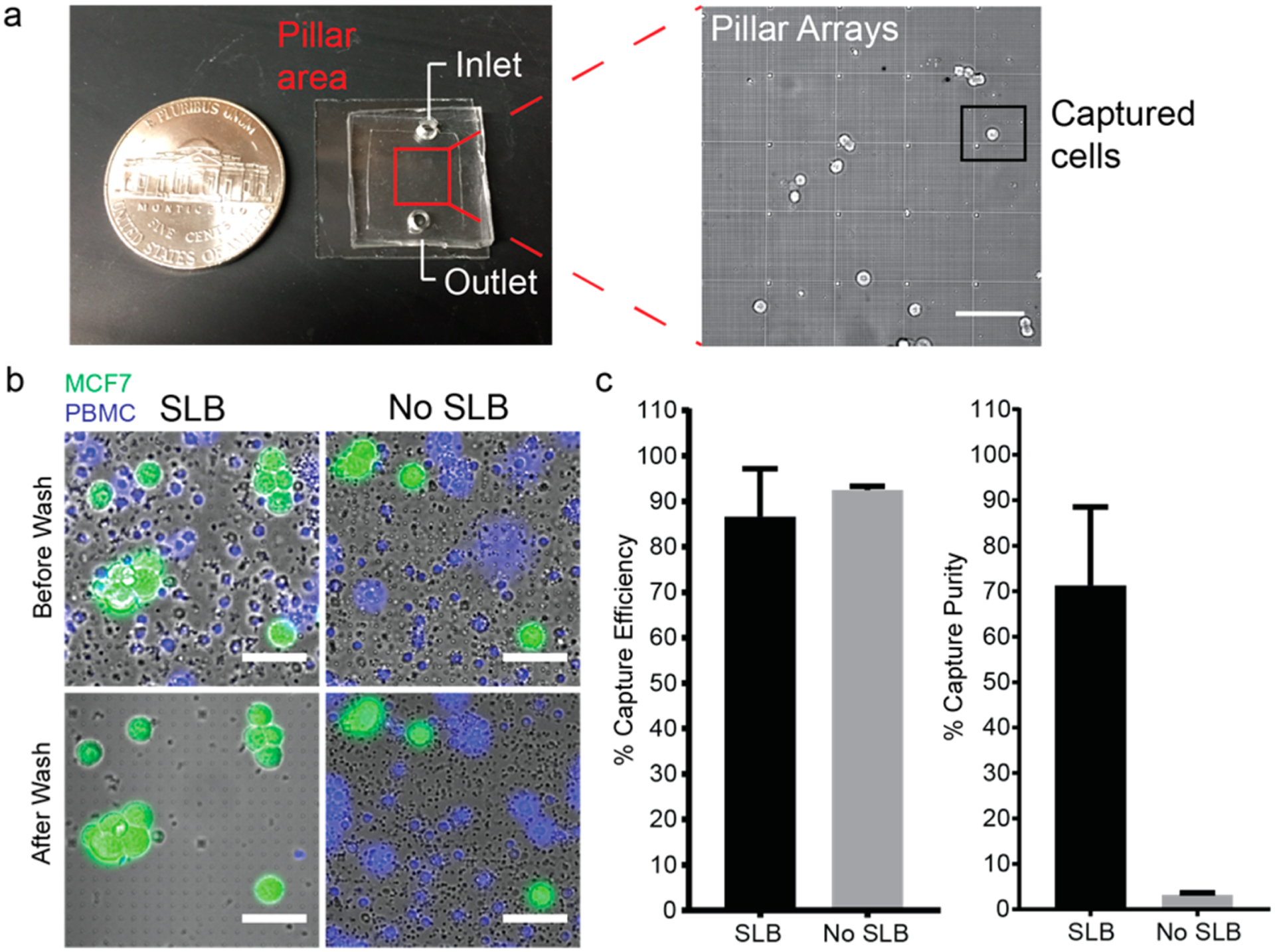
Figure 4.

Antibody-functionalized supported lipid bilayer selectively captured spiked MCF7 cells in PBMCs on quartz nanopillar arrays. (a, Left) Snapshot of the nanopillar chip. The nanopillar chip is covered with PDMS to create a chamber for both surface functionalization and cell capture. The pillar arrays are located in the center of the chip (marked by a red square). (Right) Zone-in picture of nanopillar arrays with captured MCF7 cells. (Scale bar 100 μm.) (b) Anti-EpCAM-lipid-functionalized nanopillar array-captured MCF7 stained with Celcein-AM (green), preventing the nonspecific adhesion of PBMCs stained with Hoechst 33342 (blue). (Scale bars 50 μm.) (c) Capture efficiency (left) and capture purity (right) of spiked MCF7 cells in isolated PBMCs on supported lipid bilayer-functionalized quartz nanopillar arrays and bare quartz nanopillar arrays. The capture efficiencies of supported lipid bilayer-functionalized nanopillar arrays and bare quartz nanopillar arrays are 86.6 ± 10.6 and 92.5 ± 0.8%, respectively. The purities of both surfaces are 71.3 ± 17.2 and 3.2 ± 0.4%, respectively. (Every experiment was repeated three times (n = 3), and error bars represent the standard deviation (SD).
