Abstract
Salinatis (antigenic formula, 4,12:d:eh:enz15) is a rare Salmonella serotype currently designated a triphasic variant of the diphasic serotype Duisburg (1,4,12,27:d:enz15) (underlining indicates that the O antigen is determined by phage lysogenization). Salinatis could also be related to serotype Sandiego (4,[5],12:eh:enz15), from which it might have been derived by loss of H-d flagellin genes. Nineteen Salmonella strains of serotypes Salinatis, Duisburg, and Sandiego were examined by biotyping, PvuII and SmaI ribotyping, IS200 fingerprinting, and pulsed-field gel electrophoretic profiling. Results from these methods, used alone or together, indicate that serotype Salinatis is more likely to be related to serotype Sandiego than to serotype Duisburg. For future lists of serotype names, it is recommended that Salinatis be considered a variant of Sandiego.
Serotypes of Salmonella enterica are named (in subspecies enterica) or designated by antigenic formulae (in other subspecies of S. enterica and in S. bongori) on the basis of serological tests with O and H typing sera (18, 23, 25). Most serotypes of Salmonella (other than the subspecies arizonae and houtenae and S. bongori) show diphasic variation of H antigens, alternately expressing two antigenically different kinds of flagella (18, 29). Unusual serotypes are occasionally found in nature that express three, or even more, H antigen types—for example, Montgomery, Rubislaw, and Salinatis (11, 13, 14, 30).
Salinatis (synonym: S. salinatis) is a rare serotype of Salmonella which, since it was first cultured in 1942 from rat feces in Salinas, Calif. (13), has been isolated infrequently. For example, only 10 isolations were reported worldwide from 1948 to 1964, and most of these were made in the United States and Australia (16). Salinatis has not been isolated in the United Kingdom or France in recent years (2, 17, 19).
In the most recent World Health Organization publication of salmonella serotypes (18), the name Salinatis (antigenic formula, 4,12:d:eh:enz15) was deleted because serotype Salinatis was considered to be a triphasic variant of the diphasic serotype Duisburg (1,4,12,27:d:enz15) (underlining indicates that the O antigen is determined by phage lysogenization). It should be noted, however, that culture of serotype Salinatis bacteria in the presence of H-d flagellar antiserum gave rise to d-negative variants that were biochemically and serologically identical to serotype Sandiego (4,[5],12:eh:enz15) bacteria (13). Thus, serotype Salinatis might equally well be related to serotype Sandiego.
In an attempt to resolve this controversy, the relatedness of strains of serotypes Salinatis, Duisburg, and Sandiego was explored by biotyping, ribotyping, IS200 fingerprinting, and pulsed-field gel electrophoresis (PFGE), methods that have previously provided insight into clonal relationships in diverse serotypes of Salmonella (3, 5, 7–9, 24–27).
MATERIALS AND METHODS
Bacteria.
Details of the 19 strains of Salmonella examined are shown in Table 1. The strains included the type strain of serotype Salinatis (N6247), obtained from the National Collection of Type Cultures, Central Public Health Laboratory, Colindale, London, England, and 14 strains of serotype Duisburg and 4 strains of serotype Sandiego received from the World Health Organization Collaborating Center for Reference and Research on Salmonella, Institut Pasteur, Paris, France. Cultures of these strains were stored on Dorset’s egg slopes at room temperature (18 to 20°C). When required for testing, they were plated on MacConkey agar (Oxoid) and incubated overnight in air at 37°C.
TABLE 1.
Biotype, ribotype, IS200 profiles, and PFPs of 19 Salmonella strains of serotypes Salinatis, Sandiego, and Duisburg
| Serotype and strain | Biotype | Ribotypea | IS200 profile | PFPb |
|---|---|---|---|---|
| Salinatis, N6247 | 10di | PI/SI | A | 1 |
| Sandiego | ||||
| 18K | 10di | PII/SII | B | 2 |
| CNS4.86 | 10di | PIII/SIII | C | 3 |
| CNS1.87 | 10di | PIII/SIII | C | 3 |
| CNS3.86 | 10di | PIIIvar/SIIIvarc | D | 4 |
| Duisburg | ||||
| 6.86 | 9i | PIV/SIV | —d | 5 |
| 15.86 | 9i | PIV/SIV | — | 5 |
| 2.87 | 9i | PIV/SIV | — | 5 |
| 11.87 | 9i | PIV/SIV | — | 5 |
| 14.87 | 9i | PIV/SIV | — | 5 |
| 8.86 | 9i | PIV/SIV | — | 6 |
| 9.86 | 9i | PIV/SIV | — | 6 |
| 3.87 | 9i | PIV/SIV | — | 6 |
| 4.87 | 9i | PIV/SIV | — | 6 |
| 8.87 | 9i | PIV/SIV | — | 6 |
| 10.87 | 9i | PIV/SIV | — | 6 |
| 12.87 | 9i | PIV/SIV | — | 6 |
| 13.86 | 9i | PIV/SIV | — | 7 |
| 11.86 | 10di | PV/SV | E | 8 |
After digestion of DNA with restriction enzymes PvuII and SmaI (P/S).
After digestion of DNA with restriction enzyme XbaI.
See text.
—, no IS200 present.
Biotyping.
Each strain was assigned to 1 of 32 primary biotypes according to its reactions at 37°C in five primary biotyping tests, by methods detailed elsewhere (12). For biotyping, a single colony from a fresh overnight culture was tested for fermentation of m-inositol and l-rhamnose in peptone water and d-xylose in Bitter’s minimal medium and for utilization of d- and m-tartaric acids. Subtypes within primary biotypes were distinguished by reactions in a further 10 secondary biotyping tests, including those for utilization of l-tartaric acid at 37°C and fermentation of m-inositol at 25°C. Full biotypes (e.g., 9i) were designated by numbers which indicate primary biotypes, followed by letters indicating subtypes of the primary biotypes (12).
Extraction and digestion of cellular DNA.
One colony from an overnight culture on MacConkey agar was inoculated in 10 ml of L broth (21) in a disposable 20-ml screw-capped plastic tube and incubated with shaking for 18 h at 37°C. The culture was cooled on ice and centrifuged at 3,000 × g for 20 min. Bacterial DNA, extracted by the cetyltrimethylammonium bromide miniprep method described previously (36), was dissolved in TE buffer (10 mM Tris-HCl, 1 mM EDTA, pH 8.0) and stored at −20°C until required. Samples (ca. 2 μg) were digested to completion with PvuII or SmaI (Promega, Southampton, England), restriction enzymes known to have no recognition site within the IS200 sequence (EMBL accession no. X56834).
Southern blotting and preparation of DIG-labeled gene probes of rRNA and IS200.
Digests were electrophoresed at 2 V/cm in TBE buffer (89 mM Tris, 89 mM boric acid, 2 mM EDTA), pH 8.0, along with digoxigenin (DIG)-labeled marker III (Boehringer Mannheim), through a 0.8% (wt/vol) gel (‘Hi-Pure’ Low EEO Agarose; BioGene, Kimbolton, England) containing ethidium bromide at 0.5 μg/ml. DNA fragments were transferred to Positive Membrane (Appligene Oncor, Birtley, Chester-le-Street, England) on Trans DNA Express (Appligene Oncor) for 75 min at 5.5 × 10³ Pa with 20× SSC buffer (1× SSC is 0.15 M NaCl plus 0.015 M sodium citrate) (28) and fixed by exposure to UV light (312 nm) for 5 min. The DNA probe of Escherichia coli rRNA was the 3.8-kb PvuII restriction fragment of pT711 (8). The IS200 gene probe was generated by PCR from Salmonella serotype Typhimurium strain LT2 genomic DNA with primers IS200-L2 (5′-CCT AAC AGG CGC ATA CGA TC-3′) and IS200-R2 (5′-ACA TCT TGC GGT CTG GCA AC-3′) as described elsewhere (5); the 556-bp PCR product was excised from the gel after electrophoresis through 0.5% (wt/vol) Gibco BRL Ultrapure Low Melting Point Agarose (Life Technologies, Paisley, Scotland) in TBE buffer and purified with Geneclean II (Anachem, Luton, England). Probes were labeled with DIG–11-dUTP by random priming (Boehringer Mannheim).
Hybridization with DIG-labeled probes and detection of hybrids.
Prehybridizations (for ≥2 h), hybridizations (for ≥16 h), and detection of hybrids by enhanced chemiluminescence with anti-DIG–alkaline phosphatase and CSPD were carried out as recommended by the manufacturer (Boehringer Mannheim), except that boiled, sheared salmon sperm DNA (50 μg/ml) was included in the prehybridization and hybridization solutions. Hyperfilm MP (Amersham International) was exposed to membranes for 1 to 10 min at room temperature and developed in a Kodak X-Omat Processor. Two 15-min washes at 37°C in prewarmed stripping solution (0.2 M NaOH, 0.1% [wt/vol] sodium dodecyl sulfate) were used to remove bound probe from the membrane before hybridization with the second probe. Ribotypes detected after digestion with restriction endonucleases PvuII and SmaI were described as P and S ribotypes, respectively.
PFGE profiling.
Each strain was grown overnight at 37°C in 5 ml of brain heart infusion broth (Oxoid). Cells were harvested by centrifugation for 10 min at 3,600 × g and resuspended in 0.5 ml of Pett IV buffer (10 mM Tris-HCl, 1 M NaCl, pH 7.6). An aliquot (0.3 ml) of the suspension was transferred to a microcentrifuge tube, and cells were pelleted at 12,000 × g, washed twice in Pett IV buffer, resuspended in 0.5 ml of that same buffer, and placed in a heating block at 40°C. An equal volume of 2% (wt/vol) pulsed-field-certified agarose in distilled water was added to the prewarmed cells, and the contents of the tube were mixed; 100-μl volumes were allowed to solidify in plastic plug molds (Bio-Rad, Hemel Hempstead, England). Agarose plugs were transferred to fresh microcentrifuge tubes and incubated overnight at 37°C in 0.5 ml of EC lysis buffer (1 M Tris-HCl, 1 M NaCl, 0.5 M EDTA, 0.5% [wt/vol] Brij 58, 0.2% [wt/vol] sodium deoxycholate, 0.5% [wt/vol] lauryl sarcosine, pH 7.6) which contained 1-mg/ml lysozyme and a 1-mg/ml solution of RNase A at 20 μl/ml. Plugs were transferred to fresh tubes and incubated for 48 h at 56°C in 0.5 ml of ESP solution (0.5 M EDTA, 1% [wt/vol] lauryl sarcosine, 1-mg/ml proteinase K). Thereafter, they were transferred to fresh tubes, washed in TE buffer (pH 7.5) three times for 1 h each time at room temperature, washed overnight at 37°C, and stored at 4°C until required. Digestion of DNA in plugs with 20 U of restriction enzyme XbaI or SpeI was carried out overnight at 37°C as directed by the manufacturer (Life Technologies). Electrophoresis of digestion products was carried out in 1% (wt/vol) pulsed-field-certified agarose gels in a contour-clamped homogeneous electric field DRII system (Bio-Rad) with 0.5× TBE buffer (pH 8.3) at 14°C. Run conditions were 6 V/cm for 23 h with an initial switch time of 5 s and a final switch time of 60 s. Gels were stained in ethidium bromide (0.5 μg/ml in 0.5× TBE) and photographed by UV transillumination (312 nm) on Polaroid 667 film.
Analysis of typing profiles.
Ribotypes and IS200 patterns were analyzed on a Bio-Profil gel electrophoresis image analyzer (Vilber Lourmat), and fragment sizes were calculated with reference to DIG-labeled molecular weight marker III (Boehringer Mannheim). PFGE profiles (PFPs) were analyzed and dendrograms were constructed with Phoretix ID Advanced gel analysis software, version 4.00 (Phoretix International). Dice coefficients of similarity were calculated for the ribotypes and PFPs of each pair of strains in accordance with the equation D = 2m/(a + b), where m is the number of bands common to the two profiles and a + b is the total number of bands present in the two profiles (10).
RESULTS
Biotyping.
Among the 19 strains of Salmonella examined, two different biotypes were detected, namely, 9i and 10di (Table 1). Salinatis strain N6247, all four Sandiego strains, and Duisburg strain 11.86 belonged to primary biotype 10. Thus, in tests done at 37°C, they fermented d-xylose in Bitter’s medium and l-rhamnose in peptone water and utilized d-tartaric acid in peptone water but gave negative results in tests with m-inositol and m-tartrate. The secondary biotype characters d and i indicate negative results in tests with l-tartaric acid at 37°C and m-inositol at 25°C, respectively (12). The other 13 Duisburg strains gave positive results in all biotyping tests except those with m-inositol (at both 37 and 25°C); hence, they were of biotype 9i.
Ribotyping.
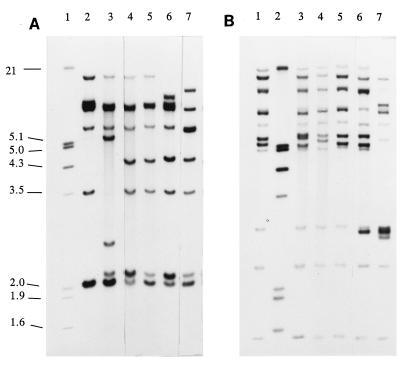
When the cellular DNAs of these 19 salmonella strains were digested with PvuII or SmaI, five ribotypes were identified (Table 1), namely, PI to PV (Fig. 1A) or SI to SV (Fig. 1B). Ribotype PI of Salinatis strain N6247 comprised six hybridized bands (ca. 10, 6.9, 6.7, 5.7, 3.5, and 2.0 kb; Fig. 1A, lane 2). Sandiego ribotypes PII and PIII contained eight bands, as follows: PII of strain 18K (ca. 10, 6.9, 6.7, 5.7, 5.3, 2.6, 2.1, and 2.0 kb; Fig. 1A, lane 3) and PIII of strains CNS4.86 (or CNS1.87) and CNS3.86 (ca. 10, 6.9, 6.7, 5.7, 4.5, 3.5, 2.1, and 2.0 kb; Fig. 1A, lanes 4 and 5). It should be noted that the ribotype of strain CNS3.86 consistently showed a difference from that of the other two PIII strains in the intensities of its 2.1- and 2.0-kb bands (Fig. 1A, lane 5); it was considered, therefore, to be a minor variant of PIII. Thirteen of the 14 Duisburg strains (Table 1) belonged to ribotype PIV with hybridized bands at ca. 7.2, 6.9, 6.7, 5.7, 4.5, 3.5, 2.1, and 2.0 kb (Fig. 1A, lane 6); the other Duisburg strain (11.86) belonged to ribotype PV, which had seven hybridized bands (ca. 8, 6.7, 5.7, 4.5, 3.5, 2.1, and 2.0 kb; Fig. 1A, lane 7).
FIG. 1.
Ribotypes of Salmonella strains of serotypes Salinatis, Sandiego, and Duisburg digested with restriction enzyme PvuII (A) or SmaI (B). (A) Lanes: 1, molecular size markers (sizes are given in kilobases on the left); 2, Salinatis N6247, ribotype PI; 3, Sandiego 18K, ribotype PII; 4 and 5, Sandiego CNS4.86 and CNS3.86, ribotypes PIII and PIIIvar, respectively; 6, Duisburg 6.86, ribotype PIV; 7, Duisburg 11.86, ribotype PV. (B) Lanes: 1, Salinatis N6247, ribotype SI; 2, molecular size markers; 3, Sandiego 18K, ribotype SII; 4 and 5, Sandiego CNS4.86 and CNS3.86, ribotypes SIII and SIIIvar, respectively; 6, Duisburg 6.86, ribotype SIV; 7, Duisburg 11.86, ribotype SV.
SmaI-generated ribotypes SI to SV (Table 1) were clearly related, with six bands in common estimated at 14, 7.1, 6.1, 3.1, 2.4, and 1.4 kb (Fig. 1B). Additional bands (ca. 18, 10, 6.9, and 5.0 kb) were common to ribotypes SI, SII, SIII, and SIIIvar, which, however, showed minor band differences in the 5.1- to 5.6-kb range, as follows: SI, 5.4 and 5.15 kb (Fig. 1B, lane 1); SII, 5.6, 5.4, and 5.1 kb (Fig. 1B, lane 3); SIII, 5.6 and 5.3 kb (Fig. 1B, lane 4); SIIIvar, 5.6, 5.5, and 5.1 kb (Fig. 1B, lane 5). Ribotype SIV of 13 of the 14 Duisburg strains (with additional bands of ca. 18, 10, 5.5, 5.1, 5.0, and 3.0 kb; Fig. 1B, lane 6) and ribotype SV of strain 11.86 (with additional bands of ca. 7.7, 7.0, 5.3, 3.0, and 2.9 kb; Fig. 1B, lane 7) showed obvious differences in band sizes in the 2.9- to 3.1-kb range. These latter ribotypes were readily distinguished, not only from each other but also from ribotypes SI to SIIIvar (Fig. 1B).
Dice values calculated for the P ribotypes of these strains (Table 2) showed that Salinatis was more likely to be related to Sandiego (relatedness value, 0.71 to 0.86) than to Duisburg (range, 0.61 to 0.71). The Dice values obtained from S ribotyping supported that conclusion, showing relatedness values in the range of 0.83 to 0.88 for Salinatis and Sandiego and 0.52 to 0.75 for Salinatis and Duisburg (Table 2).
TABLE 2.
Dice coefficients of similarity of ribotypes of Salmonella strains of serotypes Salinatis, Sandiego, and Duisburg
| Serotype | Ribotype | Strain | Dice coefficienta
|
||||
|---|---|---|---|---|---|---|---|
| N6247 | 18K | CNS4.86 | 6.86 | 11.86 | |||
| Salinatis | PI | N6247 | 1.00 | ||||
| Sandiego | PII | 18K | 0.71 | 1.00 | |||
| Sandiego | PIII | CNS4.86b | 0.86 | 0.75 | 1.00 | ||
| Duisburg | PIV | 6.86c | 0.71 | 0.62 | 0.88 | 1.00 | |
| Duisburg | PV | 11.86 | 0.61 | 0.53 | 0.80 | 0.80 | 1.00 |
| Salinatis | SI | N6247 | 1.00 | ||||
| Sandiego | SII | 18K | 0.88 | 1.00 | |||
| Sandiego | SIII | CNS4.86b | 0.83 | 0.88 | 1.00 | ||
| Duisburg | SIV | 6.86c | 0.75 | 0.80 | 0.72 | 1.00 | |
| Duisburg | SV | 11.86 | 0.52 | 0.50 | 0.52 | 0.52 | 1.00 |
Values were derived after digestion with PvuII (top) or SmaI (bottom).
Includes data for Sandiego strains CNS1.87 and CNS3.86.
Includes data for 12 other Duisburg strains of ribotype PIV (Table 1).
IS200 fingerprinting.
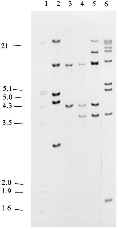
Analysis of genomic DNA showed that Salinatis strain N6247 contained five copies of IS200 on PvuII-generated fragments of ≥21, 7.5, 4.9, 4.5, and 3.0 kb (profile A; Fig. 2, lane 2). The four Sandiego strains also contained copies of IS200 and belonged to the three related profiles B, C, and D (Table 1). Profile B was the simplest; it showed PvuII fragments of 7.5 and 4.3 kb (Fig. 2, lane 3). Profile C contained the same fragments as profile B but had an additional band of 3.8 kb (Fig. 2, lane 4). Profile D contained the fragments of profile C and additional fragments of ≥21 and 11.5 kb (Fig. 2, lane 5). Of the 14 Duisburg strains examined, only 11.86 contained IS200—profile E showed nine PvuII fragments of ≥21, 19, 14, 11.5, 7.5, 5.4, 5.0, 3.8, and 1.7 kb (Fig. 2, lane 6).
FIG. 2.
IS200 profiles of Salmonella strains of serotypes Salinatis, Sandiego, and Duisburg digested with restriction enzyme PvuII. Lanes: 1, molecular size markers (sizes are given in kilobases on the left); 2, profile A of Salinatis N6247; 3 to 5, profiles B, C, and D of Sandiego strains 18K, CNS1.87, and CNS3.86, respectively; 6, profile E of Duisburg strain 11.86.
PFGE analysis.
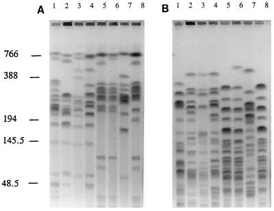
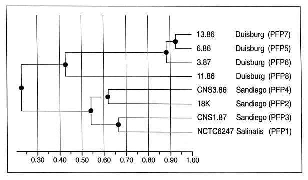
Eight PFPs, X1 to X8, generated after digestion of DNA with XbaI, were identified among the 19 salmonella strains examined: Salinatis strain N6247 had PFP X1; PFPs X2 to X4 were identified in Sandiego strains, and PFPs X5 to X8 were found in Duisburg strains (Table 1). PFP X1 of Salinatis strain N6247 was similar to PFPs X2 to X4 of Sandiego (Fig. 3A, lanes 2 to 5). Dice coefficients of similarity calculated for XbaI-generated PFPs indicated values of 0.50 to 0.67 when Salinatis PFP X1 was compared to PFPs X2 to X4 of Sandiego and 0.22 to 0.44 when Salinatis was compared to PFPs X5 to X8 of Duisburg (Table 3). A dendrogram based on the similarity coefficients of XbaI-generated PFPs showed that Salinatis and Sandiego strains formed one cluster with two branches, in each of which there were two strains (Fig. 4). Salinatis strain N6247 and Sandiego strain CNS 1.87 showed 67% similarity. The highest similarity value for Salinatis against Duisburg strains was 34%. Again, the Duisburg strains formed one cluster in which there were two branches. Comparison of Duisburg strains 13.86, 6.86, and 3.87 showed similarity values of about 90% (Fig. 4). However, Duisburg strain 11.86 was clearly an outlier, showing only 42 to 45% similarity to other Duisburg strains, 24 to 26% similarity to Sandiego strains, and 22% similarity to Salinatis (Table 1; Fig. 4).
FIG. 3.
PFPs of strains of Salmonella serotypes Salinatis, Sandiego, and Duisburg digested with restriction enzyme XbaI (A) or SpeI (B). Lanes: 1, lambda DNA concatemers (molecular sizes are given in kilobases on the left); 2, Salinatis strain N6247; 3 to 5, Sandiego strains 18K, CNS1.87, and CNS3.86, respectively; 6 to 9, Duisburg strains 6.86, 3.87, 11.86, and 13.86, respectively.
TABLE 3.
Dice coefficients of similarity of PFPs of Salmonella strains of serotypes Salinatis, Sandiego, and Duisburg
| Serotype | PFP | Strain | Dice coefficienta
|
|||||||
|---|---|---|---|---|---|---|---|---|---|---|
| N6247 | 18K | CNS1.87 | CNS3.86 | 6.86 | 3.87 | 13.86 | 11.86 | |||
| Salinatis | X1 | N6247 | 1.00 | |||||||
| Sandiego | X2 | 18K | 0.50 | 1.00 | ||||||
| Sandiego | X3 | CNS1.87 | 0.67 | 0.50 | 1.00 | |||||
| Sandiego | X4 | CNS3.86 | 0.58 | 0.62 | 0.59 | 1.00 | ||||
| Duisburg | X5 | 6.86 | 0.32 | 0.21 | 0.30 | 0.14 | 1.00 | |||
| Duisburg | X6 | 3.87 | 0.34 | 0.22 | 0.32 | 0.15 | 0.92 | 1.00 | ||
| Duisburg | X7 | 13.86 | 0.26 | 0.14 | 0.22 | 0.07 | 0.93 | 0.85 | 1.00 | |
| Duisburg | X8 | 11.86 | 0.22 | 0.24 | 0.26 | 0.25 | 0.42 | 0.45 | 0.42 | 1.00 |
| Salinatis | S1 | N6247 | 1.00 | |||||||
| Sandiego | S2 | 18K | 0.54 | 1.00 | ||||||
| Sandiego | S3 | CNS1.87 | 0.59 | 0.70 | 1.00 | |||||
| Sandiego | S4 | CNS3.86 | 0.54 | 0.60 | 0.49 | 1.00 | ||||
| Duisburg | S5 | 6.86 | 0.25 | 0.23 | 0.25 | 0.23 | 1.00 | |||
| Duisburg | S6 | 3.87 | 0.26 | 0.24 | 0.26 | 0.24 | 0.93 | 1.00 | ||
| Duisburg | S5 | 13.86 | 0.25 | 0.23 | 0.25 | 0.23 | 1.00 | 0.93 | 1.00 | |
| Duisburg | S7 | 11.86 | 0.12 | 0.16 | 0.18 | 0.16 | 0.25 | 0.21 | 0.25 | 1.00 |
Calculated from digests with XbaI (top) and SpeI (bottom).
FIG. 4.
Dendrogram based on similarity coefficients from XbaI-generated PFPs of strains of serotypes Salinatis, Sandiego, and Duisburg. The dendrogram was constructed by using the unweighted pair-group average method (Phoretix).
In further experiments, DNAs of eight strains chosen to represent XbaI-generated PFPs X1 to X8 were digested with SpeI. Strains that showed similar profiles with XbaI were also seen to be closely related when their DNAs were digested with SpeI (Fig. 3B). The seven PFPs obtained (S1 to S7) were analyzed by their Dice coefficients and dendrogram, and the above relationships suggested by XbaI analysis were confirmed (Table 3). Thus, Dice coefficients of similarity calculated for PFPs S1 to S7 indicated values of 0.54 to 0.59 when Salinatis PFP S1 was compared to PFPs S2 to S4 of Sandiego, and the corresponding values were 0.12 to 0.26 when Salinatis was compared to PFPs S5 to S7 of Duisburg (Table 3). Digestion of the DNAs of Sandiego strains with SpeI yielded PFPs that were even more similar than those obtained after digestion with XbaI, and this finding was true also for Duisburg strains. For example, with strains 6.86 and 13.86, the Dice value obtained by comparison of PFPs X5 and X7 was 0.93, but with SpeI it was 1.0, the latter result indicating that they were indistinguishable (Table 3). Thus, whereas conjoint use of biotyping, ribotyping, and IS200 profiling indicated that strain 11.86 is different from the other Duisburg strains, PFGE delineated three (SpeI) or four (XbaI) groupings among these same strains.
DISCUSSION
The Kauffmann-White serotyping scheme is the traditional method for primary characterization of salmonellae in the diagnostic setting (18). While valuable for diagnosis and epidemiology, it is considered to be inappropriate for phylogenetic purposes (4). Thus, it is necessary to explore a range of other techniques to establish genetic relationships among salmonella serotypes. In this study, biotyping provided the first evidence that Salinatis and Sandiego might be related because strains of these serotypes belonged to the same biotype (10di), which differed from that of most Duisburg strains (93%) in two biotype characters.
Although IS200 was present in Salinatis and Sandiego, fingerprints indicated differences in both location and copy number consistent with the idea of independent acquisition of IS200 by these two serotypes. The finding that only one Duisburg strain was IS200 positive confirmed that the distribution of IS200 within a salmonella serotype may be random (7, 9, 15, 27). However, in this study, the general absence of IS200 from Duisburg strains meant that IS200 fingerprinting was unhelpful for analysis of these serotypes.
Ribotyping characterizes strains on the basis of the chromosomal location and copy number of their rRNA genes, conserved sequences representing only a small proportion of the entire genome. The ribotype is stable and has been useful for long-term epidemiological studies of Salmonella (7–9). With some serotypes of Salmonella, ribotyping can be used for discrimination to the strain level; with bacteria other than Salmonella, it has indicated broader groupings that require further subdivision by other typing methods (31). In the present study, ribotyping supported the preliminary findings suggested by biotyping and indicated that the Salinatis-Sandiego relationship is probably closer than that of Salinatis and Duisburg.
Because PFGE profiles of strains reflect differences present over the entire chromosome, the information they provide differs from, and yet complements, that obtained from ribotyping. However, the diversity of types revealed by PFGE among, for example, strains of Helicobacter pylori (31)—when it is used as a primary means of strain discrimination—should not be expected to be the same when it is used for salmonellae, which have already been classified to the subspecies and serotype levels by biochemical and serological tests, respectively (18). The effectiveness of PFGE as a tool for strain discrimination in salmonellae will depend on intraserotype genetic diversity (3, 22, 33, 34) and, perhaps more importantly, on the extent to which members of a serotype have already been discriminated—for example, for strains of common serotypes such as Typhimurium or Enteritidis—by phage typing (1, 6, 35), biotyping (12), or a combination of these methods (20, 24).
Our own experience of PFGE has encouraged us to believe that it is helpful for the investigation of clonal relationships between and within serotypes. In this study, eight and seven PFPs were identified with XbaI and SpeI, respectively. Based on previously proposed guidelines (32) and regardless of the restriction enzyme used, calculations of similarity and dendrogram analysis indicated a close relationship between serotypes Salinatis and Sandiego. Both phenotypic and genotypic methods indicated that strain 11.86 was an outlier among Duisburg strains; nevertheless, it was more closely related to other Duisburg strains than to strains of Salinatis or Sandiego.
Multilocus enzyme electrophoresis has been used successfully to investigate population structure in Salmonella and has demonstrated the existence of monophyletic and polyphyletic serotypes (4, 20). An examination of strains of serotypes Salinatis, Duisburg, and Sandiego by this technique, which is expensive and not generally available, would probably be helpful in confirming relationships among them.
The loss of a large plasmid carrying H-d flagellin genes (30) by the triphasic serotype Salinatis would, of course, yield a diphasic serotype Sandiego and so account for the close phenotypic similarity long recognized between these serotypes (13). The close genetic relationship shown between serotypes Salinatis and Sandiego in this study of a few strains suggests that Salinatis should be designated a triphasic variant of Sandiego in future publications of names of salmonella serotypes. Attempts to obtain other strains of Salinatis with which to test our hypothesis have not been successful.
ACKNOWLEDGMENTS
We thank J. P. Duguid and L. Le Minor for gifts of strains and H. Mather for confirmation of their serotypes.
REFERENCES
- 1.Anderson E S, Ward L R, de Saxe M J, de Sa J D H. Bacteriophage-typing designations of Salmonella typhimurium. J Hyg. 1977;78:297–300. doi: 10.1017/s0022172400056187. [DOI] [PMC free article] [PubMed] [Google Scholar]
- 2.Anonymous. Salmonella serotypes recorded in PHLS Salmonella data sets. Communicable Disease Report (CDR Weekly Reports). London, England: Public Health Laboratory Service; 1998. [Google Scholar]
- 3.Baquar N, Burnens A, Stanley J. Comparative evaluation of molecular typing of strains from a national epidemic due to Salmonella brandenburg by rRNA gene and IS200 probes and pulsed-field gel electrophoresis. J Clin Microbiol. 1994;32:1876–1880. doi: 10.1128/jcm.32.8.1876-1880.1994. [DOI] [PMC free article] [PubMed] [Google Scholar]
- 4.Beltran P, Musser J M, Helmuth R, Farmer III J J, Frerichs W M, Wachsmuth I K, Ferris K, McWhorter A C, Wells J G, Cravioto A, Selander R K. Toward a population genetic analysis of Salmonella: genetic diversity and relationships among strains of serotypes S. choleraesuis, S. derby, S. dublin, S. enteritidis, S. heidelberg, S. infantis, S. newport, and S. typhimurium. Proc Natl Acad Sci USA. 1988;85:7753–7757. doi: 10.1073/pnas.85.20.7753. [DOI] [PMC free article] [PubMed] [Google Scholar]
- 5.Burnens A P, Stanley J, Sechter I, Nicolet J. Evolutionary origin of a monophasic Salmonella serovar, 9,12:1,v:−, revealed by IS200 profiles and restriction fragment polymorphisms of the fljB gene. J Clin Microbiol. 1996;34:1641–1645. doi: 10.1128/jcm.34.7.1641-1645.1996. [DOI] [PMC free article] [PubMed] [Google Scholar]
- 6.Callow B R. A new phage typing scheme for Salmonella typhimurium. J Hyg. 1959;57:346–359. doi: 10.1017/s0022172400020209. [DOI] [PMC free article] [PubMed] [Google Scholar]
- 7.Chisholm S A, Crichton P B, Knight H I, Old D C. Molecular typing of Salmonella serotype Thompson strains isolated from human and animal sources. Epidemiol Infect. 1999;122:33–39. doi: 10.1017/s0950268898001836. [DOI] [PMC free article] [PubMed] [Google Scholar]
- 8.Crichton P B, Old D C, Taylor A, Rankin S C. Characterisation of strains of Salmonella serotype Livingstone by multiple typing. J Med Microbiol. 1996;44:325–331. doi: 10.1099/00222615-44-5-325. [DOI] [PubMed] [Google Scholar]
- 9.Crichton P B, Old D C, Morphy H F. Distribution of insertion sequence IS200 among different clonal lines of the related Salmonella serotypes Livingstone and Eimsbuettel. J Med Microbiol. 1998;47:791–797. doi: 10.1099/00222615-47-9-791. [DOI] [PubMed] [Google Scholar]
- 10.Dice L R. Measures of the amount of ecological association between species. Ecology. 1945;26:297–302. [Google Scholar]
- 11.Douglas G W, Edwards P R. Complex flagellar phases in Salmonella. J Gen Microbiol. 1962;29:367–372. [Google Scholar]
- 12.Duguid J P, Anderson E S, Alfredsson G A, Barker R, Old D C. A new biotyping scheme for Salmonella typhimurium and its phylogenetic significance. J Med Microbiol. 1975;8:149–166. doi: 10.1099/00222615-8-1-149. [DOI] [PubMed] [Google Scholar]
- 13.Edwards P R, Bruner D W. A description of an unusual Salmonella type with specific reference to the evolution of Salmonella species. J Bacteriol. 1942;44:289–300. doi: 10.1128/jb.44.3.289-300.1942. [DOI] [PMC free article] [PubMed] [Google Scholar]
- 14.Edwards P R, Kauffmann F, Huey C R. An unusual Salmonella type (Salmonella montgomery) 11:d,a:d,e,n,z15. Acta Pathol Microbiol Scand. 1957;41:517–520. doi: 10.1111/j.1699-0463.1957.tb01718.x. [DOI] [PubMed] [Google Scholar]
- 15.Ezquerra E, Burnens A P, Frith K, Costas M, Stanley J. Molecular genotype analysis of Salmonella bovismorbificans. Mol Cell Probes. 1993;7:45–54. doi: 10.1006/mcpr.1993.1006. [DOI] [PubMed] [Google Scholar]
- 16.Kelterborn E. Salmonella species. First isolations, names and occurrences. 1967. pp. 131–132. , 332–333. Dr. W. Junk, The Hague, The Netherlands. [Google Scholar]
- 17.Le Minor L, Le Minor S. Origine et répartition en sérotypes des souches isolées en France et reçues au Centre national des Salmonella de 1977 à 1979. Rev Epidemiol Sante Publique. 1981;29:45–55. [PubMed] [Google Scholar]
- 18.Le Minor L, Popoff M Y. Antigenic formulas of the Salmonella serovars, 6th revision. Paris, France: WHO Collaborating Center for Reference and Research on Salmonella, Institut Pasteur; 1992. pp. 1–145. [Google Scholar]
- 19.Le Minor L, Le Minor S, Grimont P A D. Rapport quadriennal du Centre national des Salmonella sur l’origine et la répartition en sérotypes des souches isolées en France continentale au cours des années 1980 à 1983. Rev Epidemiol Sante Publique. 1985;33:13–21. [PubMed] [Google Scholar]
- 20.Li J, Smith N H, Nelson K, Crichton P B, Old D C, Whittam T S, Selander R K. Evolutionary origin and radiation of the avian-adapted non-motile salmonellae. J Med Microbiol. 1993;38:129–139. doi: 10.1099/00222615-38-2-129. [DOI] [PubMed] [Google Scholar]
- 21.Miller J H. Experiments in molecular genetics. Cold Spring Harbor, N.Y: Cold Spring Harbor Laboratory; 1972. p. 433. [Google Scholar]
- 22.Murase T, Okitsu T, Suzuki R, Morozumi H, Matsushima A, Nakamura A, Yamani S. Evaluation of DNA fingerprinting by PFGE as an epidemiologic tool for Salmonella infections. Microbiol Immunol. 1995;39:673–676. doi: 10.1111/j.1348-0421.1995.tb03255.x. [DOI] [PubMed] [Google Scholar]
- 23.Old D C. Nomenclature of Salmonella. J Med Microbiol. 1992;37:361–363. doi: 10.1099/00222615-37-6-361. [DOI] [PubMed] [Google Scholar]
- 24.Old D C, Barker R M. Persistent and transient clones of Salmonella typhimurium of phage type 141 recognized by biotyping. Epidemiol Infect. 1989;102:113–118. doi: 10.1017/s0950268800029745. [DOI] [PMC free article] [PubMed] [Google Scholar]
- 25.Old D C, Threlfall E J. Salmonella. In: Collier L, Balows A, Sussman M, editors. Topley and Wilson’s Microbiology and Microbial Infections. 9th ed. Vol. 2. London, England: Edward Arnold; 1998. pp. 969–997. [Google Scholar]
- 26.Olsen J E, Skov M N, Threlfall E J, Brown D J. Clonal lines of Salmonella enterica serotype Enteritidis documented by IS200-, ribo-, pulsed-field gel electrophoresis and RFLP typing. J Med Microbiol. 1994;40:15–22. doi: 10.1099/00222615-40-1-15. [DOI] [PubMed] [Google Scholar]
- 27.Pelkonen S, Romppanen E-L, Siitonen A, Pelkonen J. Differentiation of Salmonella serovar infantis isolates from human and animal sources by fingerprinting IS200 and 16S rrn loci. J Clin Microbiol. 1994;32:2128–2133. doi: 10.1128/jcm.32.9.2128-2133.1994. [DOI] [PMC free article] [PubMed] [Google Scholar]
- 28.Sambrook J, Fritsch E F, Maniatis T. Molecular cloning: a laboratory manual. 2nd ed. Vol. 3. Cold Spring Harbor, N.Y: Cold Spring Harbor Laboratory Press; 1989. p. B.13. [Google Scholar]
- 29.Silvermann M, Zieg J, Hilmen M, Simon M. Phase variation in Salmonella: genetic analysis of a recombination switch. Proc Natl Acad Sci USA. 1979;76:391–395. doi: 10.1073/pnas.76.1.391. [DOI] [PMC free article] [PubMed] [Google Scholar]
- 30.Smith N H, Selander R K. Molecular genetic basis for complex flagellar antigen expression in a triphasic serovar of Salmonella. Proc Natl Acad Sci USA. 1991;88:956–960. doi: 10.1073/pnas.88.3.956. [DOI] [PMC free article] [PubMed] [Google Scholar]
- 31.Tee W, Lambert J, Smallwood R, Schembri M, Ross B C, Dwyer B. Ribotyping of Helicobacter pylori from clinical specimens. J Clin Microbiol. 1992;30:1562–1567. doi: 10.1128/jcm.30.6.1562-1567.1992. [DOI] [PMC free article] [PubMed] [Google Scholar]
- 32.Tenover F C, Arbeit R D, Goering R V, Mickelsen P A, Murray B E, Persing D H, Swaminathan B. Interpreting chromosomal DNA restriction patterns produced by pulsed-field gel electrophoresis: criteria for bacterial strain typing. J Clin Microbiol. 1995;33:2233–2239. doi: 10.1128/jcm.33.9.2233-2239.1995. [DOI] [PMC free article] [PubMed] [Google Scholar]
- 33.Thong K-L, Cheong Y-M, Puthucheary S, Koh C-L, Pang T. Epidemiologic analysis of sporadic Salmonella typhi isolates and those from outbreaks by pulsed-field gel electrophoresis. J Clin Microbiol. 1994;32:1135–1141. doi: 10.1128/jcm.32.5.1135-1141.1994. [DOI] [PMC free article] [PubMed] [Google Scholar]
- 34.Thong K-L, Ngeow Y-F, Altwegg M, Navaratnam P, Pang T. Molecular analysis of Salmonella enteritidis by pulsed-field gel electrophoresis and ribotyping. J Clin Microbiol. 1995;33:1070–1074. doi: 10.1128/jcm.33.5.1070-1074.1995. [DOI] [PMC free article] [PubMed] [Google Scholar]
- 35.Ward L R, de Sa J D H, Rowe B. A phage-typing scheme for Salmonella enteritidis. Epidemiol Infect. 1987;99:291–304. doi: 10.1017/s0950268800067765. [DOI] [PMC free article] [PubMed] [Google Scholar]
- 36.Wilson K. Preparation of genomic DNA from bacteria. In: Ausubel F M, Brent R, Kingston R E, Moore D D, Smith J A, Seidman J A, Strehl K, editors. Current protocols in molecular biology 1987–1988. Vol. 1. New York, N.Y: Wiley; 1987. pp. 2.4.1–2.4.5. [DOI] [PubMed] [Google Scholar]


