Dear Editor,
Choroidal melanoma is a rare malignancy, and in one study from the United Kingdom, up to 30% of patients referred by the general ophthalmologist to an ocular oncology center were misdiagnosed.[1] Later, the mnemonic “To find small ocular melanoma using helpful hints daily”[2] was established to aid in the detection of this malignancy at an early point. With the rapid advancements in ocular imaging technology, quantifiable and objective tests are becoming more ubiquitous in ophthalmology. In glaucoma, standardized, automated, quantifiable, and objective optical coherence tomography (OCT) peripapillary and macular diagnostic parameters have been shown to have reliable glaucoma diagnostic capabilities[3,4] and are routinely used to diagnose glaucoma and detect progression. More recently, optical coherence tomography angiography (OCTA) parameters in vascular density have been found to be comparable to or even better than those of OCT parameters at glaucoma diagnosis.[5] Regarding uveal melanoma, Valverde-Megías et al. found that eyes with melanoma, compared with the contralateral eye, showed enlarged foveal avascular zone (FAZ) and reduced capillary vascular density (CVD).[6] Herein, we present a pilot exploration of the diagnostic capability of OCTA macular vascular parameters for choroidal melanoma.
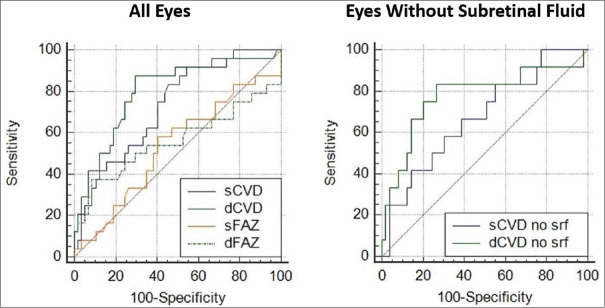
We analyzed OCTA images from the report published by Valverde-Megías et al.[6] with stricter screening criteria for OCTA scan quality. OCTA measurements of superficial (sCVD) and deep (dCVD) CVD and superficial (sFAZ) and deep (dFAZ) FAZ area were obtained. Receiver operating characteristic curves (area under the curve [AUC]) were calculated with a method proposed by DeLong et al.[7] for CVD and FAZ parameters to discriminate eyes with choroidal melanoma from nevus. Statistical significance was defined as value of P < 0.05. Statistical analyses were performed using MedCalc (MedCalc Software version 15.4, Ostend, Belgium). After excluding OCTA scans with suboptimal image quality and significant artifacts, we included 57 eyes with choroidal nevus (57 patients) and 24 eyes with choroidal melanoma (24 patients). Patient demographics and ocular features are listed [Table 1]. CVD was found to be a fair[8] choroidal melanoma diagnostic parameter (sCVD AUC 0.73 and dCVD AUC 0.80) [Table 1 and Figure 1]. FAZ was found to be a failed[8] choroidal melanoma diagnosis parameter (sFAZ AUC 0.53 and dFAZ AUC 0.56) [Table 1 and Figure 1]. The AUCs of CVD parameter was greater than that of respective FAZ parameter for both superficial (sCVD AUC 0.73 vs. sFAZ AUC 0.53; P = 0.03) and deep (dCVD AUC 0.80 vs. dFAZ AUC 0.56; P = 0.01) layers [Table 1 and Figure 1]. The parameter with the greatest AUC was dCVD (AUC = 0.80), reaching significance when compared to AUCs of sFAZ and dFAZ (0.53 and 0.56, respectively; both P < 0.02) [Table 1 and Figure 1]. Even in eyes without subretinal fluid, the dCVD parameter still has a fair choroidal melanoma diagnostic capability (AUC = 0.78) [Table 1 and Figure 1].
Table 1.
Optical Coherence Tomography Angiography for Unilateral Choroidal Nevus and Melanoma: Demographics and Ocular Features of Patients and Areas Under the Receiver Operating Characteristic Curve (AUCs) of Macular Vascular Parameters of Capillary Vascular Density and Foveal Avascular Zone to Detect Choroidal Melanoma
| Demographics and Ocular Features | Eye With Choroidal Nevus, N=57 Eyes of 57 Patients | Eye With Choroidal Melanoma, N=24 Eyes of 24 Patients | P |
|---|---|---|---|
| Age, mean (median; SD; range) | 51.7 (55; 15.8; 13-85) | 52.9 (48; 16.2; 25-77) | 0.41† |
| Sex (n=patients) n (%) | 0.27* | ||
| Female | 36 (63) | 12 (50) | |
| Male | 21 (37) | 12 (50) | |
| Involved eye (n=eyes) n (%) | 0.03* | ||
| Right | 25 (44) | 17 (71) | |
| 1eft | 32 (56) | 7 (29) | |
| Tumor location (n = eyes) n (%) | 0.92* | ||
| Macula | 26 (46) | 9 (38) | |
| Superior | 8 (14) | 5 (21) | |
| Temporal | 5 (9) | 2 (8) | |
| Inferior | 6 (11) | 2 (8) | |
| Nasal | 12 (21) | 6 (25) | |
| Thickness, mean (median; SD; range), (n=tumors), mm | 1.4 (1.4; 0.7; 0-2.4) | 4.65 (3.75; 2.6; 2-12) | <0.01† |
| Subretinal fluid in macular area (n=eyes) n (%) | 5 (9) | 12 (50) | <0.01* |
| Tumor proximity to optic disc, mean (median; SD; range) (n=tumors), mm | 2.5 (2; 2.2; 0-8) | 3.2 (2.8; 3.3; 0-12) | 0.10† |
| Tumor proximity to foveola, mean (median; SD; range) (n=tumors), mm | 2.2 (2; 2.1; 0-11) | 3.3 (2; 4.0; 0-14) | 0.06† |
| AUCs of Macular Vascular Parameters of Capillary | Superficial layer | Deep layer | |
| CVD, AUC (95% confidence interval) | 0.73 (0.62-0.82) | 0.80 (0.70-0.88)Ω | 0.12Σ |
| FAZ, AUC (95% confidence interval) | 0.53 (0.42-0.64) | 0.56 (0.45-0.67) | 0.5Σ |
| p-value | 0.03Ψ | 0.01Ψ |
*Chi-square analysis. †Student’s t-test. AUC indicates area under the curve; SD, standard deviation; CVD, capillary vascular density; FAZ, foveal avascular zone. ΩThe parameter and layer combination (deep CVD) with the greatest AUC, reached statistical significance when compared against AUCs of superficial FAZ and deep FAZ (p-value <0.01 and p-value<0.02, respectively). Stratification to only eyes without subretinal fluid the AUC of deep CVD was 0.78. ΨComparison by macular vasculature parameters (CVD vs. FAZ). ΣComparison by layers (superficial vs. deep).
Figure 1.
Melanoma diagnostic capability of macular vascular parameters: receiver operating characteristic curves comparing the AUCs (a) of CVD and FAZ (AUCs: 0.73 for sCVD, 0.80 for dCVD, 0.53 for sFAZ, and 0.56 for dFAZ) and (b) of CVD in eyes without subretinal fluid (AUCs: 0.67 for sCVD no srf and 0.78 for dCVD no srf). AUC indicates area under the receiver operating characteristic curve; sCVD, superficial capillary vascular density; dCVD, deep capillary vascular density; sFAZ, superficial foveal avascular zone; dFAZ, deep foveal avascular zone; srf, subretinal fluid
In this pilot exploration of quantitative and objective OCTA parameters for choroidal melanoma diagnosis, our results demonstrated that the dCVD parameter had a fair capability to discriminate choroidal melanoma from choroidal nevus. Choroidal melanomas have a profound need for intrinsic vascular supply, and we speculate that a relatively ischemic microenvironment could be created by the tumor to promote tumor vascularization by vascular endothelial growth factor A. The ischemic microenvironment is likely most substantial in the deeper layers of the retina, adjacent to the tumor. This exploratory report included eyes with tumors of various locations and sizes/thicknesses. Therefore, we speculate that the true diagnostic capability of CVD could be greater than presented in our study, especially for macular or paramacular tumors. Further investigation of OCTA parameters for choroidal melanoma diagnosis is warranted with a larger sample, allowing stratification of tumor based on location and size/thickness. A normalized database for the CVD “expected” for a nevus of a particular location and size can then be established, and a decrease in CVD outside of the normal range might be suggestive of melanoma. We believe this would be of interest to your readership as it poses the possibility of developing standardized, automated, quantifiable and objective screening OCTA parameters for commercially available OCTA devices, similar to those available for glaucoma, and serve as an additional tool to the “To find small ocular melanoma using helpful hints daily” mnemonic.
Sincerely,
Jason L. Chien, M. D.
Alicia Valverde-Megías, M. D.
Gwo-farn Chien, M. D.
Carol L. Shields, M. D.
Financial support and sponsorship
Eye Tumor Research Foundation, Philadelphia, PA (CLS). The funders had no role in the design and conduct of the study, in the collection, analysis, and interpretation of the data, and in the preparation, review or approval of the manuscript. Carol L. Shields, M.D. has had full access to all the data in the study and takes responsibility for the integrity of the data and the accuracy of the data analysis.
Conflicts of interest
The authors declare that there are no conflicts of interest of this paper.
References
- 1.Khan J, Damato BE. Accuracy of choroidal melanoma diagnosis by general ophthalmologists: A prospective study. Eye (Lond) 2007;21:595–7. doi: 10.1038/sj.eye.6702276. [DOI] [PubMed] [Google Scholar]
- 2.Shields CL, Furuta M, Berman EL, Zahler JD, Hoberman DM, Dinh DH, et al. Choroidal nevus transformation into melanoma: Analysis of 2514 consecutive cases. Arch Ophthalmol. 2009;127:981–7. doi: 10.1001/archophthalmol.2009.151. [DOI] [PubMed] [Google Scholar]
- 3.Chien JL, Ghassibi MP, Patthanathamrongkasem T, Abumasmah R, Rosman MS, Skaat A, et al. Glaucoma diagnostic capability of global and regional measurements of isolated ganglion cell layer and inner plexiform layer. J Glaucoma. 2017;26:208–15. doi: 10.1097/IJG.0000000000000572. [DOI] [PubMed] [Google Scholar]
- 4.Ghassibi MP, Chien JL, Patthanathamrongkasem T, Abumasmah RK, Rosman MS, Skaat A, et al. Glaucoma diagnostic capability of circumpapillary retinal nerve fiber layer thickness in circle scans with different diameters. J Glaucoma. 2017;26:335–42. doi: 10.1097/IJG.0000000000000610. [DOI] [PubMed] [Google Scholar]
- 5.Moghimi S, Hou H, Rao H, Weinreb RN. Optical coherence tomography angiography and glaucoma: A brief review. Asia Pac J Ophthalmol (Phila) 2019;8:115–25. doi: 10.22608/APO.201914. [DOI] [PubMed] [Google Scholar]
- 6.Valverde-Megías A, Say EA, Ferenczy SR, Shields CL. Differential macular features on optical coherence tomography angiography in eyes with choroidal nevus and melanoma. Retina. 2017;37:731–40. doi: 10.1097/IAE.0000000000001233. [DOI] [PubMed] [Google Scholar]
- 7.DeLong ER, DeLong DM, Clarke-Pearson DL. Comparing the areas under two or more correlated receiver operating characteristic curves: A nonparametric approach. Biometrics. 1988;44:837–45. [PubMed] [Google Scholar]
- 8.Tape TG. The Area under an ROC Curve. [Last accessed on 2020] Jan 01]. Available from: http://gim.unmc.edu/dxtests/roc3.htm .


