



Abstract
PAPP-A knock-out (KO) mice are a valuable model for investigating the effects of down-regulating localized insulin-like growth factor (IGF) action, which has been shown to extend lifespan and healthspan when the PAPP-A gene is globally deleted. Based on previous mouse models of brain-specific reduction in IGF signaling associated with longevity, we sought to generate brain-specific PAPP-A KO mice and determine effects on metabolism and lifespan. Mice with the PAPP-A gene floxed (fPAPP-A) were crossed with Nestin promoter-driven Cre recombinase transgenic mice. This cross-breeding of mice for Nestin-Cre and mice with other floxed target alleles has been used extensively to investigate brain-specific effects. Our cross-breeding generated four genotypes for study: fPAPP-A/Nestin positive (brain-specific PAPP-A KO); fPAPP-A/Nestin negative (Control for floxed PAPP-A); WT/Nestin positive (Control for Nestin-Cre); WT/Nestin negative (Wild-type Control).
The basic genotype screen of neonatal tail snip DNA clearly indicated PAPP-A gene status and the presence (pos) or absence (neg) of Nestin-Cre. We then determined tissue specificity of PAPP-A gene excision. We had expected fPAPP-A/pos mice to be relatively brain-specific for PAPP-A gene deletion and the controls (fPAPP-A/neg, WT/neg and WT/pos mice) to show no effect on PAPP-A expression in brain or other tissues. However, in fPAPP-A/neg mice we found evidence of PAPP-A excision in all tissues examined, i.e., in the presumed absence of Nestin-Cre, indicating germline recombination. We further found that fPAPP-A/pos mice showed near complete excision of the PAPP-A gene in brain, but some also showed germline recombination affecting all tissues tested.
To determine if the level of excision indicated by tissue genotyping approximated PAPP-A mRNA expression, we performed RT-qPCR. fPAPP-A/pos mice that showed markedly decreased whole brain PAPP-A mRNA expression (~80%), with little or no effect on expression in the other tissues tested, were designated as “brain-specific” PAPP-A KO. fPAPP-A/pos mice that showed germline recombination had similar decreases in PAPP-A expression in brain but also showed 40–65% decreased PAPP-A mRNA expression in other tissues as well, which was especially striking in kidney, tibia, thymus and spleen. These were designated as “non-specific” PAPP-A KO mice.
With unknown and unpredictable specificity until harvest, we chose to assess a surrogate marker of lifespan i.e., thymic involution, in 15- to 18-month-old fPAPP-A/pos and WT/pos mice, the latter an important control for a possible effect of Nestin-Cre per se. Diminished thymic involution as indicated by increased thymic weight (135%, P = 0.035) and decreased histological disruption was seen in “non-specific” PAPP-A KO mice, similar to what was previously reported in 18-month-old global PAPP-A KO mice. There was no significant difference between “brain-specific” PAPP-A KO and control mice.
This study highlights the importance of thorough characterization of assumed tissue-specific mouse models and awareness of potential germline recombination for proper data interpretation.
Keywords: PAPP-A, brain, Nestin-Cre, lifespan, thymus
1. Introduction
Reduction in insulin-like growth factor-I receptor (IGF-IR) signaling has been shown to extend lifespan in a variety of species from worms to mice1. There is also evidence that reduced IGF-IR signaling may contribute to healthy longevity in humans2–4. Mouse models that have genetic disruption of IGF-IR signaling components have been the most extensively studied. Genetic modification of upstream regulators of IGF expression, i.e., growth hormone (GH) and GH receptor (GHR), the IGF-IR itself, and downstream intracellular signaling mediators, such as insulin receptor substrate (IRS), produce mice with extended longevity compared to wild-type controls5. For the most part, these have been global homozygous or heterozygous knock-out (KO) mouse models. Tissue-specific KO mouse models have yielded little information about specific contributions to longevity, except perhaps for the brain. In worms and flies, IGF signals in the nervous system have been shown to determine lifespan in a non-cell autonomous manner6. In 16 rodent species, IGF-IR levels in brain, but not in other tissues, negatively correlated with longevity7. Kappeler et al.8 showed that lifespan can be significantly extended in mice by partial reduction of IGF-I signaling in the brain. Their data suggest that brain IGF-IR regulates GH and IGF-I secretion, which can have systemic effects on growth and energy metabolism in the periphery. On the other hand, tonic GH/IGF signaling can pose a risk for a shortened lifespan9. Reduction in brain-specific IRS-2, a major signal transduction molecule downstream of the IGF-IR (and insulin receptor), increased lifespan and altered nutrient homeostasis not involving GH. It was as effective as global IRS-2+/− in extending lifespan10. Of note, IRS-2−/− mice had decreased body growth and fatal diabetes11.
Reduction in IGF-IR signaling can also be achieved through inhibition of pregnancy-associated plasma protein-A (PAPP-A). PAPP-A is a novel zinc metalloprotease that can increase local IGF-I bioavailability through cleavage of inhibitory IGF binding proteins12. Inhibition of PAPP-A expression or its proteolytic activity represents an innovative approach to decreasing local IGF availability with moderate restraint of IGF-IR signaling. Moderate restraint is the key.
Homozygous deletion of IGF-IR in mice is peri-natal lethal13, whereas heterozygous IGF-IR KO mice have an extended lifespan14. And as noted above, IRS-2+/−, but not IRS-2−/−, mice have a longevity phenotype10. We have shown that inhibition of PAPP-A through gene deletion in mice has many beneficial effects, including a remarkable extension of lifespan by 30–40%, and a delayed onset and reduced severity of several age-related diseases in both male and female mice15. Of particular interest, PAPP-A KO mice show significantly delayed age-related thymic involution that likely plays a major role in extending lifespan16. Although these global PAPP-A KO mice are small, similar to other long-lived mice, e.g., Ames dwarf and GHR KO mice, rescue of the dwarf phenotype during development or conditional knock-down of PAPP-A in adult mice also showed lifespan extension17,18. Furthermore, these mice exhibit no secondary endocrine abnormalities (e.g., on GH secretion) and have normal circulating levels of IGF-I. Therefore, PAPP-A KO mice are a valuable model for investigating the effects of down-regulating localized IGF-I action.
As of yet, there are no tissue-specific PAPP-A KO mouse models. Based on the mouse models of brain-specific reduction in IGF signaling and effects on longevity discussed above, as well as the knowledge that PAPP-A is highly expressed in the mouse brain and that this expression increases with age19, we sought to generate brain-specific PAPP-A KO mice by crossing mice with the PAPP-A gene floxed with Nestin promoter-driven Cre recombinase (Nestin-Cre) transgenic mice. This cross-breeding scheme has been used extensively with Nestin-Cre mice and mice with other floxed target alleles to investigate brain-specific gene function. However, it turned out to be a lesson on the underappreciated and unpredictable impact of germline recombination on appropriate linking of alteration in genomic DNA to biological outcome, in this case brain-specific PAPP-A gene deletion in mice and lifespan.
2. Materials and Methods
Mice.
Male Nestin-Cre hemizygous transgenic mice (Nestin-Cre+/0; B6.Cg-Tg(Nes-Cre)1Kln/J purchased from Jackson Labs) were crossed with female homozygous loxP-flanked PAPP-A (PAPP-Aflox/flox; fPAPP-A) mice, previously established in our lab20. Effective recombination in brain is only obtained if the Nestin-Cre allele is paternally inherited8,21. This breeding yielded PAPP-Aflox/+:Nes+ and PAPP-Aflox/+:Nes0 mice. Male PAPP-Aflox/+:Nes+ were then mated to female PAPP-Aflox/+:Nes0 mice to get four genotypes for study:
PAPP-Aflox/flox: Nes+ (fPAPP-A/pos; brain-specific PAPP-A KO)
PAPP-Aflox/flox:Nes0 (fPAPP-A/neg; Control for floxed PAPP-A)
PAPP-A+/+:Nes+ (WT/pos; Control for Nes-Cre)
PAPP-A+/+:Nes0 (WT/neg; Wild-type Control)
PAPP-Aflox/flox mice are normal-sized and fertile. Nestin-Cre transgenic mice have mild hypopituitarism and growth retardation, but males were used for the breeding and did not impact the females’ ability to carry a litter to term. However, Nestin-Cre mice can have a metabolic phenotype, so it was necessary to include mice without the floxed-flanked target gene (WT/pos) to account for any effect of the Nestin-Cre, per se, on phenotype. Mice were housed as five males or five females per cage, with both controls and mutant genotypes in each cage. These cages were located in a pathogen-free mouse facility, and with ad libitum access to food and water.
All studies were approved by the Institutional Animal Care and Use Committee of Mayo Clinic.
Genotyping.
Mice were approximately 14 days old when tail snips were taken for genotyping. Primers recommended by Jackson Labs bind to the hGH sequence that is present in the Nestin-Cre transgene were: forward CCTTCCTGAAGCAGTAGAGCA, common GCCTTATTGTGGAAGGACTG, and reverse TTGCTAAAGCGCTACATAGGA. Primers for fPAPP-A were: forward GTCCCAGAGGCTCCAATAGTAGC and reverse GGAAGTTGTGATTCAGAGCACTGC. Once genotype was determined, mice were assigned a study group. Mice were re-genotyped at the end of the study to confirm the genotype at weaning. At harvest, specific tissues taken from fPAPP-A/pos mice were Proteinase K digested, purified to dsDNA and quantified via Nano Drop (Thermo Scientific). End-point PCR was used to determine efficiency of cre-excision. Primers used were: Lox 6417 TAGTTCCTCCAGCCTTTACCTTG, Lox 6683 ATTTGTCATACAGCCCCTATGT and Lox 9735 AAAATGCCATAAACTATAGGG. Thermocycler program used was: 950 for 5 minutes; then 950 for 20 sec; 540 for 30 sec for 35 cycles; then 720 for 7 min and hold at 40. The PCR product was separated on 2% agarose gels run at 100 V for 90 minutes.
RT-qPCR.
Mouse tissues were rapidly isolated and snap frozen in liquid nitrogen. Tissues were pulverized with mortar and pestle and the powdered product lysed and homogenized in Trizol (Life Technologies, Carlsbad, CA). Total RNA was isolated per manufacturer’s directions, reverse transcribed with the First-Strand Synthesis System (Life Technologies) and evaluated by real-time PCR using CFX Connect Real-Time PCR Detection System (Bio-Rad, Hercules, CA) with iTaq Universal SYBR Green Supermix (Bio-Rad). Amplification plots were analyzed with CFX Maestro Software version 4.1 (Bio-Rad). Primer sequences used for mouse PAPP-A were: forward GGATGGGTCATGGGCATTCA and reverse GAAAAAGTAGCGTGGATCTCTGT.
Aging study.
Male mice of each of the three genotypes (fPAPP-A/pos, WT/pos, WT/neg) were aged to 15- to 18-months. Mice were weighed and monitored weekly. Any signs of morbidity and imminent mortality (e.g., weight loss, lethargy, body composition score) cued euthanasia for humane reasons. At harvest, tissues were weighed and PAPP-A excision was assessed in brain, heart, skeletal muscle, kidney, thymus, spleen and tibia of fPAPP-A/pos mice to determine relative brain-specificity.
3. Results
The basic genotype screen of neonatal tail snip DNA clearly indicated PAPP-A gene status and the presence or absence of Nestin-Cre. We then determined tissue specificity of PAPP-A gene excision. We had expected fPAPP-A/pos mice to be relatively brain-specific for PAPP-A gene deletion and the controls (fPAPP-A/neg, WT/neg and WT/pos mice) to show no effect on PAPP-A expression in brain or other tissues. However, we were surprised to find patterns of PAPP-A excision in several fPAPP-A/neg mice in all tissues, i.e., in the presumed absence of Nestin-Cre (Fig. 1). Ubiquitous deletion of PAPP-A in offspring devoid of Cre recombinase indicates germline recombination mediated by Nestin-Cre22. Thus, we did not use fPAPP-A/neg mice as a negative control in this study.
Figure 1.
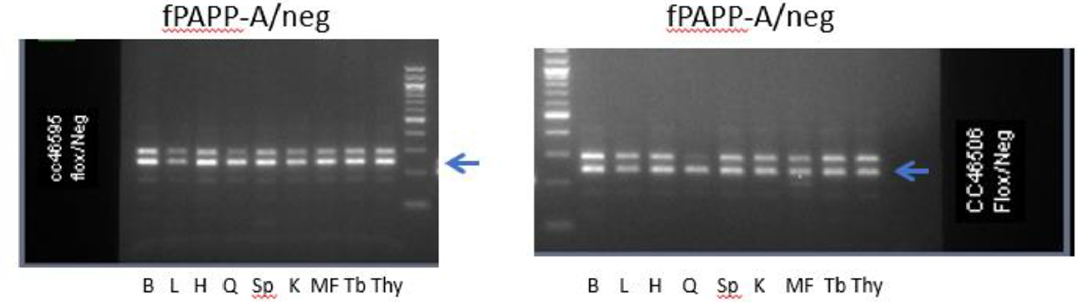
Genotyping of tissues from fPAPP-A/neg mice
B, brain; L, liver; H, heart; Q, quadriceps; Sp, spleen; K, kidney; MF, mesenteric fat; Tb; tibia; Thy, thymus
Arrow indicates the band for excised PAPP-A
If Cre expression and associated recombinase activity occur in germline cells in early embryonic life, then Cre-mediated excision of the floxed gene can occur in all cells instead of the intended tissue-specific pattern germline recombination22–25. We found that all fPAPP-A/pos mice showed near complete excision of the PAPP-A gene in brain, but many also showed germline recombination affecting all tissues tested (Fig. 2a and b). This occurred in both male and female offspring but was more prevalent in female fPAPP-A/pos mice. Mosaic recombination is indicated by the presence of both intact floxed and recombined product from one target allele (Fig. 2b). In several cases, there was global deletion of PAPP-A in tissues of fPAPP-A/pos mice (Fig. 2c). Of the mice initially tested, 9 of 16 male fPAPP-A/pos mice showed relative brain-specificity whereas only 2 of 12 female fPAPP-A/pos mice showed relative brain-specific PAPP-A gene deletion. Other tissues, especially kidney, had indications of some excision in brain-specific fPAPP-A/pos mice but not to the extent seen in brain.
Figure 2.

Genotyping of tissues from fPAPP-A/pos mice
(a) Example of brain-specific PAPP-A excision
(b) Example of non-specific PAPP-A excision – mosaic
(c) Example of non-specific PAPP-A excision -- complete
B, brain; L, liver; H, heart; Q, quadriceps; Sp, spleen; K, kidney; MF, mesenteric fat; Tb; tibia; Thy, thymus
Arrow indicates the band for excised PAPP-A
To determine if the level of excision indicated by tissue genotyping approximated PAPP-A mRNA expression, we performed RT-qPCR. “Brain-specific” fPAPP-A/pos mice showed markedly decreased (~80%) whole brain PAPP-A mRNA expression compared to Control mice (Table 1). There were no major decreases in PAPP-A expression in the other tissues evaluated (heart, skeletal muscle, tibia, thymus, spleen), although in the case of the kidney there was a 40% decrease in PAPP-A expression which fits with the extent of excision. fPAPP-A/pos mice that showed germline recombination resulting in a “non-specific” deletion pattern had similar decreases in PAPP-A expression in brain but also 40–65% decreased PAPP-A expression in other tissues as well. Difference in expression between designated “brain-specific” and “non-specific” fPAPP-A/pos mice was significant for tibia, thymus and spleen. There happened to be no fPAPP-A/pos mice with global deletion of PAPP-A in the group tested by RT-qPCR, but one could anticipate an even greater decrease of PAPP-A expression tissue-wide in these mice.
Table 1.
Tissue PAPP-A mRNA expression in fPAPP-A/pos mice (% of control)
| Brain-specific | Non-specific | |
|---|---|---|
| Brain | 15 ± 5.1 | 20 ± 8.3 |
| Heart | 68 ± 26.2 | 69 ± 23.0 |
| Quadriceps | 84 ± 21.8 | 50 ± 20.3 |
| Kidney | 59 ± 7.3 | 35 ± 11.4 |
| Tibia | 101 ± 5.1 | 34 ± 9.9* |
| Thymus | 116 ± 14.6 | 60 ± 18.0* |
| Spleen | 124 ± 25.5 | 49 ± 6.1* |
RT-qPCR results are expressed as mean ± SEM with three mice in each group (Brain-specific or Non-specific) with Control = 100.
P <0.05 Non-specific compared to Brain-specific
With an unpredictable genotype that would impact interpretation of any metabolic and longevity data, we chose to assess a surrogate marker of lifespan, thymic involution16,26,27, in fPAPP-A/pos and WT/pos mice, the latter an important control for an effect of Nestin-Cre per se. WT/neg mice were also included. Since our initial data seemed to indicate that male fPAPP-A/pos mice had a higher probability to be “brain-specific” than female mice, we chose to focus on the males. At 15–18 months of age, fPAPP-A/pos mice were analyzed for tissue specificity and thymic weight was determined for all groups of mice. Our prediction was that “non-specific” fPAPP-A/pos mice would show reduced involution, as seen in global PAPP-A KO mice16, that would be reflected in increased thymic weight relative to WT/pos. If “brain-specific” fPAPP-A/pos mice that showed no excision in immunologic tissues (thymus, spleen, tibia) also show reduced thymic involution, then we might infer an extended lifespan. Table 2 presents the data for thymus and brain weights relative to body weight of “brain-specific” fPAPP-A/pos, “non-specific” fPAPP-A/pos, WT/pos, and WT/neg mice. Over 50% of fPAPP-A/pos mice were “non-specific” with ~40% as mosaic and ~15% as global PAPP-A KOs. Only “non-specific” fPAPP-A/pos mice had a significant 135% increase in thymic weight compared to WT/pos mice. This increase in thymic weight was similar to the 125% (males) and 150% (females) increase seen in 18-month-old global PAPP-A KO versus WT mice (Table 3). There was no significant difference in brain weight relative to body weight in the 4 groups (Table 2). Similarly, there were no significant differences in weights of heart, quadriceps, kidney, tibia or spleen (data not shown).
Table 2.
Thymus and brain weights (ng/g Body weight)
| Thymus | Brain | |
|---|---|---|
| fPAPP-A/pos “brain-specific” (6) | 0.67 ± 0.049 | 1.46 ± 0.087 |
| fPAPP-A/pos “non-specific” (8) | 0.80 ± 0.049* | 1.64 ± 0.077 |
| WT/pos (13) | 0.59 ± 0.035 | 1.47 ± 0.052 |
| WT/neg (9) | 0.64 ± 0.073 | 1.35 ± 0.041 |
Male mice 15-to 18-months of age. Results are ± SEM of (n) mice.
P = 0.035
Table 3.
Thymus weights of 18-month-old WT and PAPP-A KO Mice (ng/g Body weight)
| WT | PAPP-A KO | P-value | |
|---|---|---|---|
| Males | 0.52 ± 0.023 | 0.65 ± 0.034 | 0.0130 |
| Females | 0.61 ±0.051 | 0.92 ± 0.052 | 0.0008 |
Data are from our frozen tissue repository. Results are mean ± SEM of 10–20 mice per group.
4. Discussion
PAPP-A regulates local IGF action, and global deletion of the PAPP-A gene in mice extends healthspan and lifespan12,15. Based on studies indicating that interruption of IGF signaling in the brain of mice has beneficial effects on metabolism and lifespan8,10, we sought to generate brain-specific PAPP-A KO mice by breeding loxP-flanked PAPP-A mice and Nestin-Cre recombinase transgenic mice to test the hypothesis that brain-specific PAPP-A KO mice have similar beneficial effects on metabolism and lifespan. If proven to be true, then brain PAPP-A could link neuroendocrine function to systemic aging and lifespan.
However, we failed to prove (or disprove) the hypothesis due to critical complications that can arise from the Cre-lox technology in general23–25 and the Nestin-Cre transgenic mouse model in particular22,25. Germline recombination and transient expression of Cre recombinase in the germline or during development can lead to expression in unanticipated sites, which often goes undetected using conventional genotyping methods23. Luo et al. reported on the prevalence of unexpected germline recombination in Cre driver lines designed for nervous system-specific recombination25. In this review, both Nestin-Cre transgenic lines reported germline recombination with frequencies ranging from 12.5% to 79% depending on the specific target locus. In our study, the frequency of “non-specific” mosaic recombination was ~50% for fPAPP-A/pos males and ~75% for fPAPP-A/pos females. Ubiquitous excision of the PAPP-A gene occurred in 14% of “non-specific” fPAPP-A/pos male mice and 50% in female mice. Importantly, PAPP-A gene expression reflected the extent of excision in the different tissues. Germline deletion renders the genotype of individual offspring to be unpredictable, and phenotype analysis could be seriously flawed by lack of a tissue-specific expression pattern. Phenotype of a fPAPP-A/pos mouse with hidden germline deletion of even one PAPP-A gene allele might be minimally different from that of a complete PAPP-A KO mouse, which would confound interpretation of brain PAPP-A on lifespan. Also, there could be off target effects on metabolism given PAPP-A excision in skeletal muscle and fat. Global PAPP-A KO mice are resistant to age-related thymic involution and maintain immune competence16. Delay in age-related thymic involution was also seen with other pro-longevity mechanisms26,27. We postulated that this thymic phenotype is involved in the extended lifespan of PAPP-A KO mice. Therefore, we used thymic weight as a surrogate marker of longevity in this study. We found that aged “non-specific” fPAPP-A/pos mice had significantly greater thymic weight compared to the WT/pos (and WT/neg) mice. However, “brain-specific” fPAPP-A/pos mice (complete excision in brain, with no effect on immune system tissues – thymus, spleen, tibia) did not have a significant difference in thymic weight compared to WT/pos mice. Since actual lifespan was not measured, we cannot rule out that there could be an effect on longevity. But it is clear that “non-specific” fPAPP-A/pos mice have a similar increase in thymic weight as in the global PAPP-A KO mice previously shown to be resistant to thymic atrophy16(Table 3). We used males in the current study because the frequency of germline transmission in fPAPP-A/pos mice was lower than in females. However, in 18-month-old female PAPP-A KO mice there was an even greater increase in thymic weight compared to WT (Table 3).
5. Conclusion
Unfortunately, we were unable to get a robust brain-specific PAPP-A KO mouse model with the Nestin-Cre approach. With the publication of these findings we hope to heighten awareness of potential germline recombination in Cre driver lines designed to be tissue-specific that could lead to misinterpretation of experimental data. This is in line with the directive to “highlight negative results to improve science”28,29.
Highlights.
Aim: To generate brain-specific PAPP-A KO mice and determine effect on lifespan
Floxed PAPP-A mice were crossbred with Nestin-Cre recombinase transgenic mice
Found germline recombination resulting in PAPP-A gene excision in tissues other than the brain
>50% of “brain-specific” PAPP-A KO mice had reduced expression in multiple tissues
Acknowledgements:
The authors would like to thank Hanne Lucier for her help in formatting this manuscript.
Funding:
This work was supported by the National Institutes of Health [R03AG061250].
Footnotes
Publisher's Disclaimer: This is a PDF file of an unedited manuscript that has been accepted for publication. As a service to our customers we are providing this early version of the manuscript. The manuscript will undergo copyediting, typesetting, and review of the resulting proof before it is published in its final form. Please note that during the production process errors may be discovered which could affect the content, and all legal disclaimers that apply to the journal pertain.
Declarations of interest: None
References
- 1.Kenyon C The plasticity of aging: insights from long-lived mutants. Cell. 2005.120(4):449–60. doi: 10.1016/j.cell.2005.02.002. [DOI] [PubMed] [Google Scholar]
- 2.Tazearslan C, Huang J, Barzilai N, Suh Y. Impaired IGF1R signaling in cells expressing longevity-associated human IGF1R alleles. Aging Cell. 2011.10(3):551–4. doi: 10.1111/j.1474-9726.2011.00697.x. [DOI] [PMC free article] [PubMed] [Google Scholar]
- 3.Suh Y, Atzmon G, Cho MO, Hwang D, Liu B, Leahy DJ, Barzilai N, Cohen P. Functionally significant insulin-like growth factor I receptor mutations in centenarians. Proc Natl Acad Sci U S A. 2008.105(9):3438–42. doi: 10.1073/pnas.0705467105. [DOI] [PMC free article] [PubMed] [Google Scholar]
- 4.Barbieri M, Rizzo MR, Papa M, Boccardi V, Esposito A, White MF, Paolisso G. The IRS2 Gly1057Asp variant is associated with human longevity. J Gerontol A Biol Sci Med Sci. 2010.65(3):282–6. doi: 10.1093/gerona/glp154. [DOI] [PubMed] [Google Scholar]
- 5.Bartke A Impact of reduced insulin-like growth factor-1/insulin signaling on aging in mammals: novel findings. Aging Cell. 2008.7(3):285–90. Epub 2008/03/19. doi: 10.1111/j.1474-9726.2008.00387.x. [DOI] [PubMed] [Google Scholar]
- 6.Piper MD, Selman C, McElwee JJ, Partridge L. Separating cause from effect: how does insulin/IGF signalling control lifespan in worms, flies and mice? J Intern Med. 2008.263(2):179–91. doi: 10.1111/j.1365-2796.2007.01906.x. [DOI] [PubMed] [Google Scholar]
- 7.Azpurua J, Yang JN, Van Meter M, Liu Z, Kim J, Lobo Ladd AA, Coppi AA, Gorbunova V, Seluanov A. IGF1R levels in the brain negatively correlate with longevity in 16 rodent species. Aging (Albany NY). 2013.5(4):304–14. Epub 2013/05/09. [DOI] [PMC free article] [PubMed] [Google Scholar]
- 8.Kappeler L, De Magalhaes Filho C, Dupont J, Leneuve P, Cervera P, Perin L, Loudes C, Blaise A, Klein R, Epelbaum J, Le Bouc Y, Holzenberger M. Brain IGF-1 receptors control mammalian growth and lifespan through a neuroendocrine mechanism. PLoS Biol. 2008.6(10):e254. doi: 10.1371/journal.pbio.0060254. [DOI] [PMC free article] [PubMed] [Google Scholar]
- 9.Conover CA. Discrepancies in insulin-like growth factor signaling? No, not really. Growth Horm IGF Res. 2016.30–31:42–4. doi: 10.1016/j.ghir.2016.10.005. [DOI] [PubMed] [Google Scholar]
- 10.Taguchi A, Wartschow LM, White MF. Brain IRS2 signaling coordinates life span and nutrient homeostasis. Science. 2007.317(5836):369–72. doi: 10.1126/science.1142179. [DOI] [PubMed] [Google Scholar]
- 11.Lin X, Taguchi A, Park S, Kushner JA, Li F, Li Y, White MF. Dysregulation of insulin receptor substrate 2 in beta cells and brain causes obesity and diabetes. J Clin Invest. 2004.114(7):908–16. doi: 10.1172/JCI22217. [DOI] [PMC free article] [PubMed] [Google Scholar]
- 12.Conover CA. Key questions and answers about pregnancy-associated plasma protein-A. Trends Endocrinol Metab. 2012.23(5):242–9. doi: 10.1016/j.tem.2012.02.008. [Available on 05/01/13] [DOI] [PMC free article] [PubMed] [Google Scholar]
- 13.Liu JL, Grinberg A, Westphal H, Sauer B, Accili D, Karas M, LeRoith D. Insulin-like growth factor-I affects perinatal lethality and postnatal development in a gene dosage-dependent manner: manipulation using the Cre/loxP system in transgenic mice. Mol Endocrinol. 1998.12(9):1452–62. doi: 10.1210/mend.12.9.0162. [DOI] [PubMed] [Google Scholar]
- 14.Holzenberger M, Dupont J, Ducos B, Leneuve P, Geloen A, Even PC, Cervera P, Le Bouc Y. IGF-1 receptor regulates lifespan and resistance to oxidative stress in mice. Nature. 2003.421:182–7. [DOI] [PubMed] [Google Scholar]
- 15.Conover CA, Bale LK, Mader JR, Mason MA, Keenan KP, Marler RJ. Longevity and age-related pathology of mice deficient in pregnancy-associated plasma protein-A. J Gerontol A Biol Sci Med Sci. 2010.65(6):590–9. doi: 10.1093/gerona/glq032. [DOI] [PMC free article] [PubMed] [Google Scholar]
- 16.Vallejo AN, Michel JJ, Bale LK, Lemster BH, Borghesi L, Conover CA. Resistance to age-dependent thymic atrophy in long-lived mice that are deficient in pregnancy-associated plasma protein A. Proc Natl Acad Sci USA. 2009.106(27):11252–7. doi: 10.1073/pnas.0807025106. [DOI] [PMC free article] [PubMed] [Google Scholar]
- 17.Conover CA, Bale LK, Grell JA, Mader JR, Mason MA. Longevity is not influenced by prenatal programming of body size. Aging Cell. 2010.9(4):647–9. doi: 10.1111/j.1474-9726.2010.00589.x. [DOI] [PMC free article] [PubMed] [Google Scholar]
- 18.Bale LK, West SA, Conover CA. Inducible knockdown of pregnancy-associated plasma protein-A gene expression in adult female mice extends life span. Aging Cell. 2017. doi: 10.1111/acel.12624. [DOI] [PMC free article] [PubMed] [Google Scholar]
- 19.Harstad SL, Conover CA. Tissue-specific changes in pregnancy associated plasma protein-A expression with age in mice. Exp Gerontol. 2014.57:13–7. Epub 2014/05/13. doi: 10.1016/j.exger.2014.04.011. [DOI] [PMC free article] [PubMed] [Google Scholar]
- 20.Conover CA, Bale LK, Powell DR. Inducible knock out of pregnancy-associated plasma protein-A gene expression in the adult mouse: effect on vascular injury response. Endocrinology. 2013.154(8):2734–8. Epub 2013/06/12. doi: 10.1210/en.2013-1320. [DOI] [PMC free article] [PubMed] [Google Scholar]
- 21.Dubois NC, Hofmann D, Kaloulis K, Bishop JM, Trumpp A. Nestin-Cre transgenic mouse line Nes-Cre1 mediates highly efficient Cre/loxP mediated recombination in the nervous system, kidney, and somite-derived tissues. Genesis. 2006.44(8):355–60. doi: 10.1002/dvg.20226. [DOI] [PubMed] [Google Scholar]
- 22.Zhang J, Dublin P, Griemsmann S, Klein A, Brehm R, Bedner P, Fleischmann BK, Steinhauser C, Theis M. Germ-line recombination activity of the widely used hGFAP-Cre and nestin-Cre transgenes. PLoS One. 2013.8(12):e82818. Epub 2013/12/19. doi: 10.1371/journal.pone.0082818. [DOI] [PMC free article] [PubMed] [Google Scholar]
- 23.Song AJ, Palmiter RD. Detecting and Avoiding Problems When Using the Cre-lox System. Trends Genet. 2018.34(5):333–40. Epub 2018/01/18. doi: 10.1016/j.tig.2017.12.008. [DOI] [PMC free article] [PubMed] [Google Scholar]
- 24.Harno E, Cottrell EC, White A. Metabolic pitfalls of CNS Cre-based technology. Cell Metab. 2013.18(1):21–8. Epub 2013/07/05. doi: 10.1016/j.cmet.2013.05.019. [DOI] [PubMed] [Google Scholar]
- 25.Luo L, Ambrozkiewicz MC, Benseler F, Chen C, Dumontier E, Falkner S, Furlanis E, Gomez AM, Hoshina N, Huang WH, Hutchison MA, Itoh-Maruoka Y, Lavery LA, Li W, Maruo T, Motohashi J, Pai EL, Pelkey KA, Pereira A, Philips T, Sinclair JL, Stogsdill JA, Traunmuller L, Wang J, Wortel J, You W, Abumaria N, Beier KT, Brose N, Burgess HA, Cepko CL, Cloutier JF, Eroglu C, Goebbels S, Kaeser PS, Kay JN, Lu W, Luo L, Mandai K, McBain CJ, Nave KA, Prado MAM, Prado VF, Rothstein J, Rubenstein JLR, Saher G, Sakimura K, Sanes JR, Scheiffele P, Takai Y, Umemori H, Verhage M, Yuzaki M, Zoghbi HY, Kawabe H, Craig AM. Optimizing Nervous System-Specific Gene Targeting with Cre Driver Lines: Prevalence of Germline Recombination and Influencing Factors. Neuron. 2020.106(1):37–65 e5. Epub 2020/02/07. doi: 10.1016/j.neuron.2020.01.008. [DOI] [PMC free article] [PubMed] [Google Scholar]
- 26.Yang H, Youm YH, Dixit VD. Inhibition of thymic adipogenesis by caloric restriction is coupled with reduction in age-related thymic involution. J Immunol. 2009.183(5):3040–52. doi: 10.4049/jimmunol.0900562. [DOI] [PMC free article] [PubMed] [Google Scholar]
- 27.Youm YH, Horvath TL, Mangelsdorf DJ, Kliewer SA, Dixit VD. Prolongevity hormone FGF21 protects against immune senescence by delaying age-related thymic involution. Proc Natl Acad Sci U S A. 2016.113(4):1026–31. Epub 2016/01/13. doi: 10.1073/pnas.1514511113. [DOI] [PMC free article] [PubMed] [Google Scholar]
- 28.Nature. Nurture negatives (Editorial). Nature. 2017. 511. [Google Scholar]
- 29.Mehta D Highlight negative results to improve science. Nature. 2019. Epub 2020/10/04. doi: 10.1038/d41586-019-02960-3. [DOI] [PubMed] [Google Scholar]