Fig. 1.
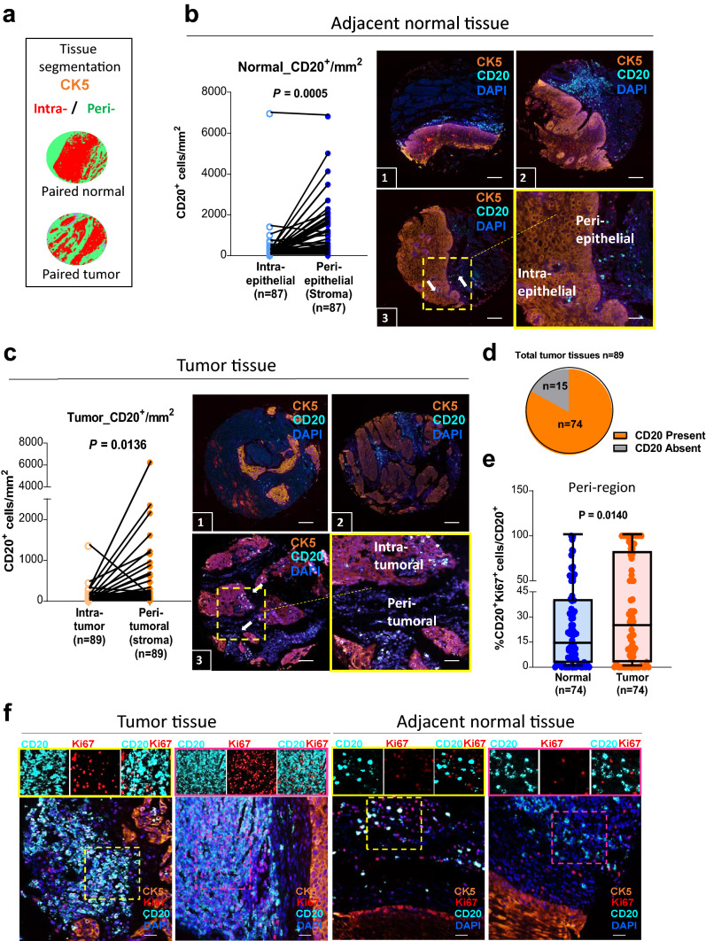
High proportions of proliferating B cells are found in the peritumoral region of ESCC tissues. a Representative image of machine learning-based image processing for tissue category classification (intra-, red; peri- green). b, c The density of CD20+ B cells as counts/mm2 in 89 paired normal (b, blue) and tumor (c, orange) tissues of ESCC. TMA stained with tumor marker (cytokeratin-5, CK5, orange), B-cell marker (CD20, turquoise), and nuclei (DAPI, blue). Scale bar: 100 µm. Three individual representative TMA cores of tumor and normal tissues are shown. Three-color overlay (top two and bottom left) and enlarged insets (border) of a representative sampling of the simultaneously acquired markers. Density of CD20+ B cells/mm2 was higher in peri-region than intra-region in both tumor (light orange vs. dark orange) and normal (light blue vs. dark blue) tissues. The sample size in normal was 87 due to deficit data on CK5 of two cores in normal tissues. d 15 out of 89 patients did not have CD20+ cells and were removed from the subsequent analysis. e Proliferating B cells (%CD20+Ki67+ cells/total CD20+ cells) were higher in the peritumoral region of esophageal cancer than in normal (n = 74). (f) Representative images of two tumor and two normal tissues are shown. Tissues stained for Ki67 (red), CD20 (turquoise) in peri-region (CK5−, orange) and counterstained with DAPI. In ESCC tumor tissues, higher amount of peritumoral B cells co-expressed with Ki67 (proliferating B cells), while in the peri-region of normal tissue, most of the B cells are not proliferative. Magnified insets with representative region of individual markers (or select combinations of markers) are shown on the side. Scale bar: 50 µm

