



Abstract
The recent genomic characterization of patient specimens has started to reveal the landscape of somatic alterations in clinical prostate cancer (CaP) and its association with disease progression and treatment resistance. The extent to which such alterations impact hallmarks of cancer is still unclear. Here, we interrogate genomic data from thousands of clinical CaP specimens that reflect progression from treatment-naïve, to castration-recurrent, and in some cases, neuroendocrine CaP for alterations in cell cycle-associated and -regulated genes, which are central to cancer initiation and progression. We evaluate gene signatures previously curated to evaluate G1-S and G2-M phase transitions or to represent the cell cycle-dependent proteome. The resulting CaP (stage)-specific overview confirmed the presence of well-known driver alterations impacting for instance the genes encoding p53 and MYC, and uncovered novel previously unrecognized mutations that affect others such as the PKMYT1 and MTBP genes. The cancer dependency and drugability of representative genomically altered cell cycle determinants was verified also. Taken together, these analyses on hundreds of often less-characterized cell cycle regulators expand considerably the scope of genomic alterations associated with CaP cell proliferation and cell cycle, and isolate such regulatory proteins as putative drivers of CaP treatment resistance and entirely novel therapeutic targets for CaP therapy.
Keywords: driver mutation, precision medicine, cell proliferation, treatment resistance
In this research letter we provide a brief report analyzing the relationship between genomic alterations and cell-cycle related genes in prostate cancer (CaP) as a companion to our review on this topic also published in this issue of Endocrine-Related Cancer. We noted that several well-characterized critical regulators of cell cycle progression are subject to genomic alterations, for instance RB loss or MYC gene amplification, both well-known to contribute to treatment resistance. Yet, the full scope of CaP cell cycle modulators and the extent to which somatic alterations impact their expression or function, and subsequently, clinical CaP progression remain unknown.
This gap in knowledge is remarkable; during the past decade thousands of clinical CaP specimens at different stages of disease progression have been analyzed via whole exome/genome sequencing and/or copy number alteration assays, and the resulting genomic data made publically available via dataportals such as the cBIO portal. These studies provided novel insights into the genomic drivers of CaP progression, treatment resistance and lethality and uncovered recurrent alterations, in a significant subset of CaP cases or at lower frequencies that follow a long-tail distribution (Armenia et al., 2018).
We performed an in-depth cBIO analysis on gene signatures previously curated to study cell cycle progression (see accompanying review). First, we examined the Molecular Signature Database Gene Ontology (GO) signatures Cell Cycle G1-S phase transition (n=303) and Cell Cycle G2-M phase transition (n=269), which assess G1-S and G2-M checkpoint transitions, respectively. We included also a signature derived from the cell cycle-dependent proteome, as defined in the human proteome atlas, which consists of 298 genes that show GO-based functional enrichment in cell cycle processes and single cell variation in their expression that is correlated to cell cycle progression. We evaluated these signatures for somatic alterations in cBIO datasets that represent different stages of CaP progression, provide genome-wide information on gene alterations (data from targeted sequencing only were not included), have consistently procured and used the same type of tissues (radical prostatectomy or CaP biopsy), and allow to assess both gene sequence and copy number alterations. Based on these selection criteria, 12 datasets were withheld and grouped based on their predominant CaP phenotype: 6 treatment-naïve localized CaP (LOC), 4 castration-recurrent CaP (CRPC) and 2 neuroendocrine CaP (NEPC) studies.
The number of genes showing genomic alterations for each cell-cycle related signature in each dataset is shown in Figure 1. Figure 2 provides more qualitative views of these changes and aligns the alterations observed for all members of the 3 gene signatures studied for LOC, CRPC and NEPC. Overall, the percentage of cases in which genomic alterations are seen increases from LOC to CRPC and NEPC and more genes are affected as CaP progresses. These observations are consistent with the general trend of more genomic heterogeneity with increasing CaP aggressiveness and with the majority of alterations (e.g. CDK2AP2, CRNN, CDK7, PKMYT1, KIF20B, PPEF1, NUCKS1) present in ≤3 percent of LOC samples (Armenia et al., 2018). Our cBIO analyses confirmed TP53, PTEN and MYC somatic alterations in a significant subset (>3%) of LOC samples, which increased in proportion in CRPC and NEPC (Figure 2). The types of alterations detected, either copy number alterations (deletions or amplification) or events impacting structural integrity (point mutations, splice isoforms, gene fusions …) were consistent with those reported before for these genes (Robinson et al., 2015), validating our approach and justifying extrapolation to the hundreds of cell cycle-related genes in the 3 gene signatures.
Figure 1. Quantitative view of genomic alteration rate among the membership of the 3 cell cycle-related gene signatures in cBio CaP datasets.
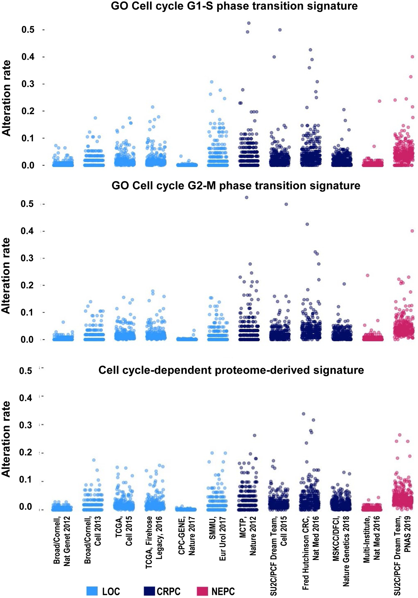
The X-axis represents the membership of 6 individual LOC, 4 individual CRCP and 2 individual NEPC cBIO datasets. Dots reflect alteration rate per gene for each gene signature and dataset. Y-axis lists the alteration rate as proportion of cases with alterations, in which 0 means that no cases show an alteration and 1 that all cases in that study do. The source data for Figure 1 were retrieved from the cBIO portal in November 2020 and are available upon request. In brief, members of each cell cycle-related gene signature (G1-S, G2-M, cell cycle-dependent proteome signature) were queried across each of the 12 cBIO studies using default cBio settings. Only data for mutations and copy number alterations were collected and shown. The percentage of alterations for each gene was retrieved using default cBIO settings. GO, Gene Ontology; LOC, localized treatment-naïve CaP; CRPC, castration-resistant CaP; NEPC, neuroendocrine CaP.
Figure 2. Qualitative overview of the genomic alteration rate per CaP stage for each gene included in the 3 cell cycle-derived signatures in cBio CaP datasets.
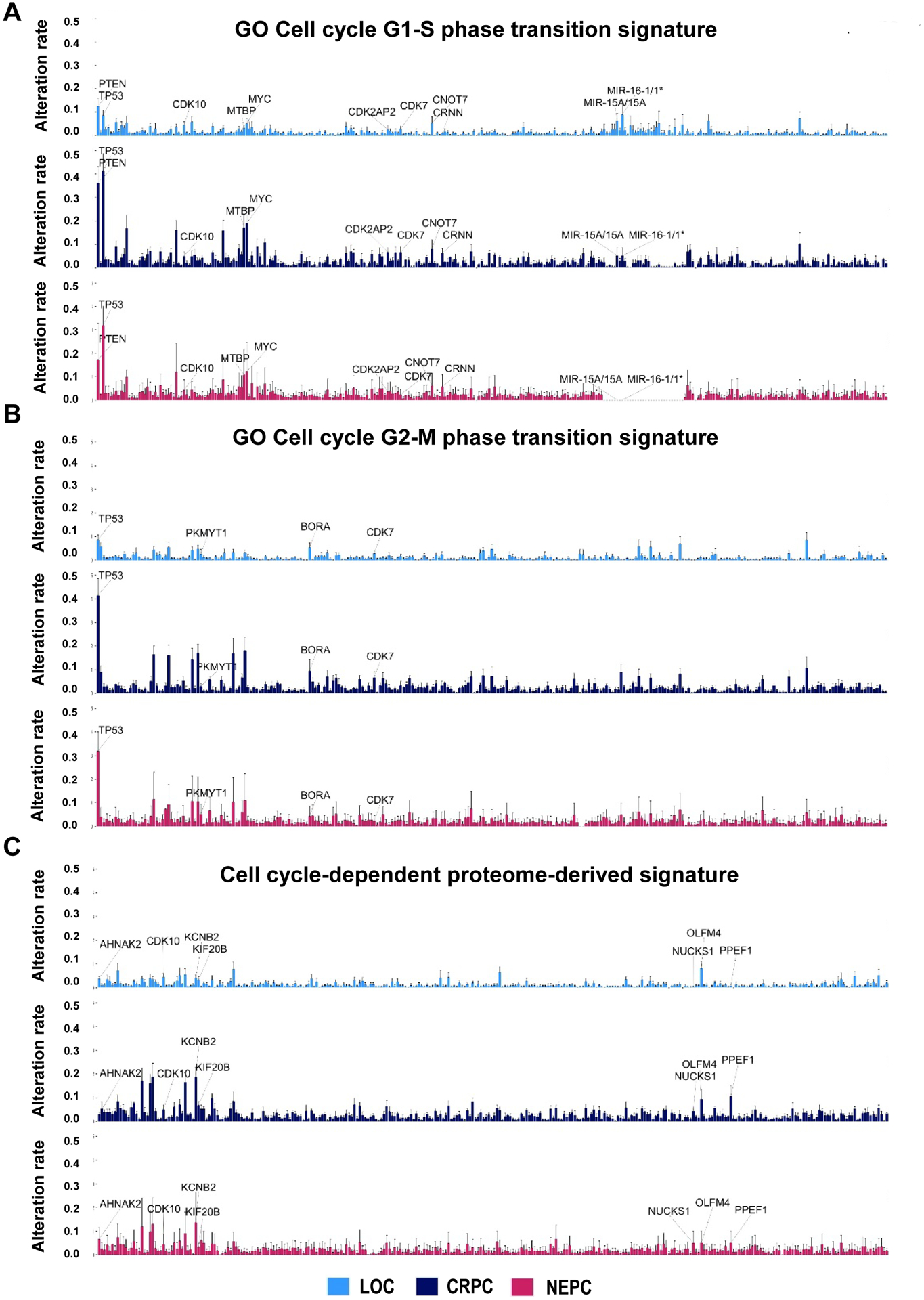
Rate of alterations in all genes derived form G1-S phase transition (A), G2-M phase transition (B), and cell cycle-dependent proteome signature (C) are presented. Somatic alterations discussed in more detail in the text are labelled. Source data was retrieved from the cBio portal in November of 2020 as described in legend to Figure 1 and are available upon request. LOC, localized treatment-naïve CaP; CRPC, castration-resistant CaP; NEPC, neuroendocrine CaP.
As representative examples, we highlight findings for a few cell cycle regulators that were present in ≥3% of LOC cases but otherwise randomly chosen from the Cell Cycle G1-S phase (Figure 2A), G2-M phase transition (Figure 2B), the cell cycle proteome-derived gene signature (Figure 2C). Several of these genes have not yet been (extensively) studied in CaP, reflecting novel information on regulation of cell cycle during disease progression. Some of the alterations highlighted demonstrate higher frequency with increased CaP aggressiveness, suggesting a driving role and/or clonal selection.
CDK10 controls ETS2 transactivation and its degradation via CDK10/Cyclin M-mediated phosphorylation (Kasten and Giordano, 2001), and may participate in the resistance of tumor cells to p53-mediated apoptosis (Maxwell and Davis, 2000). We mostly noted CDK10 deletions, the frequency of which was highest in NEPC, followed by LOC and then CRPC.
MTBP or MDM2 binding protein regulates cell cycle progression, may be involved in tumor formation and interacts with MYC to promote tumorigenesis (Grieb et al., 2014). The frequency of MTBP somatic alterations, most of which are amplifications, increases steadily during progression from LOC to CRPC and NEPC, revealing an entirely novel role for this cell cycle regulator in CaP.
BORA is an activator of Aurora A, which is required for centrosome maturation, spindle assembly, and asymmetric protein localization during mitosis. BORA is lost in about 5% of LOC cases, mostly via deletions, and this incidence decreases and increases somewhat, respectively in CRPC and NEPC. BORA has not yet been studied in CaP but its expression is a poor prognostic factor in multiple adenocarcinomas (Zhang et al., 2017).
KCNB2 or potassium voltage-gated channel subfamily B member 2, has not yet been studied in CaP but is encoded by the gain of 8p12-q24.3 region that contains also the MYC gene. Our cBIO analyses support amplification and both missense and truncating mutations impacting this gene, which we uncovered as the most frequently altered gene during late stage CaP, with more than 23% of NEPC showing KCBN2 gene alterations.
AHNAK2 or AHNAK nucleoprotein 2, an androgen-regulated gene, may play a role in calcium signaling by associating with calcium channel proteins. Hypoxia can activate the expression of AHNAK2 in HIF1α-dependent manner in a variety of human tumor cells, including the CaP cell DU145, and AHNAK2 knockdown impairs hypoxia-induced EMT and stem cell-like properties (Wang et al., 2017). We noted a prominent increase in AHNAK2 genomic alterations with CaP progression, with 9.5% of NEPC cases affected, mostly due to gene amplifications and somatic mutations.
While these results expand considerably the scope of cell cycle regulatory mechanisms that maybe altered during CaP progression, they also raise a few important questions. First, the functional implications of these newly identified alterations in cell cycle-related genes for CaP progression and acquired treatment resistance will be important to validate experimentally in models system relevant to different CaP stages. Indeed, even when gene amplifications are recognized to drive treatment resistance, whether for instance a 2-fold amplification confers the same aggressiveness as 100-fold amplification will be critical to ascertain. Similarly, whether each somatic alterations impacting a gene has the same biologic significance or the relevance of variants of unknown significance will needed to be resolved.
Second, these data suggest also that lesser known regulators drive CaP progression and constitute previously unrecognized but viable targets for therapy. To verify this possibility, we examined the potential as drug target of the example alterations belonging to the “long tail” or present in more than 3% of LOC cases highlighted above via the CanSAR database, an integrated knowledge-base that facilitates drug-discovery predictions. This analysis returned bioactive compounds for 3 of these (CDK10, CDK7 and PKMYT1). Two more entries, AHNAK2 and MTBP, were found to have ligandable structure, indicating also their potential as drug targets. That CanSAR predictions can be reliable was already verified for some of these example alterations: for CDK7 and PKMYT1, compounds such as THZ1 and fostamatinib have shown to inhibit CaP in preclinical studies (Rasool et al., 2019, Wang et al., 2020). Analysis of the cancer dependency for these genes via the DependencyMap portal returned MTBP, BORA, CDK7 and PKMYT14 as common essential genes, i.e. ranked among the top genes whose depletion impacts viability of ≥ 90 percent of cell lines. In case of PKMYT1, for which mostly gene amplifications were seen, therapeutic success of such inhibition fits with genomic alterations observed. For CDK7, however, mostly gene deletions were observed, which seems at odd with effect of inhibitor on CaP growth. Cases in which such deletions are seen may, however, represent a subtype - reminiscent of recent isolation of a class of CDK12 mutant CRPC cases that benefit form immune checkpoint immunotherapy. Supporting this possibility, in small cell lung cancer, CDK7 inhibition using a selective inhibitor, YKL-5–124, predominantly triggering immune-response signaling, provoking a robust immune surveillance program elicited by T cells, which is further enhanced by the addition of immune-checkpoint blockade (Zhang et al., 2020).
Taken together, these previously unappreciated alterations in hundreds of cell cycle-related genes during CaP progression, their drugability and cancer dependency may facilitate mechanistically novel CaP treatment strategies to target cell proliferation, a hallmark of cancer progression.
Funding:
This work was supported the National Cancer Institute (grant number CA166440), a VeloSano 5 pilot Research Award, and a Falk Medical Research Trust Catalyst Award (to HVH).
Footnotes
Declaration of interest: the authors declare no conflicting interests
Selected references
- ARMENIA J, WANKOWICZ SAM, LIU D, GAO J, KUNDRA R, REZNIK E, CHATILA WK, CHAKRAVARTY D, HAN GC, COLEMAN I, et al. 2018. The long tail of oncogenic drivers in prostate cancer. Nat Genet, 50, 645–651. [DOI] [PMC free article] [PubMed] [Google Scholar]
- GRIEB BC, GRAMLING MW, ARRATE MP, CHEN X, BEAUPARLANT SL, HAINES DS, XIAO H & EISCHEN CM 2014. Oncogenic protein MTBP interacts with MYC to promote tumorigenesis. Cancer Res, 74, 3591–602. [DOI] [PMC free article] [PubMed] [Google Scholar]
- KASTEN M & GIORDANO A 2001. Cdk10, a Cdc2-related kinase, associates with the Ets2 transcription factor and modulates its transactivation activity. Oncogene, 20, 1832–8. [DOI] [PubMed] [Google Scholar]
- MAXWELL SA & DAVIS GE 2000. Differential gene expression in p53-mediated apoptosis-resistant vs. apoptosis-sensitive tumor cell lines. Proc Natl Acad Sci U S A, 97, 13009–14. [DOI] [PMC free article] [PubMed] [Google Scholar]
- RASOOL RU, NATESAN R, DENG Q, ARAS S, LAL P, SANDER EFFRON S, MITCHELL-VELASQUEZ E, POSIMO JM, CARSKADON S, BACA SC, et al. 2019. CDK7 Inhibition Suppresses Castration-Resistant Prostate Cancer through MED1 Inactivation. Cancer Discov, 9, 1538–1555. [DOI] [PMC free article] [PubMed] [Google Scholar]
- ROBINSON D, VAN ALLEN EM, WU YM, SCHULTZ N, LONIGRO RJ, MOSQUERA JM, MONTGOMERY B, TAPLIN ME, PRITCHARD CC, ATTARD G, et al. 2015. Integrative clinical genomics of advanced prostate cancer. Cell, 161, 1215–1228. [DOI] [PMC free article] [PubMed] [Google Scholar]
- WANG J, WANG L, CHEN S, PENG H, XIAO L, DU E, LIU Y, LIN D, WANG Y, XU Y, et al. 2020. PKMYT1 is associated with prostate cancer malignancy and may serve as a therapeutic target. Gene, 744, 144608. [DOI] [PubMed] [Google Scholar]
- WANG M, LI X, ZHANG J, YANG Q, CHEN W, JIN W, HUANG YR, YANG R & GAO WQ 2017. AHNAK2 is a Novel Prognostic Marker and Oncogenic Protein for Clear Cell Renal Cell Carcinoma. Theranostics, 7, 1100–1113. [DOI] [PMC free article] [PubMed] [Google Scholar]
- ZHANG H, CHRISTENSEN CL, DRIES R, OSER MG, DENG J, DISKIN B, LI F, PAN Y, ZHANG X, YIN Y, et al. 2020. CDK7 Inhibition Potentiates Genome Instability Triggering Anti-tumor Immunity in Small Cell Lung Cancer. Cancer Cell, 37, 37–54 e9. [DOI] [PMC free article] [PubMed] [Google Scholar]
- ZHANG QX, GAO R, XIANG J, YUAN ZY, QIAN YM, YAN M, WANG ZF, LIU Q, ZHAO HD & LIU CH 2017. Cell cycle protein Bora serves as a novel poor prognostic factor in multiple adenocarcinomas. Oncotarget, 8, 43838–43852. [DOI] [PMC free article] [PubMed] [Google Scholar]