Abstract
Pulse pressure variation (PPV) and cardiac output (CO) can guide perioperative fluid management. Capstesia (Galenic App, Vitoria-Gasteiz, Spain) is a mobile application for snapshot pulse wave analysis (PWAsnap) and estimates PPV and CO using pulse wave analysis of a snapshot of the arterial blood pressure waveform displayed on any patient monitor. We evaluated the PPV and CO measurement performance of PWAsnap in adults having major abdominal surgery. In a prospective study, we simultaneously measured PPV and CO using PWAsnap installed on a tablet computer (PPVPWAsnap, COPWAsnap) and using invasive internally calibrated pulse wave analysis (ProAQT; Pulsion Medical Systems, Feldkirchen, Germany; PPVProAQT, COProAQT). We determined the diagnostic accuracy of PPVPWAsnap in comparison to PPVProAQT according to three predefined PPV categories and by computing Cohen’s kappa coefficient. We compared COProAQT and COPWAsnap using Bland-Altman analysis, the percentage error, and four quadrant plot/concordance rate analysis to determine trending ability. We analyzed 190 paired PPV and CO measurements from 38 patients. The overall diagnostic agreement between PPVPWAsnap and PPVProAQT across the three predefined PPV categories was 64.7% with a Cohen’s kappa coefficient of 0.45. The mean (± standard deviation) of the differences between COPWAsnap and COProAQT was 0.6 ± 1.3 L min− 1 (95% limits of agreement 3.1 to − 1.9 L min− 1) with a percentage error of 48.7% and a concordance rate of 45.1%. In adults having major abdominal surgery, PPVPWAsnap moderately agrees with PPVProAQT. The absolute and trending agreement between COPWAsnap with COProAQT is poor. Technical improvements are needed before PWAsnap can be recommended for hemodynamic monitoring.
Keywords: Non-invasive, Hemodynamic monitoring, Cardiovascular dynamics, Fluid management, Fluid responsiveness, Blood flow
Introduction
The assessment of fluid responsiveness using dynamic cardiac preload variables such as pulse pressure variation (PPV) and the estimation of cardiac output (CO) are mainstays of perioperative fluid management [1–3]. PPV and CO can be measured using pulse wave analysis of the arterial blood pressure waveform [4–6]. Because this technology is not included in most routine patient monitors, hemodynamic monitoring using pulse wave analysis requires advanced hemodynamic monitors. This may limit the clinical use of pulse wave analysis, especially in low-resource settings.
A promising approach to overcome the problem of additional hemodynamic monitoring equipment is the development of innovative mobile monitoring techniques using smartphones and tablet computers [7, 8]. The Capstesia application (Galenic App, Vitoria-Gasteiz, Spain), that can be installed on smartphones or tablet computers, is a mobile application for snapshot pulse wave analysis (PWAsnap). It was proposed to estimate PPV and CO by pulse wave analysis of a snapshot of the arterial blood pressure waveform displayed on any patient monitor screen. In-silico proof of concept studies demonstrated that PWAsnap is basically able to estimate PPV and CO from a waveform snapshot [9, 10]. However, the few clinical method comparison studies evaluating PWAsnap used different PPV and CO reference methods and revealed inconsistent results [11–15].
Before PWAsnap can be considered for routine hemodynamic monitoring its measurement performance needs to be investigated in comparison to established reference methods. We, therefore, performed a prospective study to compare PPV and CO estimated by PWAsnap with PPV and CO determined using invasive internally calibrated pulse wave analysis in adults having major abdominal surgery.
Materials and methods
Study design and setting
This prospective method comparison study was approved by the ethics committee (ethics committee approval number: PV5825, Ethikkommission der Ärztekammer Hamburg, Hamburg, Germany). All participants gave written informed consent. This study was performed at the University Medical Center Hamburg-Eppendorf (Hamburg, Germany) between November 2018 and October 2019.
Inclusion and exclusion criteria
Consenting patients > 18 years were eligible for study inclusion if they were scheduled for open major abdominal surgery (radical cystectomy, pancreaticoduodenectomy, ovarian cancer surgery, and partial hepatectomy) and when advanced hemodynamic monitoring using invasive internally calibrated pulse wave analysis with an arterial catheter was planned independently from the study. Exclusion criteria were a history or presence of atrial fibrillation or excessive premature atrial or ventricular contractions.
Anesthetic management
General anesthesia was induced using sufentanil, propofol, and a muscle relaxant (usually rocuronium). General anesthesia was maintained either with propofol or inhaled sevoflurane and sufentanil boluses. In addition to routine anesthetic monitoring, arterial blood pressure was continuously recorded using a 20 g radial arterial catheter. The arterial blood pressure waveform was displayed on the patient monitor routinely used in our institution (Infinity Delta monitor; Dräger Medical Deutschland, Lübeck, Germany). The tidal volume was set to 8 mL kg− 1 predicted body weight during study measurements. Predicted bodyweight was calculated as 50 + 0.91 × (height [cm] − 152.4) for male and as 45.5 + 0.91 × (height [cm] − 152.4) for female patients [16].
Study measurements and data extraction
We used PWAsnap to estimate PPV (PPVPWAsnap) and CO (COPWAsnap). For this, the Capstesia application was installed on a SPC GLOW 10.1 tablet computer with an integrated camera (Smart Products Connection S.A., Miñano, Álava, Spain). We set the sweep speed of the Infinity Delta monitor to 12.5 mm s− 1 to display at least six to eight cardiac cycles on the monitor. The Infinity Delta monitor always displays a dotted line at half of the scale. This dotted line cannot be deactivated and interferes with the PWAsnap measurement. By setting the scale to the maximum of 300 mmHg, we ensured that the dotted line on the screen did not cross the arterial blood pressure waveform displayed on the screen. By adjusting sweep speed and scale, we optimized conditions for using PWAsnap with the Infinity Delta monitor.
We took snapshots of the arterial blood pressure waveform with the integrated two megapixels camera of the tablet computer. To take a snapshot, the tablet computer had to be held parallel to the patient monitor. The snapshot was cropped to display the arterial blood pressure waveform only. Heart rate as well as systolic and diastolic arterial blood pressure were manually entered afterwards as required by PWAsnap.
We used the ProAQT system (Pulsion Medical Systems, Feldkirchen, Germany) to measure PPV (PPVProAQT) and CO (COProAQT) using invasive internally calibrated pulse wave analysis of the arterial blood pressure waveform. The sensor of the ProAQT system was put in series to the standard pressure transducer connected to the Infinity Delta monitor. The ProAQT system estimates CO based on features of the arterial blood pressure waveform and biometric data without any external calibration.
We simultaneously measured PPVPWAsnap and PPVProAQT as well as COPWAsnap and COProAQT during surgery at five time points with at least five minutes between two measurements. To obtain simultaneous measurements, we recorded PPVProAQT and COProAQT at the moment when a snapshot with PWAsnap was taken. Before each measurement, we zeroed the pressure transducer connected to the Infinity Delta monitor and ProAQT system. Measurements were performed during steady state hemodynamic conditions (i.e., no changes in vasoactive agents or anesthetic management).
Statistical analysis
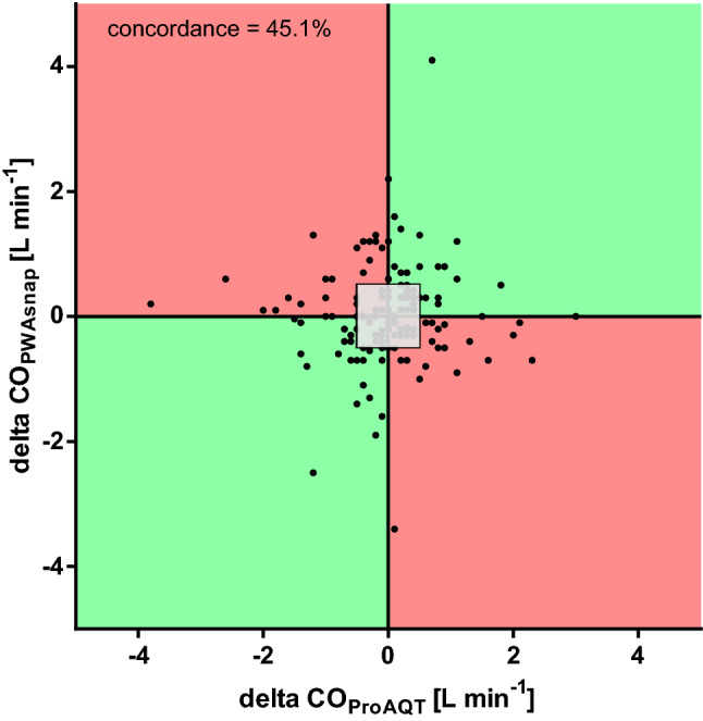
We present continuous data as mean ± standard deviation (SD) and categorical data as absolute values (n) with relative frequencies in percent. Linear regression analysis of CO values was performed and illustrated in a scatter plot. We performed Bland-Altman analysis for multiple observations per individual [17]. For Bland-Altman analysis, we subtracted PPVPWAsnap from PPVProAQT and COPWAsnap from COProAQT and calculated the mean of the differences with the accompanying upper and lower 95% limits of agreement (95%-LOA; mean of the differences ± 1.96 × standard deviation of the mean of the differences). As described previously [12], the diagnostic accuracy of the PPV measurements was determined by categorizing the PPV values in three predefined categories reflecting clinical decision making (PPV < 9%, gray zone PPV 9–13%, PPV > 13%) [12, 18]. For the assessment of the diagnostic accuracy the agreement between PPVPWAsnap and PPVProAQT across the three predefined PPV categories was calculated. Furthermore, Cohen’s kappa was computed to additionally evaluate the diagnostic accuracy [19]. For CO, we calculated the percentage error between the two methods as 1.96 × standard deviation of the mean of the differences divided by the mean CO of both methods. The percentage error threshold for clinical interchangeability was a priori set at 30% as described previously [20]. For CO, we additionally performed four-quadrant plot analysis to evaluate the trending ability [21]. For four-quadrant plot analysis, we plotted the difference of consecutive COPWAsnap values (ΔCOPWAsnap) on the y-axis and difference of consecutive COProAQT values (ΔCOProAQT) on the x-axis. A central exclusion zone of 0.5 L min− 1 was applied to exclude clinically unimportant small changes. The concordance rate is the ratio (in percent) of the ΔCO values that change in the same direction in relation to all ΔCO values [21]. A change in the same direction means that both ΔCO values either increased or decreased. For statistical analysis we used MedCalc Version 19.1.3 (MedCalc Software, Ostend, Belgium), IBM SPSS Version 25 (IBM, Armonk, NY, USA), and GraphPad PRISM (GraphPad Software, San Diego, USA).
Results
We included a total of 50 patients. Surgery was cancelled in two patients. We excluded one patient because of new-onset atrial fibrillation. Two patients were excluded because the patients were monitored with a monitoring system other than the ProAQT system. We additionally had to exclude seven patients due to unavoidable overlap between the arterial blood pressure waveform and the dotted line on the screen of the Infinity Delta monitor. This made measurements using PWAsnap impossible. We thus analyzed data of 38 patients with 190 paired PPV and CO measurements. Table 1 shows patient characteristics and intraoperative data.
Table 1.
Patient characteristics
| Demographic and biometric data | |
| Male sex, [n (%)] | 11 (29) |
| Age, mean ± SD [years] | 64 ± 11 |
| Height, mean ± SD [cm] | 168 ± 10 |
| Weight, mean ± SD [kg] | 78 ± 20 |
| Predicted body weight, mean ± SD [kg] | 61 ± 11 |
| Body Mass Index, mean ± SD [kg m− 2] | 28 ± 7 |
| Type of surgery | |
| Radical cystectomy, [n (%)] | 12 (31.6) |
| Pancreaticoduodenectomy, [n (%)] | 13 (34.2) |
| Ovarian cancer surgery, [n (%)] | 12 (31.6) |
| Partial hepatectomy, [n (%)] | 1 (2.6) |
Data are shown as mean ± standard deviation (SD) or absolute (n) and relative frequencies (%)
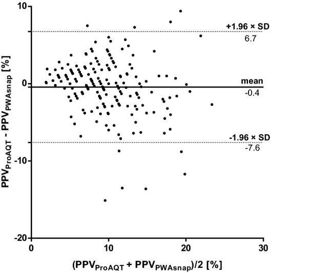
Mean PPVPWAsnap was 10.1 ± 5.1% and mean PPVProAQT was 9.7 ± 4.4%. The mean of the differences between PPVPWAsnap and PPVProAQT was − 0.4 ± 3.7% (95%-LOA 6.7 to − 7.6%) (Fig. 1). The distribution of PPVPWAsnap and PPVProAQT across the three predefined PPV categories is shown in Table 2. The overall diagnostic agreement between PPVPWAsnap and PPVProAQT across the three predefined PPV categories was 64.7% with a Cohen’s kappa coefficient of 0.45.
Fig. 1.

Bland-Altman plot comparing pulse pressure variation (PPV) measured using mobile application pulse wave analysis (PWAsnap) and invasive pulse wave analysis. The bold line represents the mean of the differences between PPV measured using the two methods. The dotted lines represent the 95%-limits of agreement. PPVPWAsnap pulse pressure variation determined with PWAsnap, PPVProAQT pulse pressure variation determined with ProAQT system
Table 2.
Distribution and diagnostic agreement of pulse pressure variation measurements across the three predefined categories
| PPVPWAsnap | Total | |||
|---|---|---|---|---|
| PPV ProAQT | < 9% | 9–13% | > 13% | |
| < 9% | 63 (72%) | 17 (20%) | 7 (8%) | 87 (100%) |
| 9–13% | 23 (33%) | 33 (48%) | 13 (19%) | 69 (100%) |
| > 13% | 0 (0%) | 7 (21%) | 27 (79%) | 34 (100%) |
PPVPWAsnap pulse pressure variation measured with mobile application pulse wave analysis, PPVProAQT pulse pressure variation measured with ProAQT system, bold measurement pairs in concordant category, italic measurement pairs in opposite category
Percentages are calculated for each horizontal row
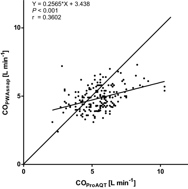
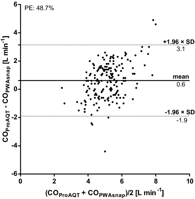
Mean ± SD COPWAsnap was 4.8 ± 0.9 L min− 1 and mean COProAQT was 5.5 ± 1.3 L min −1. The relation between COPWAsnap and COProAQT is shown in Fig. 2. The mean of the differences between COPWAsnap and COProAQT was 0.6 ± 1.3 L min− 1 (95%-LOA 3.1 to − 1.9 L min− 1) (Fig. 3). The percentage error was 48.7%. The concordance rate between changes in COPWAsnap and COProAQT was 45.1% (Fig. 4).
Fig. 2.

Scatter plot with linear regression analysis of cardiac output measured using mobile application pulse wave analysis (PWAsnap) and invasive pulse wave analysis. COPWAsnap cardiac output estimated with PWAsnap, COProAQT cardiac output estimated with ProAQT system, Y slope-intercept equation, r correlation coefficient
Fig. 3.

Bland-Altman plot comparing cardiac output (CO) measured using mobile application pulse wave analysis (PWAsnap) and invasive pulse wave analysis. The bold line represents the mean of the differences between CO measured using the two methods. The dotted lines represent the 95%-limits of agreement. COPWAsnap cardiac output estimated with PWAsnap, COProAQT cardiac output estimated with ProAQT system, PE percentage error
Fig. 4.

Four-quadrant plot to estimate the concordance rate between cardiac output measured using mobile application pulse wave analysis (PWAsnap) and invasive pulse wave analysis. The gray square is the central exclusion zone of 0.5 L min − 1. COPWAsnap cardiac output estimated with PWAsnap, COProAQT cardiac output estimated with ProAQT system
Discussion
We performed a prospective method comparison study to compare PPV and CO estimated by PWAsnap with PPV and CO determined using invasive internally calibrated pulse wave analysis in adults having major abdominal surgery. The overall diagnostic agreement between PPVPWAsnap and PPVProAQT across the three predefined PPV categories was moderate according to Cohen’s kappa [19]. The absolute agreement of COPWAsnap with COProAQT was poor and the measurements were not interchangeable. Trending ability of COPWAsnap was also poor with a low concordance rate.
An in-silico proof of concept study showed that calculating PPV and CO by analyzing snapshots of an arterial blood pressure waveform displayed on a patient monitor is basically possible using PWAsnap [10]. The analysis of these snapshots showed a good concordance with PPV and CO measurements of the raw data [10]. The measurement performance and diagnostic accuracy of PPVPWAsnap has been investigated in previous studies. An experimental study in a highly controlled simulated environment suggested that PPVPWAsnap may be a good substitute for manual PPV determination [9]. Our results on the diagnostic accuracy of PPVPWAsnap are similar to a recent study comparing PPVPWAsnap with stroke volume variation (SVV) determined using the FloTrac system (Vigileo monitor; Edwards Lifesciences, Irvine, CA, USA) in 40 patients having major abdominal surgery [12]. Like in this previous study, we used three predefined PPV categories reflecting clinical decision making according to the “gray zone” approach [18]. When comparing different methods to measure PPV it is more important to know if PPV values fall into the same category and thus result in the same treatment decisions regarding fluid therapy than if PPV values have the exact same numeric value. In contrast to the previous study, we compared PPVPWAsnap directly to PPVProAQT and not to SVV. Furthermore, the conditions to use PPV were optimized in our study (e.g., sinus rhythm and controlled mechanical ventilation) [16]. In our study, Cohen’s kappa [19] suggested moderate diagnostic agreement between PPVPWAsnap and PPVProAQT across the three predefined PPV categories in accordance with the previous study [12], but overall agreement appeared to be lower in our analysis. Notably, nearly 80% of the patients with a PPVProAQT>13% were correctly classified by PWAsnap.
The few clinical method comparison studies evaluating COPWAsnap revealed inconsistent results. COPWAsnap was compared to CO determined using invasive internally calibrated pulse wave analysis (FloTrac system) in 53 patients during major oncological surgery [14]. In this study, a percentage error of 26% suggested good agreement between COPWAsnap and FloTrac-derived CO [14]. These findings are in contrast to our study revealing wide 95%-LOA when comparing COPWAsnap with COProAQT. Furthermore, the percentage error in our analysis exceeded the threshold for clinical interchangeability of 30% [20]. In another validation study, COPWAsnap was compared to CO measured with transpulmonary thermodilution, a clinical reference method [22, 23], in 57 patients during cardiac surgery [11]. With a mean of the differences between CO measurements obtained by the two methods of 0.3 L min− 1, wide 95%-LOA of 3.3 to -2.8 L min− 1, a high percentage error of 60%, and a poor concordance rate the results of this study are consistent with our findings [11].
Previously, it was reported that close to 10% of the snapshots of the arterial blood pressure waveform cannot be analyzed by PWAsnap [11]. We did not systematically analyze the quality of the snapshots or the failure rate of PWAsnap to estimate PPV and CO. We thus can only subjectively describe our experiences of using PWAsnap in a clinical setting in the operating room. PWAsnap sometimes failed to analyze a snapshot without an apparent reason. We speculate that the poor camera performance of the tablet computer may have contributed to these difficulties in the analysis of the snapshots. Other studies that used PWAsnap installed on smartphones with high resolution cameras did not report these problems [11, 14].
Considering the results of our study, technical refinements are needed before PWAsnap can be recommended as an alternative to current monitoring methods. In general, using mobile devices in combination with hemodynamic monitoring applications instead of bulky and costly monitoring equipment is a promising approach to future hemodynamic monitoring in the operating room. Most medical personnel use smartphones making such applications almost universally available.
There are limitations of this study. We did not perform interventions such as fluid challenge or passive leg raising tests and, therefore, cannot describe the ability of PPVPWAsnap to actually predict fluid responsiveness. We compared COPWAsnap with COProAQT. Invasive internally calibrated pulse wave analysis (ProAQT system) is widely used to guide intraoperative hemodynamic therapy but is not a clinical reference method for CO measurement [22, 23]. However, the ProAQT system exhibits reasonable CO trending ability [24] and a perioperative goal-directed therapy treatment algorithm based on hemodynamic variables obtained with the ProAQT system has been shown to improve patient outcome [25]. We only included patients having major abdominal surgery. Therefore, our results are not necessarily transferable to other clinical situations in the operating room or in the intensive care unit. We did not perform study measurements at predefined time points during surgery (e.g., pre-induction, post-induction, pre-incision etc.), but all measurements were performed post-incision and with at least five minutes between two measurements.
In contrast to other studies evaluating PWAsnap [9, 11, 12], we used singular PWAsnap measurements and did not calculate a mean of three or more consecutive measurements. We think, that this approach reflects how PWAsnap would be used in clinical practice as calculating a mean of multiple measurements may be too complicated and time-consuming.
It has to be noted that PWAsnap is not commercially available at the moment and to the best of our knowledge it is unclear if a new version will be released. Nevertheless, PWAsnap remains a highly innovative approach to hemodynamic monitoring and our study comparing PWAsnap with an established reference method may provide information that is helpful for the development of future versions or new technologies for PPV and CO monitoring based on PWAsnap.
In conclusion, in adults having major abdominal surgery, PPVPWAsnap moderately agrees with PPVProAQT and the absolute and trending agreement between COPWAsnap with COProAQT is poor. Technical improvements are needed before PWAsnap can be recommended as an alternative to current monitoring methods.
Acknowledgments
Open Access funding provided by Projekt DEAL.
Funding
Support was provided solely from institutional and/or departmental funds. The Capstesia application installed on a tablet computer was provided free of charge by the manufacturer of this application AJL Ophthalmic S.A. (Alava, Spain).
Data availability
Data are available from the authors upon reasonable request.
Code availability
Not applicable.
Compliance with ethical standards
Conflict of interest
PH, FG and LB have no conflicts of interest to declare. AJ received honoraria for consulting from Edwards Lifesciences (Irvine, CA, USA), Aguettant Laboratoire (Lyon, France), and Fresenius Kabi (Bad Homburg, Germany). BS received honoraria for consulting, honoraria for giving lectures, and refunds of travel expenses from Edwards Lifesciences (Irvine, CA, USA). BS received honoraria for consulting, institutional restricted research grants, honoraria for giving lectures, and refunds of travel expenses from Pulsion Medical Systems (Feldkirchen, Germany). BS received institutional restricted research grants, honoraria for giving lectures, and refunds of travel expenses from CNSystems Medizintechnik (Graz, Austria). BS received institutional restricted research grants from Retia Medical (Valhalla, NY, USA). BS received honoraria for giving lectures from Philips Medizin Systeme Böblingen (Böblingen, Germany). BS received honoraria for consulting, institutional restricted research grants, and refunds of travel expenses from Tensys Medical (San Diego, CA, USA). BS is associate editor of the Journal of Clinical Monitoring and Computing but had no role in the handling of this manuscript.
Research involving human participants and/or animals
This method comparison study was approved by the ethics committee (Ethikkomission der Ärztekammer Hamburg, Hamburg, Germany). All procedures performed in studies involving human participants were in accordance with the ethical standards of the institutional and/or national research committee and with the 1964 Helsinki declaration and its later amendments or comparable ethical standards.
Ethical Approval
This prospective method comparison study was approved by the ethics committee (ethics committee approval number: PV5825, Ethikkommission der Ärztekammer Hamburg, Hamburg, Germany).
Informed consent
Informed consent was obtained from all individual participants included in the study.
Footnotes
Publisher's Note
Springer Nature remains neutral with regard to jurisdictional claims in published maps and institutional affiliations.
References
- 1.Saugel B, Reuter DA. Perioperative goal-directed therapy using invasive uncalibrated pulse contour analysis. Front Med. 2018;5:12. doi: 10.3389/fmed.2018.00012. [DOI] [PMC free article] [PubMed] [Google Scholar]
- 2.Monnet X, Marik PE, Teboul JL. Prediction of fluid responsiveness: an update. Ann Intensiv Care. 2016;6(1):111. doi: 10.1186/s13613-016-0216-7. [DOI] [PMC free article] [PubMed] [Google Scholar]
- 3.Benes J, Giglio M, Brienza N, Michard F. The effects of goal directed fluid therapy based on dynamic parameters on post-surgical outcome: a meta-analysis of randomized controlled trials. Crit Care (London England) 2014;18(5):584. doi: 10.1186/s13054-014-0584-z. [DOI] [PMC free article] [PubMed] [Google Scholar]
- 4.Thiele RH, Durieux ME. Arterial waveform analysis for the anesthesiologist: past, present, and future concepts. Anesth Analg. 2011;113(4):766–776. doi: 10.1213/ANE.0b013e31822773ec. [DOI] [PubMed] [Google Scholar]
- 5.Jozwiak M, Monnet X, Teboul JL. Pressure waveform analysis. Anesth Analg. 2018;126(6):1930–1933. doi: 10.1213/ANE.0000000000002527. [DOI] [PubMed] [Google Scholar]
- 6.Saugel B, Vincent JL. Cardiac output monitoring: how to choose the optimal method for the individual patient. Curr Opin Crit Care. 2018;24(3):165–172. doi: 10.1097/mcc.0000000000000492. [DOI] [PubMed] [Google Scholar]
- 7.Michard F. Smartphones and e-tablets in perioperative medicine. Korean J Anesthesiol. 2017;70(5):493–499. doi: 10.4097/kjae.2017.70.5.493. [DOI] [PMC free article] [PubMed] [Google Scholar]
- 8.Michard F, Barrachina B, Schoettker P. Is your smartphone the future of physiologic monitoring? Intensiv Care Med. 2018 doi: 10.1007/s00134-018-5419-y. [DOI] [PubMed] [Google Scholar]
- 9.Desebbe O, Joosten A, Suehiro K, Lahham S, Essiet M, Rinehart J, Cannesson M. A novel mobile phone application for pulse pressure variation monitoring based on feature extraction technology: a method comparison study in a simulated environment. Anesth Analg. 2016;123(1):105–113. doi: 10.1213/ANE.0000000000001282. [DOI] [PubMed] [Google Scholar]
- 10.Barrachina B, Cobos R, Mardones N, Castaneda A, Vinuesa C. Assessment of a smartphone app (Capstesia) for measuring pulse pressure variation: agreement between two methods: a Cross-sectional study. Eur J Anaesthesiol. 2017;34(2):75–80. doi: 10.1097/EJA.0000000000000569. [DOI] [PubMed] [Google Scholar]
- 11.Joosten A, Boudart C, Vincent JL, Vanden Eynden F, Barvais L, Van Obbergh L, Rinehart J, Desebbe O. Ability of a new smartphone pulse pressure variation and cardiac output application to predict fluid responsiveness in patients undergoing cardiac surgery. Anesth Analg. 2018 doi: 10.1213/ANE.0000000000003652. [DOI] [PubMed] [Google Scholar]
- 12.Joosten A, Jacobs A, Desebbe O, Vincent JL, Sarah S, Rinehart J, Van Obbergh L, Hapfelmeier A, Saugel B. Monitoring of pulse pressure variation using a new smartphone application (Capstesia) versus stroke volume variation using an uncalibrated pulse wave analysis monitor: a clinical decision making study during major abdominal surgery. J Clin Monit Comput. 2019;33(5):787–793. doi: 10.1007/s10877-018-00241-4. [DOI] [PubMed] [Google Scholar]
- 13.Santiago-Lopez J, Leon-Ramirez V, Hernandez-Ramirez S, Vasquez-Marquez PI, Castellanos-Olivares A. Concordance in the measurement of cardiac output. Vigileo vs. Capstesia Rev Med Inst Mex Seguro Soc. 2018;56(2):136–142. [PubMed] [Google Scholar]
- 14.Shah SB, Bhargava AK, Hariharan U, Vishvakarma G, Jain CR, Kansal A. Cardiac output monitoring: a comparative prospective observational study of the conventional cardiac output monitor Vigileo and the new smartphone-based application Capstesia. Indian J Anaesth. 2018;62(8):584–591. doi: 10.4103/ija.IJA_783_17. [DOI] [PMC free article] [PubMed] [Google Scholar]
- 15.Desebbe O, Vincent JL, Saugel B, Rinehart J, Joosten A. Pulse pressure variation using a novel smartphone application (Capstesia) versus invasive pulse contour analysis in patients undergoing cardiac surgery: a secondary analysis focusing on clinical decision making. J Clin Monit Comput. 2019 doi: 10.1007/s10877-019-00300-4. [DOI] [PubMed] [Google Scholar]
- 16.Saugel B, Mair S, Gotz SQ, Tschirdewahn J, Frank J, Hollthaler J, Schmid RM, Huber W. Indexation of cardiac output to biometric parameters in critically ill patients: a systematic analysis of a transpulmonary thermodilution-derived database. J Crit Care. 2015;30(5):957–962. doi: 10.1016/j.jcrc.2015.06.011. [DOI] [PubMed] [Google Scholar]
- 17.Bland JM, Altman DG. Agreement between methods of measurement with multiple observations per individual. J Biopharm Stat. 2007;17(4):571–582. doi: 10.1080/10543400701329422. [DOI] [PubMed] [Google Scholar]
- 18.Cannesson M, Le Manach Y, Hofer CK, Goarin JP, Lehot JJ, Vallet B, Tavernier B. Assessing the diagnostic accuracy of pulse pressure variations for the prediction of fluid responsiveness: a “gray zone” approach. Anesthesiology. 2011;115(2):231–241. doi: 10.1097/ALN.0b013e318225b80a. [DOI] [PubMed] [Google Scholar]
- 19.Landis JR, Koch GG. The measurement of observer agreement for categorical data. Biometrics. 1977;33(1):159–174. doi: 10.2307/2529310. [DOI] [PubMed] [Google Scholar]
- 20.Critchley LA, Critchley JA. A meta-analysis of studies using bias and precision statistics to compare cardiac output measurement techniques. J Clin Monit Comput. 1999;15(2):85–91. doi: 10.1023/A:1009982611386. [DOI] [PubMed] [Google Scholar]
- 21.Saugel B, Grothe O, Wagner JY. Tracking changes in cardiac output: statistical considerations on the 4-quadrant plot and the polar plot methodology. Anesth Analg. 2015;121(2):514–524. doi: 10.1213/ANE.0000000000000725. [DOI] [PubMed] [Google Scholar]
- 22.De Backer D, Bakker J, Cecconi M, Hajjar L, Liu DW, Lobo S, Monnet X, Morelli A, Myatra SN, Perel A, Pinsky MR, Saugel B, Teboul JL, Vieillard-Baron A, Vincent JL. Alternatives to the Swan-Ganz catheter. Intensiv care Med. 2018;44(6):730–741. doi: 10.1007/s00134-018-5187-8. [DOI] [PubMed] [Google Scholar]
- 23.Teboul JL, Saugel B, Cecconi M, De Backer D, Hofer CK, Monnet X, Perel A, Pinsky MR, Reuter DA, Rhodes A, Squara P, Vincent JL, Scheeren TW. Less invasive hemodynamic monitoring in critically ill patients. Intensiv Care Med. 2016;42(9):1350–1359. doi: 10.1007/s00134-016-4375-7. [DOI] [PubMed] [Google Scholar]
- 24.Monnet X, Vaquer S, Anguel N, Jozwiak M, Cipriani F, Richard C, Teboul JL. Comparison of pulse contour analysis by Pulsioflex and Vigileo to measure and track changes of cardiac output in critically ill patients. Br J Anaesth. 2015;114(2):235–243. doi: 10.1093/bja/aeu375. [DOI] [PubMed] [Google Scholar]
- 25.Salzwedel C, Puig J, Carstens A, Bein B, Molnar Z, Kiss K, Hussain A, Belda J, Kirov MY, Sakka SG, Reuter DA. Perioperative goal-directed hemodynamic therapy based on radial arterial pulse pressure variation and continuous cardiac index trending reduces postoperative complications after major abdominal surgery: a multi-center, prospective, randomized study. Critical care. 2013;17(5):R191. doi: 10.1186/cc12885. [DOI] [PMC free article] [PubMed] [Google Scholar]
Associated Data
This section collects any data citations, data availability statements, or supplementary materials included in this article.
Data Availability Statement
Data are available from the authors upon reasonable request.
Not applicable.


