Abstract
Hydrogels have been used to design synthetic matrices that capture salient features of matrix microenvironments to study and control cellular functions. Recent advances in understanding of both extracellular matrix biology and biomaterial design have shown that biophysical cues are powerful mediators of cell biology, especially that of mesenchymal stromal cells (MSCs). MSCs have been tested in many clinical trials because of their ability to modulate immune cells in different pathological conditions. While roles of biophysical cues in MSC biology have been studied in the context of multilineage differentiation, their significance in regulating immunomodulatory functions of MSCs is just beginning to be elucidated. This review first describes design principles behind how biophysical cues in native microenvironments influence the ability of MSCs to regulate immune cell production and functions. We will then discuss how biophysical cues can be leveraged to optimize cell isolation, priming, and delivery, which can help improve the success of MSC therapy for immunomodulation. Finally, a perspective is presented on how implementing biophysical cues in MSC potency assay can be important in predicting clinical outcomes.
Keywords: Biomaterials, Mechanotransduction, Immunomodulation, Mechanomedicine, Mesenchymal stromal cells
Graphical Abstract
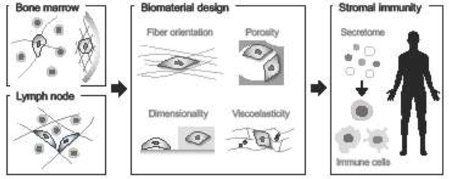
1. Introduction
Mesenchymal stromal cells (MSCs) were first identified in bone marrow in the 1970s [1, 2] as cells with multilineage potential that can differentiate in vitro into bone, cartilage and fat [3, 4]. As resident cells in bone marrow where blood and immune cells are produced, MSCs were also postulated to regulate immune cell functions. In early 2000s, preclinical in vivo studies showed that injection of MSCs promotes skin graft survival [5] and reduces graft-versus-host disease (GvHD) by suppressing T-cell activation [6]. Since then, the number of clinical trials to test therapeutic efficacy of MSCs in various acute and chronic inflammatory pathologies has grown to over one thousand [7]. In addition to T-cells, MSCs were shown to regulate other immune cell lineages, including natural killer cells [8], B-cells [9], dendritic cells [10, 11], and macrophages [12]. The predominant mode of action by which MSCs modulate immune cells is thought to be by the stimulation of MSCs with inflammatory signals from microenvironments, followed by downstream production and paracrine secretion of immunomodulatory factors [13].
Most of the studies on the role of MSCs in immunity were based on either MSCs on plastic culture in vitro or direct adoptive transfer of MSCs in vivo. However, MSCs in tissue microenvironments receive different types of signals, ranging from soluble to insoluble cues. Indeed, advances in recombinant protein engineering have enabled investigations into roles of soluble cues such as inflammatory cytokines in regulating immunomodulatory functions of MSCs. However, the contribution of insoluble cues, especially the extracellular matrix (ECM), to MSC-mediated immunomodulation has only begun to be appreciated recently. The insoluble cues from microenvironments can be classified further into biochemical and biophysical components. While proteomic approaches have defined biochemical components in the ECM, which are implicated in regulating MSC functions [14], advances in biomaterial design have enabled investigations into how matrix biophysical cues impact MSC functions by controlling mechanical properties of the matrix independently of biochemical cues [15]. Most studies have so far focused on the role of matrix mechanics in mechanotransduction and multilineage differentiation of MSCs. However, emerging studies have highlighted roles of matrix biophysical cues in regulating immunomodulatory functions of MSCs.
In this review, we will first summarize the current understanding on the role of MSCs in hematopoietic system and discuss how biophysical signals from the microenvironment regulate the ability of MSCs to modulate the immune system. We will also discuss potential factors and challenges that may impact the success of MSC therapy in immunomodulation and how biomaterial strategies can help implement MSC-based mechanomedicine for immunomodulation.
2. Stromal cells of mesenchymal origin in hematopoietic and immune organs
Various immune cells that serve specific functions in the immune response are generated and matured [16] in a number of hematopoietic and immune organs, including bone marrow, lymph nodes, spleen and thymus [17]. In this section, we focus on bone marrow and lymph nodes, where stromal cells of mesenchymal origin are most well-characterized in terms of their roles in immunity. These cells play essential roles in maintaining immunity in large part by secreting cytokines to communicate with immune cells.
In bone marrow where immune cells are produced from hematopoietic stem cells (HSCs) and progenitors, most MSCs are present as pericytes in the vasculature [18], but some MSC subpopulations are also localized near the endosteal surface [19]. Subcutaneous implantation of CD146+ MSCs alone is sufficient to create a new bone marrow ossicle, suggesting that MSCs play critical roles in generating hematopoietic microenvironments [20]. MSCs serve as niche cells for HSCs [21–23], since the number of quiescent HSCs is reduced when MSCs are genetically depleted [24]. In addition, MSCs regulate trafficking and production of myeloid lineages in bone marrow. MSCs secrete CC-chemokine ligand (CCL)-2 in response to systemic inflammation to promote monocyte egress from bone marrow into circulation in vivo [25]. MSCs are also able to generate regulatory dendritic cells from HSCs through Notch signaling [26, 27]. Moreover, MSCs support the survival of neutrophils in bone marrow [28, 29]. In terms of lymphoid lineages, MSCs are known to maintain a pool of B cell progenitors [30], while their role in producing T cell and natural killer cells still remains to be elucidated.
In lymph nodes, various stromal cell types of mesenchymal origin have been identified and are collectively called fibroblastic reticular cells (FRCs) [31]. FRCs are known to play important roles in coordinating adaptive immunity. Lymph nodes are formed during embryonic development when hematopoietic lymphoid tissue inducer cells interact with mesenchymal precursors that differentiate into lymphoid tissue organizer cells, which eventually give rise to different types of FRCs [32]. Among FRC subsets, T cell-zone reticular cells enwrap a network of the extracellular matrix and form a porous conduit network, which undergoes dynamic swelling and regulates lymph flow during inflammation [33]. During this process, these reticular cells secrete chemokines, including CCL19 and CCL21 to recruit T cells and dendritic cells [34, 35]. In contrast, B cell-zone reticular cells promote B cell survival, thereby maintaining humoral immunity by secreting B-cell survival factors, such as B-cell activating factor and CXC-chemokine ligand (CXCL)-13 [36]. Follicular dendritic cells have been identified within the B cell areas of the lymph node cortex and play critical roles in the maintenance of germinal centers [37]. While other types of stromal cells have been discovered, such as marginal reticular cells [38] and pericyte FRCs [39], their direct roles in mediating immunity remain to be determined. The summary of the effects of stromal cells on regulating various types of immune cells in marrow and lymph nodes, and potential mediators in these processes are shown in Table 1.
Table 1.
Examples of immune cell regulation by stromal cells of mesenchymal origin in bone marrow and lymph nodes.
| Organs | Effector cells | Target immune cells | Mediators from stromal cells | Effects | Refs |
|---|---|---|---|---|---|
| Bone marrow | MSCs | Monocytes | CCL2 | Essential for monocyte emigration from marrow upon infection | 25 |
| Dendritic cells | Notch pathway | Facilitate dendritic cell differentiation from HSCs | 26, 27 | ||
| Neutrophils | GM-CSF, IL6, IL8 | Support neutrophil survival, recruitment, and phagocytic activity | 28, 29 | ||
| B cells | CXCL12 | Maintain a B cell progenitor pool in marrow | 30 | ||
| Lymph node | TRCs | T-cells, dendritic cells | CCL19, CCL21 | From a conduit network to enwrap the matrix; Regulate lymph flow; Recruitment of T-cells and dendritic cells during inflammation | 34, 35 |
| BRCs | B-cells | BAFF, CXCL13 | Essential for B cell survival and humoral immunity | 36 | |
| FDCs | Essential for germinal center maintenance | 37 |
MSCs: Mesenchymal stromal cells; FRCs: Fibroblastic reticular cells; TRCs: T-cell zone reticular cells; BRCs: B-cell zone reticular cells; FDCs: Follicular dendritic cells; CCL: CC-chemokine ligand; CXCL: CXC-chemokine ligand; IL: Interleukin; GM-CSF: Granulocyte and monocyte-colony stimulating factor; BAFF: B-cell activating factor.
3. Biophysical properties of bone marrow and lymph node microenvironments and their biological implications in stromal cell-mediated immunomodulation
Understanding biophysical properties of tissue microenvironments will inform the design of biomaterials with physiologically relevant cues that can be used to better understand and control cellular functions. Bone marrow and lymph nodes represent compartments where fluid and solid environments interface with each other, thereby providing opportunities to understand the effect of diverse biophysical cues on stromal cells of mesenchymal origin and functions in the context of immunomodulation. The biophysical cues in tissues can be generally classified into (1) intrinsic cues that are encoded within tissues under steady-state, such as viscoelasticity, and (2) extrinsic cues that undergo change in response to movement, such as strain, pressure, and fluid flow.
Bone marrow as a reservoir of diverse biophysical cues.
From a biophysical point of view, bone marrow is a soft tissue that interfaces with rigid bone [40]. Various macroscale measurements including rheology and indentation show that overall elastic modulus (E) values can vary significantly from one marrow sample to another [41]. Recent analysis by atomic force microscopy (AFM) revealed biophysical complexity of the marrow environment at the microscale [42]. The E of marrow is ~0.1 kPa, while the regions closer to the inner bone surface after washing away marrow show different peaks at 2, 30, and 100 kPa, which likely represent E values of nascently secreted matrix, osteoid matrix organized into fibers [43], and the mineralized matrix [44], respectively. Adding to this complexity, matrix compositions are known to vary across different marrow regions where collagen-I is localized in the endosteal area, collagen-IV and laminin are near vessels, and fibronectin is localized throughout marrow [45]. To date, the majority of MSC subpopulations have been identified as pericytes that interface with the vasculature within the marrow region [21, 22, 46], which could provide a strategic advantage for MSCs to regulate immune cell trafficking between marrow and blood via paracrine signaling, as shown in the context of systemic infection [25]. In contrast, some MSC subpopulations have been identified near the endosteal region and contribute to bone homeostasis [19], although the contribution of MSCs in this region in immunomodulation remains unclear. A recent study shows that softer matrices increase the ability of MSCs to respond to inflammatory signals and synthesize paracrine molecules to regulate monocyte production and trafficking [47], which is consistent with the notion that vascular region properties promote immunomodulation. In addition to elastic modulus, it is known that marrow [48] exhibits viscoelastic properties. A general viscosity gradient has been reported from the ~40cP at distal to ~400cP at central regions of the marrow [49], although microscale viscoelasticity in marrow remains to be characterized. Indeed, a previous study shows that substrates with a higher damping factor increase the ability of MSCs to promote hematopoietic recovery after injury [50]. Future investigations are warranted to understand combinatorial roles of matrix biophysical cues and matrix compositions [51] in MSC-mediated immunomodulation.
Bone protects marrow from exogenous strain and pressure. However, marrow is highly vascularized and hence subject to regulation by blood flow, which is altered in response to cardiac output as a result of habitual movement. Marrow receives fluid primarily from the periosteal arteries, which connect to the central arteries and arterioles within the marrow. Within marrow, fluid collects within the central sinus and leaves by eventually connecting with venous circulation. Between these extremes, sinusoidal circulatory systems made up of capillary systems exist to evenly distribute nutrients and collect blood flow for venous circulation [52]. Fluid within the arterial regions exhibits a higher shear rate at ~2000 s−1, with a steady decrease in velocity towards venous regions to a shear rate at ~120 s−1 [53]. Intriguingly, MSC subpopulations near the arterioles are known to support lymphoid-biased HSCs and lymphoid progenitors [54], and are activated upon exercise via the mechanosensitive ion channel Piezo1 [55]. In contrast, MSC subpopulations near the sinusoids regulate the production of myeloid lineages, where granulocyte progenitors and monocyte/dendritic progenitors are localized at spatially distinct regions near the vessels [56]. Whether fluid shear directly impacts the ability of MSCs to regulate lineage decisions for adaptive versus innate immune cells in marrow remains to be investigated. Interestingly, ex vivo application of wall shear stress is known to promote anti-inflammatory effects of MSCs by upregulating prostaglandin E2 [57]. Consistent with this observation, exercise is known to reduce inflammatory cell production in an MSC-dependent manner in vivo [58]. Together, the in vivo studies suggest that blood flow is an important determinant of MSC-mediated immunomodulation in marrow.
Lymph node as a dynamic fibrous network.
A lymph node is a secondary lymphoid organ that is strategically localized between vascular and lymphatic branching points to function as filters for antigens and to promote infiltration of immune cells during immune response [59]. These filters are characterized by the conduit network in the parenchyma, which is a porous mesh of bundled and aligned matrix fibers enwrapped by FRCs [60]. The matrix fibers in the conduit network primarily consist of collagen-III and collagen-I [61, 62], which are mainly produced by FRCs [63]. While chronic tissue inflammation often results in aberrant matrix deposition and crosslinking, fibrosis rarely occurs within the lymph node [64] despite the frequent occurrence inflammatory episodes there. Interestingly, a recent study shows that matrix deposition by FRCs is reduced during inflammation to enable swelling of the conduit network [65]. In addition, the contractility of FRCs is also reduced as they interact with mature dendritic cells that present antigens [66, 67]. During lymph node swelling, FRCs are known to play dual roles in controlling T-cell responses. On one hand, FRCs, just like bone marrow-derived MSCs, secrete molecules to restrain T-cell proliferation in response to interferon-γ (IFNγ) [68]. On the other hand, interleukin-6 (IL6) from activated FRCs promotes T-cell fitness [69]. While microscale biophysical properties of the conduit network remain to be further characterized, these studies highlight the potential of the lymph node as an inspiration for a unique material system with dynamic properties at the interface of network swelling, stromal-stromal cell adhesion, matrix remodeling, and stromal cell-mediated immunomodulation.
Interstitial fluid drains into lymph vessels and enters lymph nodes via afferent lymphatics. Lymph transport lacks connection with the circulatory system, but instead relies on smooth muscle contraction, which operates under a low contraction rate (10–20 contractions per min) and low pressure (2–18 cmH2O) [70]. As a result, lymph flow is generally slow, but exercise or external massage can increase the velocity [71]. In addition, lymph flow will likely increase in some cancer or fibrotic conditions where interstitial fluid pressure becomes higher and the interstitial matrix undergoes stiffening, thereby increasing pressure gradients to drive the flow [72]. Like MSCs under fluid flow in marrow [55], FRCs in the conduit network are highly sensitive to fluid flow via Piezo1, which is essential for lymphocyte migration and antibody responses in vivo [73]. Slower fluid flow is known to increase the secretion of the chemokine CCL21 from FRCs, and blocking the flow through the lymph node inhibits CCL21 expression, suggesting that lymph flow is essential for the ability of FRCs to recruit mature dendritic cells and naïve T-cells [74]. While lymph flow likely drives conduit network swelling, which can subsequently stretch FRCs, these studies also highlight the direct role of fluid flow in regulating stromal cell-mediated immunity.
4. Roles of cellular mechanotransduction in regulating fundamental processes related to immunomodulation
Immune cells are known to communicate with each other through receptor-antigen interaction on the cell surface or secretion of humoral mediators, such as cytokines. In understanding how mechanotransduction impacts the ability of stromal cells to communicate with immune cells, it will be important to consider fundamental mechanisms behind how biophysical forces impact biological processes that mediate intercellular communications, including receptor activation and exocytosis/endocytosis, all of which can be influenced by biophysical regulation of the plasma membrane (Fig. 1).
Figure 1. Fundamental mechanisms that impact biological processes related to stromal cell-mediated immunomodulation.
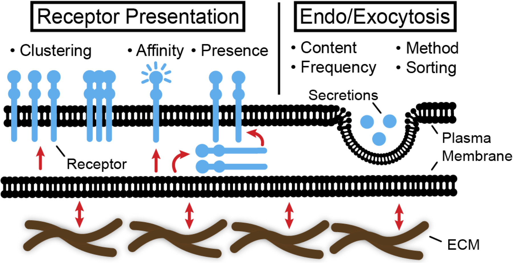
Biophysical properties such as elasticity, density and viscoelasticity of extracellular matrix (ECM) impact the immunomodulatory properties of stromal cells through outside-in signaling by regulating the receptor presentation, clustering, the affinity to ligands and endocytosis, as well as though inside-out signaling including exocytosis to control the release of secreted factors.
At the molecular level, external force is sufficient to increase affinities of B-cell receptors [75] and T-cell receptors [76] to antigens. Consistent with these results, 2D substrate rigidity is known to increase T-cell activation [77, 78], B-cell activation [79], and dendritic cell activation of T-cells in an actin polymerization-dependent manner [80]. Thus, in cell suspension without the matrix or on 2D substrates, it appears that increased biophysical forces enhance activation and surface presentation of immune receptors. However, in 3D environments, a recent study showed that tumor necrosis factor (TNF) receptor clustering on the plasma membrane of MSCs is enhanced in a softer 3D matrix in response to TNFα, thereby increasing the downstream production of monocyte factors [47]. Unlike 2D environments, spatial confinement is an important determinant of cell spreading in 3D [81–83], which may also impact plasma membrane dynamics, and subsequently receptor activation. Thus, the effect of matrix degradation and viscoelasticity needs to be considered in future studies to understand how 3D matrix mechanics impacts the activation of cytokine receptors in MSCs as well as the juxtacrine interactions between MSCs and immune cells.
Earlier studies showed that cell spreading on rigid plastic requires the addition of cell membrane, thereby driving biological processes that lead to increased membrane fusion, such as exocytosis [84, 85]. These studies have led to a physical model where cells with increased intracellular tension activate biological processes that decrease tension to maintain homeostasis by adding more membrane, as occurs in exocytosis [86]. This model seems to be consistent with the finding that receptors undergo more internalization on softer 2D substrates through activation of endocytosis [87], while lysosomal secretion by exocytosis is increased by stiffer substrates [88]. However, a recent study showed that MSCs increase secretion of immunomodulatory factors on softer substrates in an actomyosin-dependent manner [89], although it is possible that this phenomenon is due to changes in protein synthesis, rather than exocytosis per se. Indeed, a recent study shows that matrix stiffness does not impact constitutive protein secretion, but increased production of secreted proteins in softer substrates is due to increased transcription in response to inflammatory activation, though it occurs in a myosin-II independent manner [47]. In addition, increasing homotypic cell-cell interactions of MSCs by encapsulation in scaffolds with a larger porosity [90] or by forming spheroids [91] promotes paracrine factor secretions. While cell-cell interactions enhance tissue-scale tension [92], which could then result in increased exocytosis [86], the contribution of cell-cell interactions to protein exocytosis/endocytosis vs. synthesis remains to be dissected. Future studies will likely address roles of matrix mechanics in regulating different membrane vesicle trafficking mechanisms that impact protein secretions, most notably, extracellular vesicles, which have emerged as carriers of therapeutic cargo [93] that can transport through the matrix [94].
5. Biomaterial strategies to study roles of matrix biophysical cues on MSC-based immunomodulation
Many previous studies used biomaterial strategies to reveal the sensitivity of blood and immune cells to biophysical cues, a topic which has been reviewed extensively [40, 95–97]. In general, material biophysical cues are known to mediate fundamental processes of immune response, including deformation, adhesion, and trafficking of cells [98, 99], cell-cell interactions [76, 100], and antigen affinity [101, 102]. Recent studies have shown that some of these regulatory processes for blood and immune cells can also be relevant to understanding biophysical regulation of MSC-based immunomodulation. Emerging studies show that biophysical properties of biomaterials, including topography, porosity, dimensionality, and viscoelasticity may play important roles in MSC-based immunomodulation (Fig. 2).
Figure 2. Biologically inspired design of materials to study biophysical regulation of stromal cell-mediated immunomodulation.
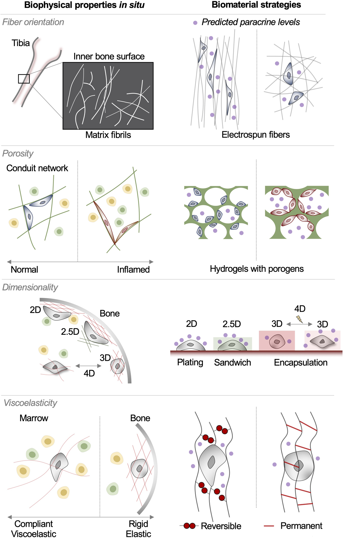
A deeper understanding of matrix organization and stromal cell-matrix interactions in immune organs, including bone marrow and lymph nodes, enables investigators to pursue appropriate biomaterial strategies to tune different matrix biophysical parameters, including fiber orientation, porosity, dimensionality, and viscoelasticity. Recent studies show the effect of these parameters on the level of paracrine secretions by stromal cells to mediate immunomodulation.
Matrix fibers are visible at the interface between inner bone surface and marrow, where they appear to exhibit different orientations [42]. To test the biological significance of matrix fiber orientation in stromal cell-mediated immunity, topography of substrates for cell adhesion can be controlled by various microfabrication approaches. In the context of studying stromal cell functions, soft lithography, electrospinning, and stereolithography-based printing have been employed. Soft lithography has been widely used to generate patterns of adhesive substrates at the microscale, including microgrooves and microposts [103]. In general, a mask with desired micropatterns is used to fabricate a photoresist mold, on which softer materials, such as polydimethylsiloxane (PDMS) can be casted. Patterned PDMS substrates can be precisely tuned in terms of mechanical properties and functionalization with matrix molecules so that they can be used to interface with MSCs. While this approach has been used extensively to study mechanical regulation of MSCs [104, 105], it remains to be leveraged to understand biophysical regulation of stromal cell-based immunomodulation. Recent studies employed different approaches to fabricate more physiologically relevant microenvironments with controlled substate topography to study their impact on stromal cell-based immunomodulation. Electrospinning can be used to generate fibrous matrices with independent control of architecture and mechanics [106]. Using this approach, it was shown that MSCs increase the secretion of immunomodulatory factors when cultured on aligned fibers than on random fibers in a Yes-associated protein (YAP) and focal adhesion kinase (FAK) dependent manner [107], suggesting the potential importance of matrix fiber orientation in regulating stromal cell-mediated immunity. In contrast to electrospinning, stereolithography-based printing offers more precise control of where matrix fibers can be placed in a given space, at the microscale [108]. This approach can also expand the repertoire of materials that can be used to control topography beyond PDMS-based materials, such as hydrogels that can be cured by light. By leveraging this approach, it was shown that varying the placement and intrinsic properties of discrete matrix signals differentially impacts cell volume and YAP-based mechanotransduction of MSCs, which could in turn impact MSC-based immunomodulation [109].
Tissues need to be porous to accommodate immune cell production and trafficking. However, hydrogels are generally nanoporous. Porosity of nanoporous hydrogels is often inversely correlated with stiffness, although stiffness of alginate hydrogels can be tuned independently of porosity based on an egg-box model [110]. Increasing porosity in hydrogels will likely provide cells with less spatially confined environments, which could facilitate cell spreading, migration and intercellular interactions. The most convenient way to achieve this goal is to use freeze-drying of crosslinked hydrogels where ice crystals create the macroporous voids, followed by reconstitution and seeding of cells [111]. Using this approach, a previous study showed that MSCs in macroporous alginate-based hydrogels increase the production of growth factors, such as vascular endothelial growth factor (VEGF) due to N-cadherin mediated cell-cell interactions as opposed to MSCs in nanoporous alginate hydrogels, while the microscale material stiffness is kept constant [90]. Consistent with this observation, MSC spheroids in alginate gels show higher VEGF secretion compared to dissociated cells [91]. However, a freeze-drying approach results in a broad pore size distribution. To overcome this limitation, encapsulation of degradable microgels has recently been used to form monodisperse pores in hydrogels independently of intrinsic material properties—in this context, the pore-forming microgels have been created either by an aerosol-based method [112] or a droplet microfluidic approach [113]. In addition, electrospinning or printing can be used to fabricate fibrous matrices with defined porosity [114]. These recent approaches will help advance our understanding of how microscale porosity impacts MSC-mediated immunity in conjunction with dynamic fluid flow as seen in lymph nodes.
Some stromal cells in tissues are present either on matrix fibers (2D) as in the conduit network, while others can be surrounded by the matrix as in marrow (3D). Substate dimensionality is generally controlled by seeding of cells on top of pre-formed materials (2D) or encapsulating cells in materials (3D) [115]. It is also possible to create an intermediate (‘2.5D’) condition where cells are sandwiched between two hydrogel layers [116], which can simulate the condition where cells are placed at the interface between two different regions, such as marrow and inner bone surface. A previous study showed that MSCs secrete less pro-inflammatory cytokines at the basal level in 3D compared to 2D in a non-hydrogel polymer scaffold [117]. Supporting this observation, another study reported that the expression of tryptophan 2,3-dioxygenase, which is associated with an immunosuppressive effect, is reduced when MSCs are encapsulated in 3D alginate-based hydrogels [118]. However, whether these effects are due to substrate dimensionality per se or other factors such as increased spatial confinement in 3D compared to 2D remains to be investigated by using strategies to selectively control porosity. With recent advances in temporal control of material properties [119], biophysical regulation of stromal cell-mediated immunity can also be studied over time (a ‘4th dimension’). The ‘4D’ material systems will be useful to understand how immunomodulatory properties of MSCs may change under temporal pathological conditions where tissues undergo stiffening over time such as in fibrosis and cancer, and the potential roles of mechanical memory [120] in regulating this process.
To simulate microscale variations in mechanical properties of natural marrow (Section 3), it is possible to leverage hydrogel systems with tunable matrix viscoelasticity. Matrix elasticity is a function of polymer crosslinking and can be controlled independently of ligand density and porosity either by conjugating a ligand to a polymer backbone [43, 121, 122] or interpenetrating a natural polymeric ligand with a synthetic polymer that is used to control elasticity [123]. In general, softer matrices have shown to be beneficial in enhancing basal secretion of different cytokines and growth factors by MSCs [124] and the responsiveness of MSCs to TNFα to increase the production of monocyte regulatory factors [47]. Since most tissues exhibit viscoelastic behaviors due to energy dissipation, efforts have been made to develop hydrogels with tunable viscoelasticity, which can be characterized by stress relaxation (decreased stress under constant strain), creep (increased strain under constant stress) or loss tangent (ratio between viscous modulus and elastic modulus). This has generally been achieved by employing reversible chemical bonds to crosslink hydrogels. For instance, alginate hydrogels undergo faster stress relaxation when they are crosslinked with ionic bonds vs. covalent bonds [125]. Decreasing molecular weight of alginate and introducing steric hinderance by PEG conjugation can further accelerate stress relaxation [81]. For non-ionic hydrogels, variation of monomer ratios [126], host-guest chemistry [127] and dynamic covalent crosslinking [128] have been used to introduce viscoelasticity. A recent study leveraged an interpenetrating network of collagen-I and alginate to show that MSCs in an ionically crosslinked hydrogel produce a higher level of anti-inflammatory factors in response to inflammatory challenge than MSCs in a covalently crosslinked hydrogel [129]. Together, these studies highlight the importance of both elastic and viscous material properties in regulating MSC-mediated immunomodulation.
6. Relevance of material biophysical cues to isolation and delivery of MSCs for immunomodulation
Understanding biophysical regulation of cell-matrix interactions may inform better strategies to isolate and deliver stromal cells of mesenchymal origin for immunomodulation (Fig. 3). Endogenous MSC populations in marrow are rare (~0.05%) [130]. Thus, most investigators have been using MSCs that are derived from plastic adherence followed by culture in low glucose medium to remove any adherent hematopoietic cells and to expand MSCs. This process can take more than one month to obtain sufficient cells for downstream applications. However, it is known that MSCs have mechanical memory [120]. Thus, prolonging culture time on plastic may impair the ability of MSCs to sense softer substrates, which might negatively impact the corresponding sensitivity of inflammatory activation [47] or the constitutive production of immunomodulatory factors [89]. To address this issue, it may be beneficial to expand MSCs in natural or biologically-inspired substrates. For instance, collagen-based scaffolds were shown to promote MSC seeding and survival after isolation [131], to facilitate MSC proliferation [132], and to preserve MSC phenotypes [133]. Culturing MSCs in spheroids within Arg-Gly-Asp (RGD)-conjugated alginate hydrogels could also increase MSC survival [91].
Figure 3. Biomaterials with tunable matrix biophysical properties improve the therapeutic potential of stromal cells.
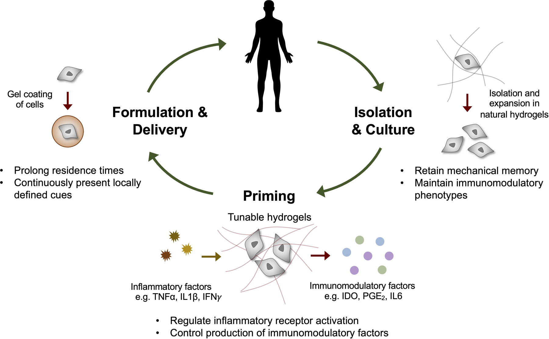
With proper design of material properties, stromal cell isolation and culture can be improved. Biophysical cues also impact the immunomodulatory properties of stromal cells by regulating the receptor expression, sensitivity and the production of secreted factors by stromal cells. Encapsulating stromal cells in biomaterials can enhance in vivo residence of donor cells by preventing direct contact between donor cells and the host defense, and enable continuous presentation of specifically defined cues to donor cells.
While MSCs modulate immune cells, they are not resistant to immune clearance. In fact, most donor MSCs are cleared from the host within 24~48 hours [134], unless they are delivered directly to the tissue of origin as shown by long-term engraftment studies in bone marrow [47, 135]. In addition, it is likely that MSCs delivered in circulation respond to shear stress, which can impact their survival and functions [57]. While MSCs are known to express low Class I Major Histocompatibility Complex (MHC) and no class II MHC, both receptors can undergo upregulation upon inflammatory activation, which in turn could trigger a host defense mechanism, including foreign body response (FBR) to remove donor MSCs after administration [136]. In addition, MSCs express a low level of CD47, ‘marker of self’, compared to blood cells [137], thereby making them susceptible to potential phagocytosis by macrophages [138]. While some biomaterials are also susceptible to FBR, MSCs themselves are known to reduce FBR of biomaterials by reducing macrophage activation [139]. It is also possible to modify the surface of biomaterials to reduce FBR as shown by introducing zwitterionic groups [140] or triazole analogs to alginate hydrogels, the latter of which prolongs allogeneic islet transplantation in primates [141]. However, the utility of these approaches in prolonging delivery of MSCs remains unclear. Interestingly, coating individual MSCs with a thin alginate gel by droplet-based microfluidics prolongs the residence time of MSCs after intravenous injection [142], which is further enhanced by modifying the alginate coating with adsorption of poly-l-lysine [143]. Whether these observations are also applicable to other routes of administration will likely depend on the immune milieu of the administration site. The ability to precisely tune material properties of the gel coating around single MSCs [144, 145] will not only help control the biodistribution of MSCs, but also allow local cue specification to donor MSCs for optimal efficacy.
7. Potential roles of biomaterials in evaluating MSCs as therapeutic products
Like other therapeutic products, MSCs as therapeutic cells must be evaluated in terms of potency, cooperativity, and efficacy (Fig. 4). In particular, potency assays that capture the relevant biological activity of therapeutic products are essential requirements to submit an investigational new drug application to U.S. Food and Drug Administration to use an MSC product as an immunotherapy [146]. Investigations into MSC mechanotransduction by using biomaterial design have elucidated the effects of microenvironmental properties on MSC phenotypes. However, conventional MSC potency assays use standard tissue culture plastic, which does not capture the sensitivity of MSCs to matrix biophysical cues in different physiological or pathological conditions. To best consider the effects of microenvironmental cues on the therapeutic potential of MSCs, it will be important to more completely define these effects in terms of the established framework of pharmacodynamics for small molecules and biologics [147] (potency, cooperativity, and efficacy), and consider how biomaterial design can impact each parameter.
Figure 4. Pharmacodynamics of cells.
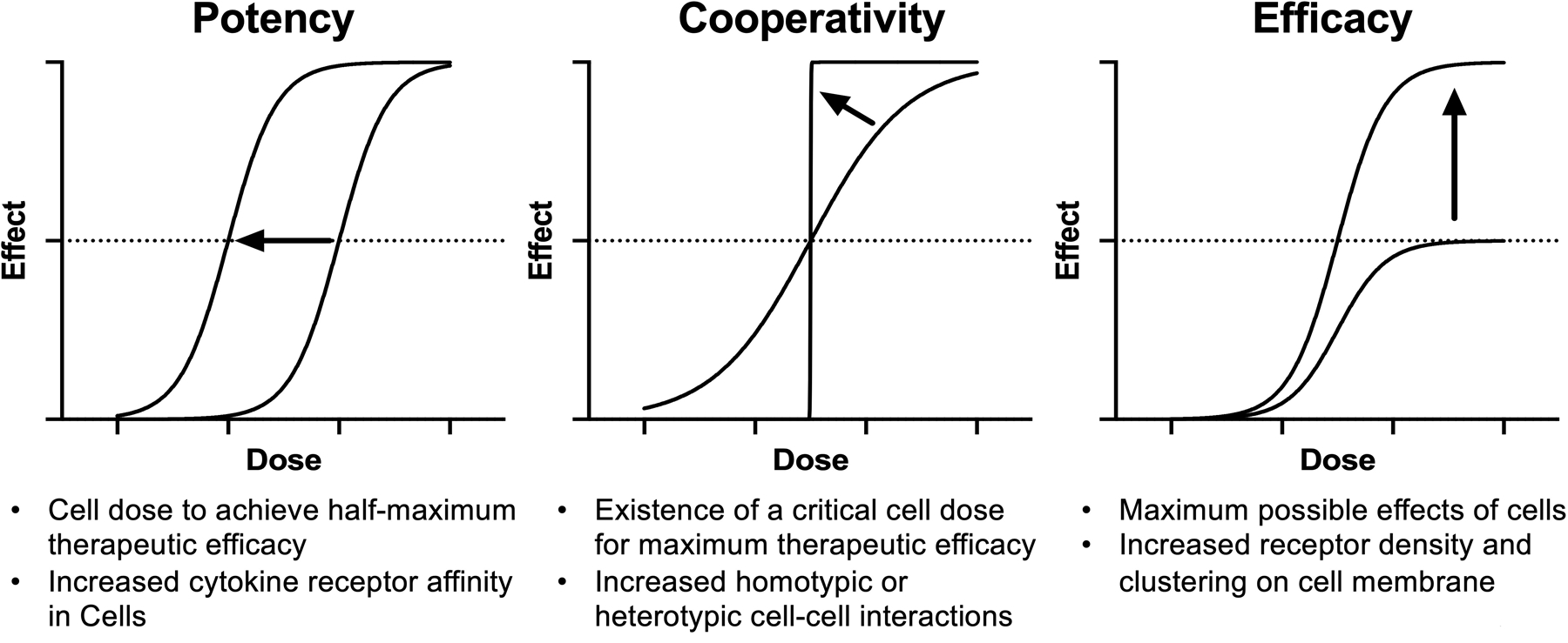
Dose response of cells is determined by three key parameters, including potency, cooperativity and efficacy as defined below each graph. Factors that can enhance each parameter are also described. Biomaterials can be designed to better inform each pharmacodynamic parameter of cells.
To determine the potency of MSCs as therapies, in vivo dose response studies can be performed to determine the cell dose that results in a half-maximum therapeutic response in an animal model. Since the primary mode of action by which MSCs modulate immune cells requires the exposure to inflammatory signals in the host [13], one way to predict in vivo dose response is to test the sensitivity of MSC products to inflammatory activation in vitro [148]. In pharmacology, the potency of receptor activation is influenced by intrinsic affinity or conformation of receptors, which is indeed influenced by substrate stiffness as demonstrated in immune cells [77–80]. Thus, understanding how different biomaterial properties impact the potency of inflammatory activation will be a key to better evaluating MSC potency for disease indications where mechanical properties of tissues vary. In addition, biomaterials may inform how a certain inflammatory ligand can be presented to MSCs for optimal immunomodulatory effects. For instance, some receptors, such as c-kit, can bind to ligands in an insoluble form at a lower concentration than the soluble form [149]. In this case, biomaterials can potentially be delivered along with MSCs to incorporate and present ligands from the host to MSCs to increase the potency of inflammatory activation.
Cooperativity indicates whether the intended effect is increased gradually with an increasing dose or as an “on/off” switch at a specific threshold dose, which is indicated by the slope of a dose response curve. The on/off response was previously reported in the context of MSC therapy where a systemic cytokine upregulation in the host could be observed only when a certain threshold dose of donor MSCs was used [150]. At the cellular level, positive cooperativity will likely suggest that the therapeutic activity of MSCs is amplified with a higher MSC dose due to homotypic interaction among MSC populations to enhance the production of immunomodulatory factors. In addition, positive cooperativity may indicate that when a single target immune cell is modulated by a single MSC, this causes other immune cells to become better influenced by other MSCs through heterotypic interactions. Thus, biomaterials that can tune cell-cell interactions [90] or spheroid formation [91] will inform the cooperativity of a given MSC product. The ability to precisely direct single cells by biomaterials [109, 144], and assemble single cells into clusters in a bottom-up manner [151–153] will also improve our ability to predict MSC cooperativity.
Efficacy of MSCs refers to the maximum possible effect of a given MSC product. Unlike potency, efficacy is influenced by receptor density and clustering on the cell membrane. Thus, membrane fluidity, endocytosis and exocytosis are likely important determinants to determine the efficacy of MSCs. Indeed, a recent study showed that matrix stiffness impacts the maximum response of TNFα activation in MSCs by regulating TNF receptor clustering [47]. However, the same study showed that unlike TNF receptor activation, matrix stiffness does not impact IFN receptor activation. Thus, mapping the effect of different matrix biophysical parameters on paracrine secretions of immunomodulatory factors will be an important goal in the field, which can be facilitated by combining single cell RNA sequencing [154] with single cell encapsulation approaches [142, 144], both of which are based on droplet-based microfluidics. In addition, both secretion and recycling of paracrine factors will likely impact the maximum net release of immunomodulatory factors from MSCs. At the cellular level, the ability of donor MSCs to interact with the host and gain access to immune cells is likely an important determinant of MSC efficacy, since MSCs need to undergo substantial mechanical squeezing to permeate biological tissues, while immune cells can do so much more readily [155, 156]. To address this issue, biomaterial design can be combined with microfabrication approaches [157] to predict the ability of MSCs to traffic through spatial confinement and hence interact with immune cells.
8. Potential roles of material biophysical cues in improving MSC-based immunotherapy for clinical applications
One major class of clinical indications for which MSCs have been tested is immunological rejection due to allogeneic transplantation or autoimmunity. In the context of bone marrow transplantation, T-cells in allogeneic donor marrow recognize the host as foreign due to human leukocyte antigen mismatches and attack the host within 3 months, leading to acute GvHD [158]. The clinical use of MSCs for acute GvHD has been approved in some countries for patients that are resistant to anti-inflammatory steroids [159]. However, the clinical outcomes have been variable [160]. In fact, two distinct phase 3 clinical trials failed to show beneficial outcomes by MSCs in steroid-resistant GvHD patients [161]. Similarly, MSCs have been tested to treat autoimmune disorders, most notably, the Crohn’s disease, leading to approval in Europe to treat fistulas, a common complication of Crohn’s disease [162]. However, a phase 3 trial of MSCs in Crohn’s disease was not successful in the U.S [163]. While a number of factors may have contributed to variable clinical outcomes in these cases, one plausible possibility is that patients are administered anti-inflammatory corticosteroids, which are important clinical interventions to temporarily alleviate the symptoms, but also likely reduce the level of inflammation necessary to activate MSCs to produce paracrine factors that confer immune tolerance [164]. Priming MSCs on soft [47, 124], viscoelastic [129] materials with aligned fiber orientation [107] can potentially help increase the sensitivity of MSCs to inflammatory activation even when inflammation in the host is alleviated by steroid treatment. In addition, material strategies to prolong the residence time of MSCs [142, 143] will enable the integration of attenuated inflammatory signals from the host by steroids.
MSCs have also been tested to treat acute tissue injuries. In particular, MSCs have shown to be efficacious in preclinical models of acute respiratory distress syndrome (ARDS) in part due to polarization of alveolar macrophages [165], which leads to increased phagocytosis [166] and restoration of vascular permeability [167] by secreting paracrine factors or extracellular vesicles. The number of clinical trials to test MSCs in ARDS have increased dramatically in the past year due to the COVID-19 pandemic [168], since ARDS is a major symptom of COVID-19 [169]. While MSCs were shown to be well-tolerated in phase 1 studies, their efficacy was shown to be unclear in a recent phase 2a clinical trial [170, 171]. Since pulmonary functions are significantly compromised in ARDS, it will be important to control the dose of MSCs to avoid occurrence of pulmonary embolism. Thus, material strategies to increase the sensitivity of MSCs to inflammatory activation or to delay the clearance of MSCs will likely help reduce the number of effective MSC doses needed for the treatment of ARDS.
In devising treatment strategies for chronic tissue injuries, it is not only important to consider inflammation but also subsequent aberrant tissue remodeling processes, eventually leading to fibrosis [172], which stiffens tissues due to increased collagen production and crosslinking [173]. For example, chronic GvHD leads to fibrosis in different organs, which contribute to long-term morbidity and mortality [174]. In addition, a subset of ARDS survivors can develop lung fibrosis later in life [175]. In this context, MSCs have shown to be potentially beneficial for treatment of myocardial infarction based on some phase 2 clinical studies, although a large randomized controlled trial remains to be completed [176]. A landmark preclinical study showed that the efficacy of MSCs in myocardial infarction requires the secretion of TNF-stimulated gene-6 (TSG6) [177], which has also been attributed to the efficacy of MSCs in preventing skin fibrosis in a preclinical model [178]. MSCs were also shown to be beneficial to prevent preclinical models of fibrotic lung injury and chronic obstructive pulmonary disease when administrated at early stages, but not later stages [179], highlighting the current limitations of MSC therapy in treating chronic tissue injuries. These limitations can be attributed by a couple of factors. First, inflammation subsides by the time fibrosis is diagnosed, thereby limiting inflammatory activation of MSCs to synthesize therapeutic factors once delivered to the host [13]. Second, significant biophysical changes in fibrotic microenvironments may influence donor MSCs to further adopt fibrotic phenotypes [180], thereby potentially limiting therapeutic efficacy or even exacerbating fibrosis as previously observed in a preclinical study of myocardial infarction [181]. To overcome these limitations, a recent study leveraged a conformal gel coating as a means to provide donor MSCs with locally specified biophysical and biochemical signals—using this approach, it was shown that MSCs singly coated with a soft gel that continuously presents recombinant TNFα facilitate the reversal of fibrotic lung injury in a preclinical model when delivered at later time points [145]. Thus, biomaterial design can be leveraged to design MSC-based therapeutics for chronic tissue injuries by providing control over how donor cells interact with specifically engineered environments versus those within the host.
9. Conclusion and Future Directions: Towards a single cell-level control for precision mechanomedicine
The contribution of matrix biophysical cues to MSC-based immunomodulation is an important yet largely unexplored area. Advances in biomaterials will enable better recapitulation of the physiological microenvironment to study MSC functions in various immune organs and disease contexts, as well as enable improvement of MSC priming, isolation, and delivery. With precise control of biophysical properties, emerging studies show that biomaterial systems can be tailored to direct MSC secretomes as potential therapeutics to treat immunological rejections and tissue injuries.
One major challenge of understanding the impact of matrix biophysical cues on MSC-based immunomodulation is heterogeneity of stromal cell populations, as recently demonstrated by single-cell RNAseq analysis [51, 182]. Supporting this challenge, a recent study showed that distinct MSC subpopulations exhibit differential mechanosensitivity to varied matrix elasticity, which influences their differentiation potential [183]. Since droplet-based microfluidic approaches have been used to profile cytokine secretions from single immune cells [184, 185], these approaches can potentially be combined with single cell encapsulation strategies to advance the field of single stromal cell mechanoimmunology. This line of approach can also be used to screen for individual MSC clones in engineered gel coatings with desired paracrine secretion activities, and subsequently deliver them to the host for therapeutic purposes.
The ability to miniaturize biomaterials down to the single cell level and specify their properties will likely advance the field of MSC-based immunotherapy. As shown in the airways [145], it will be possible to deliver MSCs along with locally specified synthetic microenvironments to tissues with a narrow space where immune cells reside via various routes of administration. Gel-coated MSCs can also be incorporated into larger tissue constructs or used as basic units to assemble into tissues, thereby potentially conferring immunomodulatory properties on engineered tissues. One of the biggest challenges in translating cell therapy is that mode of action is either poorly defined or considerably complex to immediately understand. However, as the clinical success of chimeric antigen receptor-T cell therapy has taught us [186], one way to overcome this challenge is to precisely define the input that confers a predictable therapeutic activity on engineered cells. Together with advances in synthetic biology [187], physical approaches to biomaterial design provide opportunities to specify microenvironmental cues around MSCs, to understand their impact on immunomodulation, and to use these cues directly to tailor MSCs for different disease indications.
The future of MSC-based immunotherapy remains optimistic and with a high ceiling for advancement by combining MSCs and biomaterials. With research efforts focused on investigating MSC mechanotransduction and developing novel biomaterial systems, the therapeutic potential of MSCs to control disorders of the immune system can be improved by leveraging biophysical cues.
Statement of Significance.
Stromal cells of mesenchymal origin are known to direct immune cell functions in vivo by secreting paracrine mediators. This property has been leveraged in developing mesenchymal stromal cell (MSC)-based therapeutics by adoptive transfer to treat immunological rejection and tissue injuries, which have been tested in over one thousand clinical trials to date, but with mixed success. Advances in biomaterial design have enabled precise control of biophysical cues based on how stromal cells interact with the extracellular matrix in microenvironments in situ. Investigators have begun to use this approach to understand how different matrix biophysical parameters, such as fiber orientation, porosity, dimensionality, and viscoelasticity impact stromal cell-mediated immunomodulation. The insights gained from this effort can potentially be used to precisely define the microenvironmental cues for isolation, priming, and delivery of MSCs, which can be tailored based on different disease indications for optimal therapeutic outcomes.
Acknowledgments
This work was supported by National Institutes of Health Grants R01-GM141147 (J.-W.S.), R01-HL141255 (J.-W.S.), R00-HL125884 (J.-W.S.), the Chicago Biomedical Consortium with support from the Searle Funds at the Chicago Community Trust (J.-W.S.), K99-AR079561 (S.W.W.), T32-HL07829 (S.L.), and American Heart Association Grant 19PRE34380087 (S.L.).
Footnotes
Publisher's Disclaimer: This is a PDF file of an unedited manuscript that has been accepted for publication. As a service to our customers we are providing this early version of the manuscript. The manuscript will undergo copyediting, typesetting, and review of the resulting proof before it is published in its final form. Please note that during the production process errors may be discovered which could affect the content, and all legal disclaimers that apply to the journal pertain.
Declaration of interests
The authors declare that they have no known competing financial interests or personal relationships that could have appeared to influence the work reported in this paper.
References
- [1].Friedenstein AJ, Chailakhjan RK, Lalykina KS, The development of fibroblast colonies in monolayer cultures of guinea-pig bone marrow and spleen cells, Cell Tissue Kinet 3(4) (1970) 393–403. [DOI] [PubMed] [Google Scholar]
- [2].Owen M, Friedenstein AJ, Stromal stem cells: marrow-derived osteogenic precursors, Ciba Found Symp 136 (1988) 42–60. [DOI] [PubMed] [Google Scholar]
- [3].Caplan AI, Mesenchymal stem cells, Journal of orthopaedic research : official publication of the Orthopaedic Research Society 9(5) (1991) 641–50. [DOI] [PubMed] [Google Scholar]
- [4].Pittenger MF, Mackay AM, Beck SC, Jaiswal RK, Douglas R, Mosca JD, Moorman MA, Simonetti DW, Craig S, Marshak DR, Multilineage potential of adult human mesenchymal stem cells, Science 284(5411) (1999) 143–7. [DOI] [PubMed] [Google Scholar]
- [5].Bartholomew A, Sturgeon C, Siatskas M, Ferrer K, McIntosh K, Patil S, Hardy W, Devine S, Ucker D, Deans R, Moseley A, Hoffman R, Mesenchymal stem cells suppress lymphocyte proliferation in vitro and prolong skin graft survival in vivo, Exp Hematol 30(1) (2002) 42–8. [DOI] [PubMed] [Google Scholar]
- [6].Maitra B, Szekely E, Gjini K, Laughlin MJ, Dennis J, Haynesworth SE, Koc ON, Human mesenchymal stem cells support unrelated donor hematopoietic stem cells and suppress T-cell activation, Bone Marrow Transplant 33(6) (2004) 597–604. [DOI] [PubMed] [Google Scholar]
- [7].Pittenger MF, Discher DE, Peault BM, Phinney DG, Hare JM, Caplan AI, Mesenchymal stem cell perspective: cell biology to clinical progress, NPJ Regen Med 4 (2019) 22. [DOI] [PMC free article] [PubMed] [Google Scholar]
- [8].Spaggiari GM, Capobianco A, Abdelrazik H, Becchetti F, Mingari MC, Moretta L, Mesenchymal stem cells inhibit natural killer-cell proliferation, cytotoxicity, and cytokine production: role of indoleamine 2,3-dioxygenase and prostaglandin E2, Blood 111(3) (2008) 1327–33. [DOI] [PubMed] [Google Scholar]
- [9].Corcione A, Benvenuto F, Ferretti E, Giunti D, Cappiello V, Cazzanti F, Risso M, Gualandi F, Mancardi GL, Pistoia V, Uccelli A, Human mesenchymal stem cells modulate B-cell functions, Blood 107(1) (2006) 367–72. [DOI] [PubMed] [Google Scholar]
- [10].English K, Barry FP, Mahon BP, Murine mesenchymal stem cells suppress dendritic cell migration, maturation and antigen presentation, Immunol Lett 115(1) (2008) 50–8. [DOI] [PubMed] [Google Scholar]
- [11].Spaggiari GM, Abdelrazik H, Becchetti F, Moretta L, MSCs inhibit monocyte-derived DC maturation and function by selectively interfering with the generation of immature DCs: central role of MSC-derived prostaglandin E2, Blood 113(26) (2009) 6576–83. [DOI] [PubMed] [Google Scholar]
- [12].Nemeth K, Leelahavanichkul A, Yuen PS, Mayer B, Parmelee A, Doi K, Robey PG, Leelahavanichkul K, Koller BH, Brown JM, Hu X, Jelinek I, Star RA, Mezey E, Bone marrow stromal cells attenuate sepsis via prostaglandin E(2)-dependent reprogramming of host macrophages to increase their interleukin-10 production, Nat Med 15(1) (2009) 42–9. [DOI] [PMC free article] [PubMed] [Google Scholar]
- [13].Wang Y, Chen X, Cao W, Shi Y, Plasticity of mesenchymal stem cells in immunomodulation: pathological and therapeutic implications, Nat Immunol 15(11) (2014) 1009–16. [DOI] [PubMed] [Google Scholar]
- [14].Ragelle H, Naba A, Larson BL, Zhou F, Prijic M, Whittaker CA, Del Rosario A, Langer R, Hynes RO, Anderson DG, Comprehensive proteomic characterization of stem cell-derived extracellular matrices, Biomaterials 128 (2017) 147–159. [DOI] [PMC free article] [PubMed] [Google Scholar]
- [15].Vining KH, Mooney DJ, Mechanical forces direct stem cell behaviour in development and regeneration, Nature reviews. Molecular cell biology 18(12) (2017) 728–742. [DOI] [PMC free article] [PubMed] [Google Scholar]
- [16].Zhang Y, Gao S, Xia J, Liu F, Hematopoietic Hierarchy - An Updated Roadmap, Trends Cell Biol 28(12) (2018) 976–986. [DOI] [PubMed] [Google Scholar]
- [17].Riether C, Schurch CM, Ochsenbein AF, Regulation of hematopoietic and leukemic stem cells by the immune system, Cell Death Differ 22(2) (2015) 187–98. [DOI] [PMC free article] [PubMed] [Google Scholar]
- [18].Ding L, Saunders TL, Enikolopov G, Morrison SJ, Endothelial and perivascular cells maintain haematopoietic stem cells, Nature 481(7382) (2012) 457–62. [DOI] [PMC free article] [PubMed] [Google Scholar]
- [19].Park D, Spencer JA, Koh BI, Kobayashi T, Fujisaki J, Clemens TL, Lin CP, Kronenberg HM, Scadden DT, Endogenous bone marrow MSCs are dynamic, fate-restricted participants in bone maintenance and regeneration, Cell stem cell 10(3) (2012) 259–72. [DOI] [PMC free article] [PubMed] [Google Scholar]
- [20].Sacchetti B, Funari A, Michienzi S, Di Cesare S, Piersanti S, Saggio I, Tagliafico E, Ferrari S, Robey PG, Riminucci M, Bianco P, Self-renewing osteoprogenitors in bone marrow sinusoids can organize a hematopoietic microenvironment, Cell 131(2) (2007) 324–36. [DOI] [PubMed] [Google Scholar]
- [21].Mendez-Ferrer S, Michurina TV, Ferraro F, Mazloom AR, Macarthur BD, Lira SA, Scadden DT, Ma’ayan A, Enikolopov GN, Frenette PS, Mesenchymal and haematopoietic stem cells form a unique bone marrow niche, Nature 466(7308) (2010) 829–34. [DOI] [PMC free article] [PubMed] [Google Scholar]
- [22].Kunisaki Y, Bruns I, Scheiermann C, Ahmed J, Pinho S, Zhang D, Mizoguchi T, Wei Q, Lucas D, Ito K, Mar JC, Bergman A, Frenette PS, Arteriolar niches maintain haematopoietic stem cell quiescence, Nature 502(7473) (2013) 637–43. [DOI] [PMC free article] [PubMed] [Google Scholar]
- [23].Asada N, Kunisaki Y, Pierce H, Wang Z, Fernandez NF, Birbrair A, Ma’ayan A, Frenette PS, Differential cytokine contributions of perivascular haematopoietic stem cell niches, Nat Cell Biol 19(3) (2017) 214–223. [DOI] [PMC free article] [PubMed] [Google Scholar]
- [24].Omatsu Y, Sugiyama T, Kohara H, Kondoh G, Fujii N, Kohno K, Nagasawa T, The essential functions of adipo-osteogenic progenitors as the hematopoietic stem and progenitor cell niche, Immunity 33(3) (2010) 387–99. [DOI] [PubMed] [Google Scholar]
- [25].Shi C, Jia T, Mendez-Ferrer S, Hohl TM, Serbina NV, Lipuma L, Leiner I, Li MO, Frenette PS, Pamer EG, Bone marrow mesenchymal stem and progenitor cells induce monocyte emigration in response to circulating toll-like receptor ligands, Immunity 34(4) (2011) 590–601. [DOI] [PMC free article] [PubMed] [Google Scholar]
- [26].Liu X, Ren S, Ge C, Cheng K, Zenke M, Keating A, Zhao RC, Sca-1+Lin-CD117-mesenchymal stem/stromal cells induce the generation of novel IRF8-controlled regulatory dendritic cells through Notch-RBP-J signaling, J Immunol 194(9) (2015) 4298–308. [DOI] [PubMed] [Google Scholar]
- [27].Zhang B, Liu R, Shi D, Liu X, Chen Y, Dou X, Zhu X, Lu C, Liang W, Liao L, Zenke M, Zhao RC, Mesenchymal stem cells induce mature dendritic cells into a novel Jagged-2-dependent regulatory dendritic cell population, Blood 113(1) (2009) 46–57. [DOI] [PubMed] [Google Scholar]
- [28].Cassatella MA, Mosna F, Micheletti A, Lisi V, Tamassia N, Cont C, Calzetti F, Pelletier M, Pizzolo G, Krampera M, Toll-like receptor-3-activated human mesenchymal stromal cells significantly prolong the survival and function of neutrophils, Stem cells 29(6) (2011) 1001–11. [DOI] [PubMed] [Google Scholar]
- [29].Raffaghello L, Bianchi G, Bertolotto M, Montecucco F, Busca A, Dallegri F, Ottonello L, Pistoia V, Human mesenchymal stem cells inhibit neutrophil apoptosis: a model for neutrophil preservation in the bone marrow niche, Stem cells 26(1) (2008) 151–62. [DOI] [PubMed] [Google Scholar]
- [30].Tokoyoda K, Egawa T, Sugiyama T, Choi BI, Nagasawa T, Cellular niches controlling B lymphocyte behavior within bone marrow during development, Immunity 20(6) (2004) 707–18. [DOI] [PubMed] [Google Scholar]
- [31].Fletcher AL, Acton SE, Knoblich K, Lymph node fibroblastic reticular cells in health and disease, Nature reviews. Immunology 15(6) (2015) 350–61. [DOI] [PMC free article] [PubMed] [Google Scholar]
- [32].Benezech C, White A, Mader E, Serre K, Parnell S, Pfeffer K, Ware CF, Anderson G, Caamano JH, Ontogeny of stromal organizer cells during lymph node development, Journal of immunology 184(8) (2010) 4521–30. [DOI] [PMC free article] [PubMed] [Google Scholar]
- [33].Bajenoff M, Egen JG, Koo LY, Laugier JP, Brau F, Glaichenhaus N, Germain RN, Stromal cell networks regulate lymphocyte entry, migration, and territoriality in lymph nodes, Immunity 25(6) (2006) 989–1001. [DOI] [PMC free article] [PubMed] [Google Scholar]
- [34].Link A, Vogt TK, Favre S, Britschgi MR, Acha-Orbea H, Hinz B, Cyster JG, Luther SA, Fibroblastic reticular cells in lymph nodes regulate the homeostasis of naive T cells, Nature immunology 8(11) (2007) 1255–65. [DOI] [PubMed] [Google Scholar]
- [35].Luther SA, Tang HL, Hyman PL, Farr AG, Cyster JG, Coexpression of the chemokines ELC and SLC by T zone stromal cells and deletion of the ELC gene in the plt/plt mouse, Proceedings of the National Academy of Sciences of the United States of America 97(23) (2000) 12694–9. [DOI] [PMC free article] [PubMed] [Google Scholar]
- [36].Fletcher AL, Lukacs-Kornek V, Reynoso ED, Pinner SE, Bellemare-Pelletier A, Curry MS, Collier AR, Boyd RL, Turley SJ, Lymph node fibroblastic reticular cells directly present peripheral tissue antigen under steady-state and inflammatory conditions, The Journal of experimental medicine 207(4) (2010) 689–97. [DOI] [PMC free article] [PubMed] [Google Scholar]
- [37].Wang X, Cho B, Suzuki K, Xu Y, Green JA, An J, Cyster JG, Follicular dendritic cells help establish follicle identity and promote B cell retention in germinal centers, The Journal of experimental medicine 208(12) (2011) 2497–510. [DOI] [PMC free article] [PubMed] [Google Scholar]
- [38].Katakai T, Suto H, Sugai M, Gonda H, Togawa A, Suematsu S, Ebisuno Y, Katagiri K, Kinashi T, Shimizu A, Organizer-like reticular stromal cell layer common to adult secondary lymphoid organs, Journal of immunology 181(9) (2008) 6189–200. [DOI] [PubMed] [Google Scholar]
- [39].Mueller SN, Matloubian M, Clemens DM, Sharpe AH, Freeman GJ, Gangappa S, Larsen CP, Ahmed R, Viral targeting of fibroblastic reticular cells contributes to immunosuppression and persistence during chronic infection, Proceedings of the National Academy of Sciences of the United States of America 104(39) (2007) 15430–5. [DOI] [PMC free article] [PubMed] [Google Scholar]
- [40].Wong SW, Lenzini S, Shin JW, Perspective: Biophysical regulation of cancerous and normal blood cell lineages in hematopoietic malignancies, APL Bioeng 2(3) (2018) 031802. [DOI] [PMC free article] [PubMed] [Google Scholar]
- [41].Jansen LE, Birch NP, Schiffman JD, Crosby AJ, Peyton SR, Mechanics of intact bone marrow, Journal of the mechanical behavior of biomedical materials 50 (2015) 299–307. [DOI] [PMC free article] [PubMed] [Google Scholar]
- [42].Ivanovska IL, Swift J, Spinler K, Dingal D, Cho S, Discher DE, Cross-linked matrix rigidity and soluble retinoids synergize in nuclear lamina regulation of stem cell differentiation, Molecular biology of the cell 28(14) (2017) 2010–2022. [DOI] [PMC free article] [PubMed] [Google Scholar]
- [43].Engler AJ, Sen S, Sweeney HL, Discher DE, Matrix elasticity directs stem cell lineage specification, Cell 126(4) (2006) 677–89. [DOI] [PubMed] [Google Scholar]
- [44].Alexander B, Daulton TL, Genin GM, Lipner J, Pasteris JD, Wopenka B, Thomopoulos S, The nanometre-scale physiology of bone: steric modelling and scanning transmission electron microscopy of collagen-mineral structure, J R Soc Interface 9(73) (2012) 1774–86. [DOI] [PMC free article] [PubMed] [Google Scholar]
- [45].Nilsson SK, Debatis ME, Dooner MS, Madri JA, Quesenberry PJ, Becker PS, Immunofluorescence characterization of key extracellular matrix proteins in murine bone marrow in situ, The journal of histochemistry and cytochemistry : official journal of the Histochemistry Society 46(3) (1998) 371–7. [DOI] [PubMed] [Google Scholar]
- [46].Zhou BO, Yue R, Murphy MM, Peyer JG, Morrison SJ, Leptin-receptor-expressing mesenchymal stromal cells represent the main source of bone formed by adult bone marrow, Cell stem cell 15(2) (2014) 154–68. [DOI] [PMC free article] [PubMed] [Google Scholar]
- [47].Wong SW, Lenzini S, Cooper MH, Mooney DJ, Shin JW, Soft extracellular matrix enhances inflammatory activation of mesenchymal stromal cells to induce monocyte production and trafficking, Sci Adv 6(15) (2020) eaaw0158. [DOI] [PMC free article] [PubMed] [Google Scholar]
- [48].Zhong Z, Akkus O, Effects of age and shear rate on the rheological properties of human yellow bone marrow, Biorheology 48(2) (2011) 89–97. [DOI] [PubMed] [Google Scholar]
- [49].Gurkan UA, Akkus O, The mechanical environment of bone marrow: a review, Annals of biomedical engineering 36(12) (2008) 1978–91. [DOI] [PubMed] [Google Scholar]
- [50].Liu FD, Pishesha N, Poon Z, Kaushik T, Van Vliet KJ, Material Viscoelastic Properties Modulate the Mesenchymal Stem Cell Secretome for Applications in Hematopoietic Recovery, ACS biomaterials science & engineering 3(12) (2017) 3292–3306. [DOI] [PubMed] [Google Scholar]
- [51].Baryawno N, Przybylski D, Kowalczyk MS, Kfoury Y, Severe N, Gustafsson K, Kokkaliaris KD, Mercier F, Tabaka M, Hofree M, Dionne D, Papazian A, Lee D, Ashenberg O, Subramanian A, Vaishnav ED, Rozenblatt-Rosen O, Regev A, Scadden DT, A Cellular Taxonomy of the Bone Marrow Stroma in Homeostasis and Leukemia, Cell 177(7) (2019) 1915–1932 e16. [DOI] [PMC free article] [PubMed] [Google Scholar]
- [52].Marenzana M, Arnett TR, The Key Role of the Blood Supply to Bone, Bone Res 1(3) (2013) 203–15. [DOI] [PMC free article] [PubMed] [Google Scholar]
- [53].Bixel MG, Kusumbe AP, Ramasamy SK, Sivaraj KK, Butz S, Vestweber D, Adams RH, Flow Dynamics and HSPC Homing in Bone Marrow Microvessels, Cell reports 18(7) (2017) 1804–1816. [DOI] [PMC free article] [PubMed] [Google Scholar]
- [54].Pinho S, Marchand T, Yang E, Wei Q, Nerlov C, Frenette PS, Lineage-Biased Hematopoietic Stem Cells Are Regulated by Distinct Niches, Developmental cell 44(5) (2018) 634–641 e4. [DOI] [PMC free article] [PubMed] [Google Scholar]
- [55].Shen B, Tasdogan A, Ubellacker JM, Zhang J, Nosyreva ED, Du L, Murphy MM, Hu S, Yi Y, Kara N, Liu X, Guela S, Jia Y, Ramesh V, Embree C, Mitchell EC, Zhao YC, Ju LA, Hu Z, Crane GM, Zhao Z, Syeda R, Morrison SJ, A mechanosensitive peri-arteriolar niche for osteogenesis and lymphopoiesis, Nature 591(7850) (2021) 438–444. [DOI] [PMC free article] [PubMed] [Google Scholar]
- [56].Zhang J, Wu Q, Johnson CB, Pham G, Kinder JM, Olsson A, Slaughter A, May M, Weinhaus B, D’Alessandro A, Engel JD, Jiang JX, Kofron JM, Huang LF, Prasath VBS, Way SS, Salomonis N, Grimes HL, Lucas D, In situ mapping identifies distinct vascular niches for myelopoiesis, Nature 590(7846) (2021) 457–462. [DOI] [PMC free article] [PubMed] [Google Scholar]
- [57].Diaz MF, Vaidya AB, Evans SM, Lee HJ, Aertker BM, Alexander AJ, Price KM, Ozuna JA, Liao GP, Aroom KR, Xue H, Gu L, Omichi R, Bedi S, Olson SD, Cox CS Jr., Wenzel PL, Biomechanical Forces Promote Immune Regulatory Function of Bone Marrow Mesenchymal Stromal Cells, Stem cells 35(5) (2017) 1259–1272. [DOI] [PMC free article] [PubMed] [Google Scholar]
- [58].Frodermann V, Rohde D, Courties G, Severe N, Schloss MJ, Amatullah H, McAlpine CS, Cremer S, Hoyer FF, Ji F, van Koeverden ID, Herisson F, Honold L, Masson GS, Zhang S, Grune J, Iwamoto Y, Schmidt SP, Wojtkiewicz GR, Lee IH, Gustafsson K, Pasterkamp G, de Jager SCA, Sadreyev RI, MacFadyen J, Libby P, Ridker P, Scadden DT, Naxerova K, Jeffrey KL, Swirski FK, Nahrendorf M, Exercise reduces inflammatory cell production and cardiovascular inflammation via instruction of hematopoietic progenitor cells, Nature medicine 25(11) (2019) 1761–1771. [DOI] [PMC free article] [PubMed] [Google Scholar]
- [59].Krishnamurty AT, Turley SJ, Lymph node stromal cells: cartographers of the immune system, Nature immunology 21(4) (2020) 369–380. [DOI] [PubMed] [Google Scholar]
- [60].Hayakawa M, Kobayashi M, Hoshino T, Direct contact between reticular fibers and migratory cells in the paracortex of mouse lymph nodes: a morphological and quantitative study, Arch Histol Cytol 51(3) (1988) 233–40. [DOI] [PubMed] [Google Scholar]
- [61].Kramer RH, Rosen SD, McDonald KA, Basement-membrane components associated with the extracellular matrix of the lymph node, Cell and tissue research 252(2) (1988) 367–75. [DOI] [PubMed] [Google Scholar]
- [62].Sobocinski GP, Toy K, Bobrowski WF, Shaw S, Anderson AO, Kaldjian EP, Ultrastructural localization of extracellular matrix proteins of the lymph node cortex: evidence supporting the reticular network as a pathway for lymphocyte migration, BMC Immunol 11 (2010) 42. [DOI] [PMC free article] [PubMed] [Google Scholar]
- [63].Sixt M, Kanazawa N, Selg M, Samson T, Roos G, Reinhardt DP, Pabst R, Lutz MB, Sorokin L, The conduit system transports soluble antigens from the afferent lymph to resident dendritic cells in the T cell area of the lymph node, Immunity 22(1) (2005) 19–29. [DOI] [PubMed] [Google Scholar]
- [64].Mano R, Becerra MF, Carver BS, Bosl GJ, Motzer RJ, Bajorin DF, Feldman DR, Sheinfeld J, Clinical Outcome of Patients with Fibrosis/Necrosis at Post-Chemotherapy Retroperitoneal Lymph Node Dissection for Advanced Germ Cell Tumors, J Urol 197(2) (2017) 391–397. [DOI] [PMC free article] [PubMed] [Google Scholar]
- [65].Martinez VG, Pankova V, Krasny L, Singh T, Makris S, White IJ, Benjamin AC, Dertschnig S, Horsnell HL, Kriston-Vizi J, Burden JJ, Huang PH, Tape CJ, Acton SE, Fibroblastic Reticular Cells Control Conduit Matrix Deposition during Lymph Node Expansion, Cell reports 29(9) (2019) 2810–2822 e5. [DOI] [PMC free article] [PubMed] [Google Scholar]
- [66].Acton SE, Farrugia AJ, Astarita JL, Mourao-Sa D, Jenkins RP, Nye E, Hooper S, van Blijswijk J, Rogers NC, Snelgrove KJ, Rosewell I, Moita LF, Stamp G, Turley SJ, Sahai E, Reis e Sousa C, Dendritic cells control fibroblastic reticular network tension and lymph node expansion, Nature 514(7523) (2014) 498–502. [DOI] [PMC free article] [PubMed] [Google Scholar]
- [67].Astarita JL, Cremasco V, Fu J, Darnell MC, Peck JR, Nieves-Bonilla JM, Song K, Kondo Y, Woodruff MC, Gogineni A, Onder L, Ludewig B, Weimer RM, Carroll MC, Mooney DJ, Xia L, Turley SJ, The CLEC-2-podoplanin axis controls the contractility of fibroblastic reticular cells and lymph node microarchitecture, Nature immunology 16(1) (2015) 75–84. [DOI] [PMC free article] [PubMed] [Google Scholar]
- [68].Lukacs-Kornek V, Malhotra D, Fletcher AL, Acton SE, Elpek KG, Tayalia P, Collier AR, Turley SJ, Regulated release of nitric oxide by nonhematopoietic stroma controls expansion of the activated T cell pool in lymph nodes, Nature immunology 12(11) (2011) 1096–104. [DOI] [PMC free article] [PubMed] [Google Scholar]
- [69].Brown FD, Sen DR, LaFleur MW, Godec J, Lukacs-Kornek V, Schildberg FA, Kim HJ, Yates KB, Ricoult SJH, Bi K, Trombley JD, Kapoor VN, Stanley IA, Cremasco V, Danial NN, Manning BD, Sharpe AH, Haining WN, Turley SJ, Fibroblastic reticular cells enhance T cell metabolism and survival via epigenetic remodeling, Nature immunology 20(12) (2019) 1668–1680. [DOI] [PMC free article] [PubMed] [Google Scholar]
- [70].Moore JE Jr., Bertram CD, Lymphatic System Flows, Annu Rev Fluid Mech 50 (2018) 459–482. [DOI] [PMC free article] [PubMed] [Google Scholar]
- [71].Havas E, Parviainen T, Vuorela J, Toivanen J, Nikula T, Vihko V, Lymph flow dynamics in exercising human skeletal muscle as detected by scintography, J Physiol 504 (Pt 1) (1997) 233–9. [DOI] [PMC free article] [PubMed] [Google Scholar]
- [72].Swartz MA, Lund AW, Lymphatic and interstitial flow in the tumour microenvironment: linking mechanobiology with immunity, Nat Rev Cancer 12(3) (2012) 210–9. [DOI] [PubMed] [Google Scholar]
- [73].Chang JE, Buechler MB, Gressier E, Turley SJ, Carroll MC, Mechanosensing by Peyer’s patch stroma regulates lymphocyte migration and mucosal antibody responses, Nature immunology 20(11) (2019) 1506–1516. [DOI] [PMC free article] [PubMed] [Google Scholar]
- [74].Tomei AA, Siegert S, Britschgi MR, Luther SA, Swartz MA, Fluid flow regulates stromal cell organization and CCL21 expression in a tissue-engineered lymph node microenvironment, Journal of immunology 183(7) (2009) 4273–83. [DOI] [PubMed] [Google Scholar]
- [75].Natkanski E, Lee WY, Mistry B, Casal A, Molloy JE, Tolar P, B cells use mechanical energy to discriminate antigen affinities, Science 340(6140) (2013) 1587–90. [DOI] [PMC free article] [PubMed] [Google Scholar]
- [76].Liu B, Chen W, Evavold BD, Zhu C, Accumulation of dynamic catch bonds between TCR and agonist peptide-MHC triggers T cell signaling, Cell 157(2) (2014) 357–368. [DOI] [PMC free article] [PubMed] [Google Scholar]
- [77].O’Connor RS, Hao X, Shen K, Bashour K, Akimova T, Hancock WW, Kam LC, Milone MC, Substrate rigidity regulates human T cell activation and proliferation, Journal of immunology 189(3) (2012) 1330–9. [DOI] [PMC free article] [PubMed] [Google Scholar]
- [78].Saitakis M, Dogniaux S, Goudot C, Bufi N, Asnacios S, Maurin M, Randriamampita C, Asnacios A, Hivroz C, Different TCR-induced T lymphocyte responses are potentiated by stiffness with variable sensitivity, eLife 6 (2017). [DOI] [PMC free article] [PubMed] [Google Scholar]
- [79].Shaheen S, Wan Z, Li Z, Chau A, Li X, Zhang S, Liu Y, Yi J, Zeng Y, Wang J, Chen X, Xu L, Chen W, Wang F, Lu Y, Zheng W, Shi Y, Sun X, Li Z, Xiong C, Liu W, Substrate stiffness governs the initiation of B cell activation by the concerted signaling of PKCbeta and focal adhesion kinase, eLife 6 (2017). [DOI] [PMC free article] [PubMed] [Google Scholar]
- [80].Blumenthal D, Chandra V, Avery L, Burkhardt JK, Mouse T cell priming is enhanced by maturation-dependent stiffening of the dendritic cell cortex, eLife 9 (2020). [DOI] [PMC free article] [PubMed] [Google Scholar]
- [81].Chaudhuri O, Gu L, Klumpers D, Darnell M, Bencherif SA, Weaver JC, Huebsch N, Lee HP, Lippens E, Duda GN, Mooney DJ, Hydrogels with tunable stress relaxation regulate stem cell fate and activity, Nature materials 15(3) (2016) 326–34. [DOI] [PMC free article] [PubMed] [Google Scholar]
- [82].Khetan S, Guvendiren M, Legant WR, Cohen DM, Chen CS, Burdick JA, Degradation-mediated cellular traction directs stem cell fate in covalently crosslinked three-dimensional hydrogels, Nature materials 12(5) (2013) 458–65. [DOI] [PMC free article] [PubMed] [Google Scholar]
- [83].Caliari SR, Vega SL, Kwon M, Soulas EM, Burdick JA, Dimensionality and spreading influence MSC YAP/TAZ signaling in hydrogel environments, Biomaterials 103 (2016) 314–323. [DOI] [PMC free article] [PubMed] [Google Scholar]
- [84].Raucher D, Sheetz MP, Cell spreading and lamellipodial extension rate is regulated by membrane tension, The Journal of cell biology 148(1) (2000) 127–36. [DOI] [PMC free article] [PubMed] [Google Scholar]
- [85].Gauthier NC, Fardin MA, Roca-Cusachs P, Sheetz MP, Temporary increase in plasma membrane tension coordinates the activation of exocytosis and contraction during cell spreading, Proceedings of the National Academy of Sciences of the United States of America 108(35) (2011) 14467–72. [DOI] [PMC free article] [PubMed] [Google Scholar]
- [86].Diz-Munoz A, Fletcher DA, Weiner OD, Use the force: membrane tension as an organizer of cell shape and motility, Trends Cell Biol 23(2) (2013) 47–53. [DOI] [PMC free article] [PubMed] [Google Scholar]
- [87].Du J, Chen X, Liang X, Zhang G, Xu J, He L, Zhan Q, Feng XQ, Chien S, Yang C, Integrin activation and internalization on soft ECM as a mechanism of induction of stem cell differentiation by ECM elasticity, Proceedings of the National Academy of Sciences of the United States of America 108(23) (2011) 9466–71. [DOI] [PMC free article] [PubMed] [Google Scholar]
- [88].Wang G, Nola S, Bovio S, Bun P, Coppey-Moisan M, Lafont F, Galli T, Biomechanical Control of Lysosomal Secretion Via the VAMP7 Hub: A Tug-of-War between VARP and LRRK1, iScience 4 (2018) 127–143. [DOI] [PMC free article] [PubMed] [Google Scholar]
- [89].Ji Y, Li J, Wei Y, Gao W, Fu X, Wang Y, Substrate stiffness affects the immunosuppressive and trophic function of hMSCs via modulating cytoskeletal polymerization and tension, Biomater Sci 7(12) (2019) 5292–5300. [DOI] [PubMed] [Google Scholar]
- [90].Qazi TH, Mooney DJ, Duda GN, Geissler S, Biomaterials that promote cell-cell interactions enhance the paracrine function of MSCs, Biomaterials 140 (2017) 103–114. [DOI] [PubMed] [Google Scholar]
- [91].Ho SS, Murphy KC, Binder BY, Vissers CB, Leach JK, Increased Survival and Function of Mesenchymal Stem Cell Spheroids Entrapped in Instructive Alginate Hydrogels, Stem cells translational medicine 5(6) (2016) 773–81. [DOI] [PMC free article] [PubMed] [Google Scholar]
- [92].Charras G, Yap AS, Tensile Forces and Mechanotransduction at Cell-Cell Junctions, Current biology : CB 28(8) (2018) R445–R457. [DOI] [PubMed] [Google Scholar]
- [93].Wiklander OPB, Brennan MA, Lotvall J, Breakefield XO, El Andaloussi S, Advances in therapeutic applications of extracellular vesicles, Science translational medicine 11(492) (2019). [DOI] [PMC free article] [PubMed] [Google Scholar]
- [94].Lenzini S, Bargi R, Chung G, Shin JW, Matrix mechanics and water permeation regulate extracellular vesicle transport, Nat Nanotechnol 15(3) (2020) 217–223. [DOI] [PMC free article] [PubMed] [Google Scholar]
- [95].Kim JK, Shin YJ, Ha LJ, Kim DH, Kim DH, Unraveling the Mechanobiology of the Immune System, Advanced healthcare materials 8(4) (2019) e1801332. [DOI] [PMC free article] [PubMed] [Google Scholar]
- [96].Zhang X, Kim TH, Thauland TJ, Li H, Majedi FS, Ly C, Gu Z, Butte MJ, Rowat AC, Li S, Unraveling the mechanobiology of immune cells, Curr Opin Biotechnol 66 (2020) 236–245. [DOI] [PMC free article] [PubMed] [Google Scholar]
- [97].Huse M, Mechanical forces in the immune system, Nature reviews. Immunology 17(11) (2017) 679–690. [DOI] [PMC free article] [PubMed] [Google Scholar]
- [98].Thelen M, Stein JV, How chemokines invite leukocytes to dance, Nat Immunol 9(9) (2008) 953–9. [DOI] [PubMed] [Google Scholar]
- [99].Alon R, Ley K, Cells on the run: shear-regulated integrin activation in leukocyte rolling and arrest on endothelial cells, Curr Opin Cell Biol 20(5) (2008) 525–32. [DOI] [PMC free article] [PubMed] [Google Scholar]
- [100].Das DK, Feng Y, Mallis RJ, Li X, Keskin DB, Hussey RE, Brady SK, Wang JH, Wagner G, Reinherz EL, Lang MJ, Force-dependent transition in the T-cell receptor beta-subunit allosterically regulates peptide discrimination and pMHC bond lifetime, Proc Natl Acad Sci U S A 112(5) (2015) 1517–22. [DOI] [PMC free article] [PubMed] [Google Scholar]
- [101].Zeng Y, Yi J, Wan Z, Liu K, Song P, Chau A, Wang F, Chang Z, Han W, Zheng W, Chen YH, Xiong C, Liu W, Substrate stiffness regulates B-cell activation, proliferation, class switch, and T-cell-independent antibody responses in vivo, Eur J Immunol 45(6) (2015) 1621–34. [DOI] [PubMed] [Google Scholar]
- [102].Wan Z, Zhang S, Fan Y, Liu K, Du F, Davey AM, Zhang H, Han W, Xiong C, Liu W, B cell activation is regulated by the stiffness properties of the substrate presenting the antigens, J Immunol 190(9) (2013) 4661–75. [DOI] [PubMed] [Google Scholar]
- [103].Qin D, Xia Y, Whitesides GM, Soft lithography for micro- and nanoscale patterning, Nature protocols 5(3) (2010) 491–502. [DOI] [PubMed] [Google Scholar]
- [104].Fu J, Wang YK, Yang MT, Desai RA, Yu X, Liu Z, Chen CS, Mechanical regulation of cell function with geometrically modulated elastomeric substrates, Nature methods 7(9) (2010) 733–6. [DOI] [PMC free article] [PubMed] [Google Scholar]
- [105].Yim EK, Pang SW, Leong KW, Synthetic nanostructures inducing differentiation of human mesenchymal stem cells into neuronal lineage, Experimental cell research 313(9) (2007) 1820–9. [DOI] [PMC free article] [PubMed] [Google Scholar]
- [106].Baker BM, Trappmann B, Wang WY, Sakar MS, Kim IL, Shenoy VB, Burdick JA, Chen CS, Cell-mediated fibre recruitment drives extracellular matrix mechanosensing in engineered fibrillar microenvironments, Nature materials 14(12) (2015) 1262–1268. [DOI] [PMC free article] [PubMed] [Google Scholar]
- [107].Wan S, Fu X, Ji Y, Li M, Shi X, Wang Y, FAK- and YAP/TAZ dependent mechanotransduction pathways are required for enhanced immunomodulatory properties of adipose-derived mesenchymal stem cells induced by aligned fibrous scaffolds, Biomaterials 171 (2018) 107–117. [DOI] [PubMed] [Google Scholar]
- [108].Gauvin R, Chen YC, Lee JW, Soman P, Zorlutuna P, Nichol JW, Bae H, Chen S, Khademhosseini A, Microfabrication of complex porous tissue engineering scaffolds using 3D projection stereolithography, Biomaterials 33(15) (2012) 3824–34. [DOI] [PMC free article] [PubMed] [Google Scholar]
- [109].Devine D, Vijayakumar V, Wong SW, Lenzini S, Newman P, Shin JW, Hydrogel Micropost Arrays with Single Post Tunability to Study Cell Volume and Mechanotransduction, Adv Biosyst 4(11) (2020) e2000012. [DOI] [PMC free article] [PubMed] [Google Scholar]
- [110].Lee KY, Mooney DJ, Alginate: properties and biomedical applications, Progress in polymer science 37(1) (2012) 106–126. [DOI] [PMC free article] [PubMed] [Google Scholar]
- [111].Stokols S, Tuszynski MH, The fabrication and characterization of linearly oriented nerve guidance scaffolds for spinal cord injury, Biomaterials 25(27) (2004) 5839–46. [DOI] [PubMed] [Google Scholar]
- [112].Huebsch N, Lippens E, Lee K, Mehta M, Koshy ST, Darnell MC, Desai RM, Madl CM, Xu M, Zhao X, Chaudhuri O, Verbeke C, Kim WS, Alim K, Mammoto A, Ingber DE, Duda GN, Mooney DJ, Matrix elasticity of void-forming hydrogels controls transplanted-stem-cell-mediated bone formation, Nature materials 14(12) (2015) 1269–77. [DOI] [PMC free article] [PubMed] [Google Scholar]
- [113].Highley CB, Song KH, Daly AC, Burdick JA, Jammed Microgel Inks for 3D Printing Applications, Adv Sci (Weinh) 6(1) (2019) 1801076. [DOI] [PMC free article] [PubMed] [Google Scholar]
- [114].Annabi N, Nichol JW, Zhong X, Ji C, Koshy S, Khademhosseini A, Dehghani F, Controlling the porosity and microarchitecture of hydrogels for tissue engineering, Tissue Eng Part B Rev 16(4) (2010) 371–83. [DOI] [PMC free article] [PubMed] [Google Scholar]
- [115].Baker BM, Chen CS, Deconstructing the third dimension: how 3D culture microenvironments alter cellular cues, Journal of cell science 125(Pt 13) (2012) 3015–24. [DOI] [PMC free article] [PubMed] [Google Scholar]
- [116].Rehfeldt F, Brown AE, Raab M, Cai S, Zajac AL, Zemel A, Discher DE, Hyaluronic acid matrices show matrix stiffness in 2D and 3D dictates cytoskeletal order and myosin-II phosphorylation within stem cells, Integr Biol (Camb) 4(4) (2012) 422–30. [DOI] [PubMed] [Google Scholar]
- [117].Valles G, Bensiamar F, Crespo L, Arruebo M, Vilaboa N, Saldana L, Topographical cues regulate the crosstalk between MSCs and macrophages, Biomaterials 37 (2015) 124–33. [DOI] [PMC free article] [PubMed] [Google Scholar]
- [118].Duggal S, Fronsdal KB, Szoke K, Shahdadfar A, Melvik JE, Brinchmann JE, Phenotype and gene expression of human mesenchymal stem cells in alginate scaffolds, Tissue engineering. Part A 15(7) (2009) 1763–73. [DOI] [PubMed] [Google Scholar]
- [119].Brown TE, Anseth KS, Spatiotemporal hydrogel biomaterials for regenerative medicine, Chem Soc Rev 46(21) (2017) 6532–6552. [DOI] [PMC free article] [PubMed] [Google Scholar]
- [120].Yang C, Tibbitt MW, Basta L, Anseth KS, Mechanical memory and dosing influence stem cell fate, Nature materials 13(6) (2014) 645–52. [DOI] [PMC free article] [PubMed] [Google Scholar]
- [121].Lo CM, Wang HB, Dembo M, Wang YL, Cell movement is guided by the rigidity of the substrate, Biophysical journal 79(1) (2000) 144–52. [DOI] [PMC free article] [PubMed] [Google Scholar]
- [122].Kong HJ, Polte TR, Alsberg E, Mooney DJ, FRET measurements of cell-traction forces and nano-scale clustering of adhesion ligands varied by substrate stiffness, Proceedings of the National Academy of Sciences of the United States of America 102(12) (2005) 4300–5. [DOI] [PMC free article] [PubMed] [Google Scholar]
- [123].Chaudhuri O, Koshy ST, Branco da Cunha C, Shin JW, Verbeke CS, Allison KH, Mooney DJ, Extracellular matrix stiffness and composition jointly regulate the induction of malignant phenotypes in mammary epithelium, Nature materials 13(10) (2014) 970–8. [DOI] [PubMed] [Google Scholar]
- [124].Rao VV, Vu MK, Ma H, Killaars AR, Anseth KS, Rescuing mesenchymal stem cell regenerative properties on hydrogel substrates post serial expansion, Bioeng Transl Med 4(1) (2019) 51–60. [DOI] [PMC free article] [PubMed] [Google Scholar]
- [125].Zhao X, Huebsch N, Mooney DJ, Suo Z, Stress-relaxation behavior in gels with ionic and covalent crosslinks, Journal of applied physics 107(6) (2010) 63509. [DOI] [PMC free article] [PubMed] [Google Scholar]
- [126].Cameron AR, Frith JE, Cooper-White JJ, The influence of substrate creep on mesenchymal stem cell behaviour and phenotype, Biomaterials 32(26) (2011) 5979–93. [DOI] [PubMed] [Google Scholar]
- [127].Loebel C, Mauck RL, Burdick JA, Local nascent protein deposition and remodelling guide mesenchymal stromal cell mechanosensing and fate in three-dimensional hydrogels, Nature materials 18(8) (2019) 883–891. [DOI] [PMC free article] [PubMed] [Google Scholar]
- [128].Accardo JV, Kalow JA, Reversibly tuning hydrogel stiffness through photocontrolled dynamic covalent crosslinks, Chem Sci 9(27) (2018) 5987–5993. [DOI] [PMC free article] [PubMed] [Google Scholar]
- [129].Vining KH, Stafford A, Mooney DJ, Sequential modes of crosslinking tune viscoelasticity of cell-instructive hydrogels, Biomaterials 188 (2019) 187–197. [DOI] [PMC free article] [PubMed] [Google Scholar]
- [130].Pinho S, Frenette PS, Haematopoietic stem cell activity and interactions with the niche, Nature reviews. Molecular cell biology 20(5) (2019) 303–320. [DOI] [PMC free article] [PubMed] [Google Scholar]
- [131].Niemeyer P, Krause U, Fellenberg J, Kasten P, Seckinger A, Ho AD, Simank HG, Evaluation of mineralized collagen and alpha-tricalcium phosphate as scaffolds for tissue engineering of bone using human mesenchymal stem cells, Cells Tissues Organs 177(2) (2004) 68–78. [DOI] [PubMed] [Google Scholar]
- [132].Chen XD, Dusevich V, Feng JQ, Manolagas SC, Jilka RL, Extracellular matrix made by bone marrow cells facilitates expansion of marrow-derived mesenchymal progenitor cells and prevents their differentiation into osteoblasts, J Bone Miner Res 22(12) (2007) 1943–56. [DOI] [PubMed] [Google Scholar]
- [133].Han S, Zhao Y, Xiao Z, Han J, Chen B, Chen L, Dai J, The three-dimensional collagen scaffold improves the stemness of rat bone marrow mesenchymal stem cells, J Genet Genomics 39(12) (2012) 633–41. [DOI] [PubMed] [Google Scholar]
- [134].Parekkadan B, Milwid JM, Mesenchymal stem cells as therapeutics, Annu Rev Biomed Eng 12 (2010) 87–117. [DOI] [PMC free article] [PubMed] [Google Scholar]
- [135].Tormin A, Li O, Brune JC, Walsh S, Schutz B, Ehinger M, Ditzel N, Kassem M, Scheding S, CD146 expression on primary nonhematopoietic bone marrow stem cells is correlated with in situ localization, Blood 117(19) (2011) 5067–77. [DOI] [PMC free article] [PubMed] [Google Scholar]
- [136].Ankrum JA, Ong JF, Karp JM, Mesenchymal stem cells: immune evasive, not immune privileged, Nature biotechnology 32(3) (2014) 252–60. [DOI] [PMC free article] [PubMed] [Google Scholar]
- [137].Subramanian S, Parthasarathy R, Sen S, Boder ET, Discher DE, Species- and cell type-specific interactions between CD47 and human SIRPalpha, Blood 107(6) (2006) 2548–56. [DOI] [PMC free article] [PubMed] [Google Scholar]
- [138].Hayes BH, Tsai RK, Dooling LJ, Kadu S, Lee JY, Pantano D, Rodriguez PL, Subramanian S, Shin JW, Discher DE, Macrophages show higher levels of engulfment after disruption of cis interactions between CD47 and the checkpoint receptor SIRPalpha, Journal of cell science 133(5) (2020). [DOI] [PMC free article] [PubMed] [Google Scholar]
- [139].Swartzlander MD, Blakney AK, Amer LD, Hankenson KD, Kyriakides TR, Bryant SJ, Immunomodulation by mesenchymal stem cells combats the foreign body response to cell-laden synthetic hydrogels, Biomaterials 41 (2015) 79–88. [DOI] [PMC free article] [PubMed] [Google Scholar]
- [140].Zhang L, Cao Z, Bai T, Carr L, Ella-Menye JR, Irvin C, Ratner BD, Jiang S, Zwitterionic hydrogels implanted in mice resist the foreign-body reaction, Nature biotechnology 31(6) (2013) 553–6. [DOI] [PubMed] [Google Scholar]
- [141].Vegas AJ, Veiseh O, Doloff JC, Ma M, Tam HH, Bratlie K, Li J, Bader AR, Langan E, Olejnik K, Fenton P, Kang JW, Hollister-Locke J, Bochenek MA, Chiu A, Siebert S, Tang K, Jhunjhunwala S, Aresta-Dasilva S, Dholakia N, Thakrar R, Vietti T, Chen M, Cohen J, Siniakowicz K, Qi M, McGarrigle J, Graham AC, Lyle S, Harlan DM, Greiner DL, Oberholzer J, Weir GC, Langer R, Anderson DG, Combinatorial hydrogel library enables identification of materials that mitigate the foreign body response in primates, Nature biotechnology 34(3) (2016) 345–52. [DOI] [PMC free article] [PubMed] [Google Scholar]
- [142].Mao AS, Shin JW, Utech S, Wang H, Uzun O, Li W, Cooper M, Hu Y, Zhang L, Weitz DA, Mooney DJ, Deterministic encapsulation of single cells in thin tunable microgels for niche modelling and therapeutic delivery, Nature materials 16(2) (2017) 236–243. [DOI] [PMC free article] [PubMed] [Google Scholar]
- [143].Mao AS, Ozkale B, Shah NJ, Vining KH, Descombes T, Zhang L, Tringides CM, Wong SW, Shin JW, Scadden DT, Weitz DA, Mooney DJ, Programmable microencapsulation for enhanced mesenchymal stem cell persistence and immunomodulation, Proceedings of the National Academy of Sciences of the United States of America 116(31) (2019) 15392–15397. [DOI] [PMC free article] [PubMed] [Google Scholar]
- [144].Wong SW, Lenzini S, Bargi R, Feng Z, Macaraniag C, Lee JC, Peng Z, Shin JW, Controlled Deposition of 3D Matrices to Direct Single Cell Functions, Adv Sci (Weinh) 7(20) (2020) 2001066. [DOI] [PMC free article] [PubMed] [Google Scholar]
- [145].Wong SW, Tamatam CR, Cho IS, Toth PT, Bargi R, Belvitch P, Lee JC, Rehman J, Reddy SP, Shin JW, Inhibition of aberrant tissue remodelling by mesenchymal stromal cells singly coated with soft gels presenting defined chemomechanical cues, Nature biomedical engineering (2021). [DOI] [PMC free article] [PubMed] [Google Scholar]
- [146].de Wolf C, van de Bovenkamp M, Hoefnagel M, Regulatory perspective on in vitro potency assays for human mesenchymal stromal cells used in immunotherapy, Cytotherapy 19(7) (2017) 784–797. [DOI] [PubMed] [Google Scholar]
- [147].D.E.W. C, V.D.B. M, Hoefnagel M, Regulatory perspective on in vitro potency assays for human dendritic cells used in anti-tumor immunotherapy, Cytotherapy 20(11) (2018) 1289–1308. [DOI] [PubMed] [Google Scholar]
- [148].Chinnadurai R, Rajan D, Qayed M, Arafat D, Garcia M, Liu Y, Kugathasan S, Anderson LJ, Gibson G, Galipeau J, Potency Analysis of Mesenchymal Stromal Cells Using a Combinatorial Assay Matrix Approach, Cell Rep 22(9) (2018) 2504–2517. [DOI] [PMC free article] [PubMed] [Google Scholar]
- [149].Horiuchi K, Morioka H, Takaishi H, Akiyama H, Blobel CP, Toyama Y, Ectodomain shedding of FLT3 ligand is mediated by TNF-alpha converting enzyme, Journal of immunology 182(12) (2009) 7408–14. [DOI] [PMC free article] [PubMed] [Google Scholar]
- [150].Lee OJ, Luk F, Korevaar SS, Koch TG, Baan CC, Merino A, Hoogduijn MJ, The Importance of Dosing, Timing, and (in)Activation of Adipose Tissue-Derived Mesenchymal Stromal Cells on Their Immunomodulatory Effects, Stem Cells Dev 29(1) (2020) 38–48. [DOI] [PubMed] [Google Scholar]
- [151].Hu Y, Mao AS, Desai RM, Wang H, Weitz DA, Mooney DJ, Controlled self-assembly of alginate microgels by rapidly binding molecule pairs, Lab on a chip 17(14) (2017) 2481–2490. [DOI] [PMC free article] [PubMed] [Google Scholar]
- [152].Zhang L, Chen K, Zhang H, Pang B, Choi CH, Mao AS, Liao H, Utech S, Mooney DJ, Wang H, Weitz DA, Microfluidic Templated Multicompartment Microgels for 3D Encapsulation and Pairing of Single Cells, Small 14(9) (2018). [DOI] [PubMed] [Google Scholar]
- [153].Todhunter ME, Jee NY, Hughes AJ, Coyle MC, Cerchiari A, Farlow J, Garbe JC, LaBarge MA, Desai TA, Gartner ZJ, Programmed synthesis of three-dimensional tissues, Nature methods 12(10) (2015) 975–81. [DOI] [PMC free article] [PubMed] [Google Scholar]
- [154].Macosko EZ, Basu A, Satija R, Nemesh J, Shekhar K, Goldman M, Tirosh I, Bialas AR, Kamitaki N, Martersteck EM, Trombetta JJ, Weitz DA, Sanes JR, Shalek AK, Regev A, McCarroll SA, Highly Parallel Genome-wide Expression Profiling of Individual Cells Using Nanoliter Droplets, Cell 161(5) (2015) 1202–1214. [DOI] [PMC free article] [PubMed] [Google Scholar]
- [155].Shin JW, Spinler KR, Swift J, Chasis JA, Mohandas N, Discher DE, Lamins regulate cell trafficking and lineage maturation of adult human hematopoietic cells, Proceedings of the National Academy of Sciences of the United States of America 110(47) (2013) 18892–7. [DOI] [PMC free article] [PubMed] [Google Scholar]
- [156].Harada T, Swift J, Irianto J, Shin JW, Spinler KR, Athirasala A, Diegmiller R, Dingal PC, Ivanovska IL, Discher DE, Nuclear lamin stiffness is a barrier to 3D migration, but softness can limit survival, The Journal of cell biology 204(5) (2014) 669–82. [DOI] [PMC free article] [PubMed] [Google Scholar]
- [157].Lautscham LA, Kammerer C, Lange JR, Kolb T, Mark C, Schilling A, Strissel PL, Strick R, Gluth C, Rowat AC, Metzner C, Fabry B, Migration in Confined 3D Environments Is Determined by a Combination of Adhesiveness, Nuclear Volume, Contractility, and Cell Stiffness, Biophysical journal 109(5) (2015) 900–13. [DOI] [PMC free article] [PubMed] [Google Scholar]
- [158].Zeiser R, Blazar BR, Acute Graft-versus-Host Disease - Biologic Process, Prevention, and Therapy, The New England journal of medicine 377(22) (2017) 2167–2179. [DOI] [PMC free article] [PubMed] [Google Scholar]
- [159].Cyranoski D, Canada approves stem cell product, Nature biotechnology 30 (2012) 571. [Google Scholar]
- [160].Amorin B, Alegretti AP, Valim V, Pezzi A, Laureano AM, da Silva MA, Wieck A, Silla L, Mesenchymal stem cell therapy and acute graft-versus-host disease: a review, Hum Cell 27(4) (2014) 137–50. [DOI] [PMC free article] [PubMed] [Google Scholar]
- [161].Galipeau J, The mesenchymal stromal cells dilemma--does a negative phase III trial of random donor mesenchymal stromal cells in steroid-resistant graft-versus-host disease represent a death knell or a bump in the road?, Cytotherapy 15(1) (2013) 2–8. [DOI] [PubMed] [Google Scholar]
- [162].Scott LJ, Darvadstrocel: A Review in Treatment-Refractory Complex Perianal Fistulas in Crohn’s Disease, BioDrugs 32(6) (2018) 627–634. [DOI] [PubMed] [Google Scholar]
- [163].Allison M, Genzyme backs Osiris, despite Prochymal flop, Nature biotechnology 27(11) (2009) 966–7. [DOI] [PubMed] [Google Scholar]
- [164].Chen X, Gan Y, Li W, Su J, Zhang Y, Huang Y, Roberts AI, Han Y, Li J, Wang Y, Shi Y, The interaction between mesenchymal stem cells and steroids during inflammation, Cell death & disease 5 (2014) e1009. [DOI] [PMC free article] [PubMed] [Google Scholar]
- [165].Morrison TJ, Jackson MV, Cunningham EK, Kissenpfennig A, McAuley DF, O’Kane CM, Krasnodembskaya AD, Mesenchymal Stromal Cells Modulate Macrophages in Clinically Relevant Lung Injury Models by Extracellular Vesicle Mitochondrial Transfer, American journal of respiratory and critical care medicine 196(10) (2017) 1275–1286. [DOI] [PMC free article] [PubMed] [Google Scholar]
- [166].Masterson C, Devaney J, Horie S, O’Flynn L, Deedigan L, Elliman S, Barry F, O’Brien T, O’Toole D, Laffey JG, Syndecan-2-positive, Bone Marrow-derived Human Mesenchymal Stromal Cells Attenuate Bacterial-induced Acute Lung Injury and Enhance Resolution of Ventilator-induced Lung Injury in Rats, Anesthesiology 129(3) (2018) 502–516. [DOI] [PubMed] [Google Scholar]
- [167].Shen Y, Song J, Wang Y, Chen Z, Zhang L, Yu J, Zhu D, Zhong M, M2 macrophages promote pulmonary endothelial cells regeneration in sepsis-induced acute lung injury, Annals of translational medicine 7(7) (2019) 142. [DOI] [PMC free article] [PubMed] [Google Scholar]
- [168].Wang W, Lei W, Jiang L, Gao S, Hu S, Zhao ZG, Niu CY, Zhao ZA, Therapeutic mechanisms of mesenchymal stem cells in acute respiratory distress syndrome reveal potentials for Covid-19 treatment, J Transl Med 19(1) (2021) 198. [DOI] [PMC free article] [PubMed] [Google Scholar]
- [169].Ackermann M, Verleden SE, Kuehnel M, Haverich A, Welte T, Laenger F, Vanstapel A, Werlein C, Stark H, Tzankov A, Li WW, Li VW, Mentzer SJ, Jonigk D, Pulmonary Vascular Endothelialitis, Thrombosis, and Angiogenesis in Covid-19, The New England journal of medicine 383(2) (2020) 120–128. [DOI] [PMC free article] [PubMed] [Google Scholar]
- [170].Zhang H, Li Y, Slutsky AS, Precision medicine for cell therapy in acute respiratory distress syndrome, The Lancet. Respiratory medicine 7(4) (2019) e13. [DOI] [PubMed] [Google Scholar]
- [171].Matthay MA, Calfee CS, Zhuo H, Thompson BT, Wilson JG, Levitt JE, Rogers AJ, Gotts JE, Wiener-Kronish JP, Bajwa EK, Donahoe MP, McVerry BJ, Ortiz LA, Exline M, Christman JW, Abbott J, Delucchi KL, Caballero L, McMillan M, McKenna DH, Liu KD, Treatment with allogeneic mesenchymal stromal cells for moderate to severe acute respiratory distress syndrome (START study): a randomised phase 2a safety trial, The Lancet. Respiratory medicine 7(2) (2019) 154–162. [DOI] [PMC free article] [PubMed] [Google Scholar]
- [172].Pinet K, McLaughlin KA, Mechanisms of physiological tissue remodeling in animals: Manipulating tissue, organ, and organism morphology, Dev Biol 451(2) (2019) 134–145. [DOI] [PubMed] [Google Scholar]
- [173].Henderson NC, Rieder F, Wynn TA, Fibrosis: from mechanisms to medicines, Nature 587(7835) (2020) 555–566. [DOI] [PMC free article] [PubMed] [Google Scholar]
- [174].Zeiser R, Blazar BR, Pathophysiology of Chronic Graft-versus-Host Disease and Therapeutic Targets, The New England journal of medicine 377(26) (2017) 2565–2579. [DOI] [PubMed] [Google Scholar]
- [175].Burnham EL, Janssen WJ, Riches DW, Moss M, Downey GP, The fibroproliferative response in acute respiratory distress syndrome: mechanisms and clinical significance, Eur Respir J 43(1) (2014) 276–85. [DOI] [PMC free article] [PubMed] [Google Scholar]
- [176].Jeong H, Yim HW, Park HJ, Cho Y, Hong H, Kim NJ, Oh IH, Mesenchymal Stem Cell Therapy for Ischemic Heart Disease: Systematic Review and Meta-analysis, Int J Stem Cells 11(1) (2018) 1–12. [DOI] [PMC free article] [PubMed] [Google Scholar]
- [177].Lee RH, Pulin AA, Seo MJ, Kota DJ, Ylostalo J, Larson BL, Semprun-Prieto L, Delafontaine P, Prockop DJ, Intravenous hMSCs improve myocardial infarction in mice because cells embolized in lung are activated to secrete the anti-inflammatory protein TSG-6, Cell stem cell 5(1) (2009) 54–63. [DOI] [PMC free article] [PubMed] [Google Scholar]
- [178].Qi Y, Jiang D, Sindrilaru A, Stegemann A, Schatz S, Treiber N, Rojewski M, Schrezenmeier H, Vander Beken S, Wlaschek M, Bohm M, Seitz A, Scholz N, Durselen L, Brinckmann J, Ignatius A, Scharffetter-Kochanek K, TSG-6 released from intradermally injected mesenchymal stem cells accelerates wound healing and reduces tissue fibrosis in murine full-thickness skin wounds, J Invest Dermatol 134(2) (2014) 526–537. [DOI] [PubMed] [Google Scholar]
- [179].Cruz FF, Rocco PRM, The potential of mesenchymal stem cell therapy for chronic lung disease, Expert Rev Respir Med 14(1) (2020) 31–39. [DOI] [PubMed] [Google Scholar]
- [180].El Agha E, Kramann R, Schneider RK, Li X, Seeger W, Humphreys BD, Bellusci S, Mesenchymal Stem Cells in Fibrotic Disease, Cell stem cell 21(2) (2017) 166–177. [DOI] [PubMed] [Google Scholar]
- [181].Breitbach M, Bostani T, Roell W, Xia Y, Dewald O, Nygren JM, Fries JW, Tiemann K, Bohlen H, Hescheler J, Welz A, Bloch W, Jacobsen SE, Fleischmann BK, Potential risks of bone marrow cell transplantation into infarcted hearts, Blood 110(4) (2007) 1362–9. [DOI] [PubMed] [Google Scholar]
- [182].Buechler MB, Pradhan RN, Krishnamurty AT, Cox C, Calviello AK, Wang AW, Yang YA, Tam L, Caothien R, Roose-Girma M, Modrusan Z, Arron JR, Bourgon R, Muller S, Turley SJ, Cross-tissue organization of the fibroblast lineage, Nature 593(7860) (2021) 575–579. [DOI] [PubMed] [Google Scholar]
- [183].Brielle S, Bavli D, Motzik A, Kan-Tor Y, Sun X, Kozulin C, Avni B, Ram O, Buxboim A, Delineating the heterogeneity of matrix-directed differentiation toward soft and stiff tissue lineages via single-cell profiling, Proceedings of the National Academy of Sciences of the United States of America 118(19) (2021). [DOI] [PMC free article] [PubMed] [Google Scholar]
- [184].Eyer K, Doineau RCL, Castrillon CE, Briseno-Roa L, Menrath V, Mottet G, England P, Godina A, Brient-Litzler E, Nizak C, Jensen A, Griffiths AD, Bibette J, Bruhns P, Baudry J, Single-cell deep phenotyping of IgG-secreting cells for high-resolution immune monitoring, Nature biotechnology 35(10) (2017) 977–982. [DOI] [PubMed] [Google Scholar]
- [185].Chokkalingam V, Tel J, Wimmers F, Liu X, Semenov S, Thiele J, Figdor CG, Huck WT, Probing cellular heterogeneity in cytokine-secreting immune cells using droplet-based microfluidics, Lab on a chip 13(24) (2013) 4740–4. [DOI] [PubMed] [Google Scholar]
- [186].June CH, O’Connor RS, Kawalekar OU, Ghassemi S, Milone MC, CAR T cell immunotherapy for human cancer, Science 359(6382) (2018) 1361–1365. [DOI] [PubMed] [Google Scholar]
- [187].Lim WA, June CH, The Principles of Engineering Immune Cells to Treat Cancer, Cell 168(4) (2017) 724–740. [DOI] [PMC free article] [PubMed] [Google Scholar]


