Abstract
Dural arteriovenous fistulas (DAVF) are rare acquired lesions resulting from abnormal shunting between intracranial dural arteries and venous system. Typically arising from structural weakness of the dura and a coinciding trigger factor, DAVFs can present with similar clinical and imaging characteristics to sinus thrombosis. A 61-year-old male with a history of meningioma previously managed with subtotal resection and stereotactic radiosurgery presented with progressive right-sided vision loss and bilateral papilledema. Initial imaging suggested possible sinus occlusion. Catheter angiogram revealed a Borden-Shucart grade III DAVF of the superior sagittal sinus and elevated venous pressures and the patient subsequently underwent endovascular transarterial intervention twice. We report on the first case of a superior sagittal sinus DAVF occurring after surgical resection of a parasagittal meningioma.
Keywords: Superior sagittal sinus, Dural arteriovenous fistula, Meningioma, Endovascular therapy
INTRODUCTION
Dural arteriovenous fistulas (DAVF) are rare acquired lesions defined as an abnormal shunt between the intracranial dural arteries and the venous system, accounting for 10% to 15% of all intracranial arteriovenous malformations (AVMs) [8,10,15-17]. The etiology of DAVFs is controversial, as venous thrombosis, intracranial surgery, tumor, puerperium, trauma, hypercoagulable state, and congenital causes have been found to be associated to these lesions [16]. These lesions are thought to arise from structural weaknesses of the dura coinciding with a trigger factor, such as venous thrombosis or injury due to mass effect from a tumor or local infection causing inflammation, fibrosis, and angiogenesis with the body compensating for this by attempts at recanalization [15,16].
The natural history of DAVFs are variable, as patients may be asymptomatic at all, have benign symptomatology for many years, and a small group of cases exhibiting aggressive neurological behavior [2]. Higher-grade lesions (Borden-Shucart grade 2/3, Cognard grade IIb, IIa+b, III/IV) are more likely to become symptomatic with patients developing cranial neuropathies, visual loss, retro-orbital pain, and chemosis or proptosis with cavernous (or precavernous) and tentorial DAVFs or pulsatile tinnitus, encephalopathy, parkinsonism, and cerebellar dysfunction with transverse sigmoid sinus DAVFs [21]. Treatment of lesions are based on an analysis of the patient’s symptoms, medical comorbidities, and risk of intracranial hemorrhage and may consist of conservative management (observation or carotid compression), endovascular treatment, surgery, or radiosurgery [17,21]. With the rise of transarterial embolization with Onyx (Medtronic, Irvine, CA, USA), endovascular management has become the most popular treatment option in patients presenting with DAVF [17].
DAVF of the superior sagittal sinus (SSS) is even rarer, occurring in 4.6% to 11% of all cases [20]. As far as we are aware this is the first report of SSS DAVF caused by treatment of a parasagittal meningioma presenting as sinus thrombosis with increased intracranial pressure. Treatment with Onyx transarterial embolization cured the papilledema.
DESCRIPTION OF CASES
A 61-year-old male with a history of meningioma status-post subtotal resection with a small residual tumor nodule adjacent to the superior sagittal sinus and gamma knife (GK) radiosurgery in 2014 who had been previously followed every two years for surveillance by the neurosurgical department for his residual tumor as well as type II diabetes mellitus and atrial fibrillation presented in May 2020 with intermittently blurry vision in his right eye beginning in January 2020. The patient was evaluated by ophthalmology, who noted bilateral papilledema and magnetic resonance imaging (MRI) revealed a left parietal parafalcine meningioma with tumor extension into the superior sagittal sinus (Fig. 1). A lumbar puncture (LP) performed in March revealed an opening pressure of 29 mmHg with slightly elevated protein without abnormalities in glucose levels or cell counts. Follow-up MRI venogram (MRV) revealed occlusion of the anterior to mid superior sagittal sinus which was attributed to the invasion from the patient’s previous subtotal tumor resection (Fig. 1). He was diagnosed with sinus occlusion and resulting intracranial hypertension. Acetazolamide was trialed for medical therapy, which mildly improved the patient’s papilledema, however, visual field testing revealed enlarging blind spots.
Fig. 1.
Magnetic resonance imaging (MRI). (A), (B) Axial and coronal view of parafalcine meningioma with tumor extension into the superior sagittal sinus. (C) MRI venogram revealing anterior-to-mid superior sagittal sinus occlusion as a result of tumor invasion of the sinus.
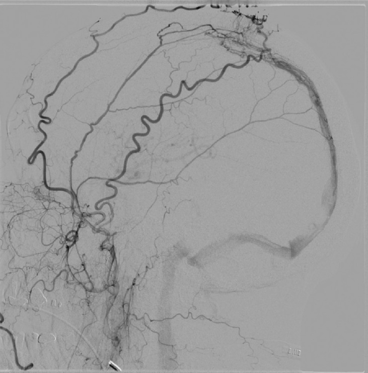
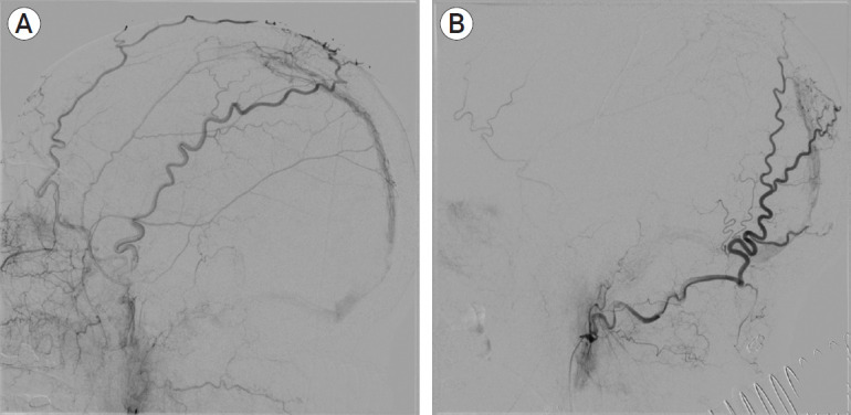
In early June, the patient underwent six-vessel diagnostic cerebral angiogram and venous pressure monitoring, which revealed a DAVF at the site of the patient’s previous tumor resection fed by scalp and dural arteries (from the right and left superficial temporal, right and left meningeal, and right occipital arteries) with a small amount of retrograde cortical venous drainage (Borden-Shucart grade III, Cognard grade III; Fig. 2). Venous pressure was 53 mmHg and 46 mmHg, 20 mmHg, and 18 mmHg at the parietal and occipital superior sagittal sinus, sinus confluence, and jugular vein, respectively. The patient returned one-week later for Onyx 18 balloon-assisted embolization of the left meningeal artery, which allowed complete embolization of the feeding arteries of the left meningeal artery and right anterior branch of the middle meningeal artery and contributing to considerable slowing of flow (Fig. 3). However, we believed there was further contribution from the right posterior branch of the middle meningeal artery and right occipital artery and the patient required re-operation in 2 weeks for further embolization. Repeat angiogram at that time revealed continued filling of the fistula from the right and left external carotid artery branches, and the right occipital artery was utilized as an embolization pedicle and check angiography revealed complete obliteration of flow through the occipital artery and inferior loop of the middle meningeal artery (Fig. 3). Notably, there was continued right-sided feeding of the fistula from the left via the occipital artery.
Fig. 2.

Digital subtraction angiography (DSA), lateral view. Superior sagittal sinus dural arteriovenous fistula with the bilateral superficial temporal, bilateral meningeal, and right occipital arterial feeding vessels and retrograde cortical venous drainage (Borden-Shucart grade III; Cognard grade III).
Fig. 3.

Digital subtraction angiography (DSA), lateral view. First embolization leading to complete obliteration of the left meningeal feeding artery and right anterior branch of the middle meningeal artery as well as slowing of flow (A). Follow-up embolization 2 weeks later was performed on the right posterior branch of the middle meningeal and right occipital artery revealing residual dural arteriovenous fistula with continued right-sided feeding from the left via the occipital artery (B).
Repeat angiogram 3 months after his initial embolization showed complete resolution of his DAVF (Fig. 4). In regard to his vision, the patient was also seen by ophthalmology, who noted resolving papilledema.
Fig. 4.
Digital subtraction angiography (DSA), lateral view in (A) arterial phase from external carotid artery injection, (B) arterial phase from internal carotid artery injection, and (C) venous phase following internal carotid artery injection. Follow-up angiography 3 months after initial embolization demonstrates complete obliteration of the dural arteriovenous fistula.
DISCUSSION
DAVFs account for 10% to 15% of all intracranial AVMs, with SSS DAVFs accounting for 4.6% to 11% of these occurring equally in males and females [8,13,15,20]. Most of these lesions are considered to be acquired and arise idiopathically, however, trauma and sinus thrombosis has been attributed as inciting factors associated with DAVF development of the SSS [13,20]. Conversely, surgical intervention has not been reported previously as a cause of SSS DAVF [20]. We present the first case of a SSS DAVF following surgical resection and subsequent stereotactic radiosurgery of an intracranial lesion.
There are several possible explanations as to why surgical resection, stereotactic radiosurgery, and residual tumor lead to dural sinus thrombosis and DAVF formation. With regard to this case in this report, the patient had surgical intervention and intracranial tumor, two known risk factors for cerebral venous obstruction [9,20]. When sinus thrombosis occurs, DAVFs may form when (1) the dural wall becomes inflamed causing thin-walled vessels to form via neovascularization leading to fistulous connections between abnormal thin-walled meningeal vessels and the venous lumen or (2) when engorged dural venous collaterals cause embryonic arteriovenous communication to occur [1,3]. Alternatively, apposition of muscle blood vessels to the dura following surgery have also been proposed to cause postoperative sinus thrombosis and ipsilateral DAVFs 2 to 6 years after operative intervention [1,9]. The other proposed mechanism in the natural history of DAVF formation is arteriovenous shunting favoring the recruitment of arterial feeders into the nidus with secondary venous hypertension [1]. A highly vascular tumor such as a meningioma may cause arteriovenous shunting via the tumor bed into the dural sinus, resulting in increased intraluminal pressure and DAVF formation [1].
In addition to dural sinus occlusion, vascular insult as a consequence of radiation therapy has been well documented and may have potentiated the formation of fistula formation in this case. In animal models receiving between 2 to 2000 Gy radiation, progressive endothelial loss occurs due to apoptosis in a dose-dependent fashion [14]. While the direct physiological consequence is disruption of the blood-brain barrier, long-term morphological changes include endothelial proliferation, basement membrane thickening, adventitial fibrosis, and vessel dilation [1]. When large doses of radiation are administered, blood vessel wall necrosis and inflammation of the vasa vasorum can increase the risk of fistula formation between affected blood vessels and neighboring structures [14]. There is evidence that radiation-induced changes from SRS are likely to occur at higher doses, increased SRS margin, and with comorbidities such as diabetes, however, there is little available data demonstrating the long-term vascular impacts of SRS [11,14,18].
Venous hypertension due to DAVF is generally recognized to determine clinical presentation and can be misdiagnosed as a dural sinus thrombosis [6]. Unique to this case was venous hypertension leading to signs of intracranial hypertension as well as initial MRI venogram findings of dural sinus thrombosis without DAVF. Proper diagnosis is critical in order to avoid contraindicated therapies, especially due to the risk of intracranial hemorrhage from anticoagulation therapy in lesions associated with retrograde venous flow and the potential for acute tonsillar herniation and death due to rapid changes in CSF dynamics in patients with DAVF [5,19]. To correctly distinguish isolated intracranial hypertension from benign intracranial hypertension and to avoid diagnostic confusion, diagnostic angiography should be strongly considered in patients with findings on noninvasive imaging consistent with dural sinus thrombosis prior to initiating therapy [5,19]. As has been previously published, MRV, which relies on the direction of flow to determine sinus patency, should not be viewed as definitive, especially when previous intervention upon the dural sinus makes fistula a possibility [19].
Therapies for DAVF include conservative monitoring, transarterial embolization, transvenous occlusion, direct surgery, and SRS [17]. Management largely depends on the grade of lesions, as patients with type I lesions do not have aggressive neurologic events in 97% to 98% of cases and can be managed conservatively with observation only or ipsilateral carotid or occipital artery compression [15,17]. In high grade lesions, such as the one presented here, there is evidence that conservative therapies such as acetazolamide does not increase cerebral blood flow due to a reduced pressure gradient between the cerebral arteries and veins, thus blunting its effects [6]. Also, higher-grade DAVFs with cortical venous reflux exhibit a higher long-term risk and intracranial hemorrhage necessitating curative treatment to achieve complete disconnection between arteries and veins [17]. In particular, SSS DAVFs with cortical venous drainage increases the risk of hemorrhage more than DAVFs in other locations and should be treated aggressively (i.e. requiring complete occlusion) [7]. Care must be taken to consider the angiographic appearance of the lesion as well as the feasibility of safe endovascular access. The transvenous approach is advantageous in large, complex DAVFs when a portion of the normal draining dural sinus is isolated from the normal venous anatomy and may be considered when the brain is well drained by other venous channels [16,21]. Alternatively, the transarterial approach is usually effective for treating small DAVFs and lesions associated with venous occlusions or significant venous stenosis that may limit a transvenous approach [21].
The location of the DAVF in this case, at the SSS, allowed for easy transarterial access via the middle meningeal artery and provides a navigable access to microcatheterization [20]. In this case, the utilization of Onyx, a liquid embolic agent, due to the ability to position the microcatheter more proximally to the fistulous point within a feeding artery, its ability to penetrate more distally to the fistula, and retrograde embolization of multiple feeder vessels, providing a success rate of 50% to 90% [16,17]. However, our patient required repeated treatment due to numerous small feeding arteries, which has been shown to result in a lower rate of complete occlusion [4,12].
CONCLUSIONS
The development of the SSS DAVF in our case was likely multifactorial and is the first reported case of an SSS DAVF following surgical intervention and SRS. Moreover, as has been previously published, MRV, which relies on the direction of flow to determine sinus patency, should not be viewed as definitive, especially when previous intervention upon the dural sinus makes fistula a possibility [19].
Footnotes
Disclosure
The authors report no conflict of interest concerning the materials or methods used in this study or the findings specified in this paper.
REFERENCES
- 1.Arnautović KI, Al-Mefty O, Angtuaco E, Phares LJ. Dural arteriovenous malformations of the transverse/sigmoid sinus acquired from dominant sinus occlusion by a tumor: report of two cases. Neurosurgery. 1998 Feb;42(2):383–8. doi: 10.1097/00006123-199802000-00112. [DOI] [PubMed] [Google Scholar]
- 2.Awad IA, Little JR, Akarawi WP, Ahl J. Intracranial dural arteriovenous malformations: factors predisposing to an aggressive neurological course. J Neurosurg. 1990 Jun;72(6):839–50. doi: 10.3171/jns.1990.72.6.0839. [DOI] [PubMed] [Google Scholar]
- 3.Borden JA, Wu JK, Shucart WA. A proposed classification for spinal and cranial dural arteriovenous fistulous malformations and implications for treatment. J Neurosurg. 1995 Feb;82(2):166–79. doi: 10.3171/jns.1995.82.2.0166. [DOI] [PubMed] [Google Scholar]
- 4.Chandra RV, Leslie-Mazwi TM, Mehta BP, Yoo AJ, Rabinov JD, Pryor JC, et al. Transarterial onyx embolization of cranial dural arteriovenous fistulas: long-term follow-up. AJNR Am J Neuroradiol. 2014 Sep;35(9):1793–7. doi: 10.3174/ajnr.A3938. [DOI] [PMC free article] [PubMed] [Google Scholar]
- 5.Cognard C, Casasco A, Toevi M, Houdart E, Chiras J, Merland JJ. Dural arteriovenous fistulas as a cause of intracranial hypertension due to impairment of cranial venous outflow. J Neurol Neurosurg Psychiatry. 1998 Sep;65(3):308–16. doi: 10.1136/jnnp.65.3.308. [DOI] [PMC free article] [PubMed] [Google Scholar]
- 6.Deguchi J, Yamada M, Kobata H, Kuroiwa T. Regional cerebral blood flow after acetazolamide challenge in patients with dural arteriovenous fistula: simple way to evaluate intracranial venous hypertension. AJNR Am J Neuroradiol. 2005 May;26(5):1101–6. [PMC free article] [PubMed] [Google Scholar]
- 7.Fukai J, Terada T, Kuwata T, Hyotani G, Raimura M, Nakagawa M, et al. Transarterial intravenous coil embolization of dural arteriovenous fistula involving the superior sagittal sinus. Surg Neurol. 2001 Jun;55(6):353–8. doi: 10.1016/s0090-3019(01)00469-4. [DOI] [PubMed] [Google Scholar]
- 8.Halbach VV, Higashida RT, Hieshima GB, Rosenblum M, Cahan L. Treatment of dural arteriovenous malformations involving the superior sagittal sinus. AJNR Am J Neuroradiol. 1988 Mar-Apr;9(2):337–43. [PMC free article] [PubMed] [Google Scholar]
- 9.Horinaka N, Nonaka Y, Nakayama T, Mori K, Wada R, Maeda M. Dural arteriovenous fistula of the transverse sinus with concomitant ipsilateral meningioma. Acta Neurochir (Wien) 2003 Jun;145(6):501–4. doi: 10.1007/s00701-003-0030-5. discussion 504. [DOI] [PubMed] [Google Scholar]
- 10.Houser OW, Baker HL, Jr, Rhoton AL, Jr, Okazaki H. Intracranial dural arteriovenous malformations. Radiology. 1972 Oct;105(1):55–64. doi: 10.1148/105.1.55. [DOI] [PubMed] [Google Scholar]
- 11.Ilyas A, Chen CJ, Ding D, Buell TJ, Raper DMS, Lee CC, et al. Radiation-induced changes after stereotactic radiosurgery for brain arteriovenous malformations: A systematic review and meta-analysis. Neurosurgery. 2018 Sep;83(3):365–76. doi: 10.1093/neuros/nyx502. [DOI] [PubMed] [Google Scholar]
- 12.Kim B, Jeon P, Kim K, Kim S, Kim H, Byun HS, Jo KI. Predictive Factors for Response of Intracranial Dural Arteriovenous Fistulas to Transarterial Onyx Embolization: Angiographic Subgroup Analysis of Treatment Outcomes. World Neurosurg. 2016 Apr;88:609–18. doi: 10.1016/j.wneu.2015.10.052. [DOI] [PubMed] [Google Scholar]
- 13.Kurl S, Saari T, Vanninen R, Hernesniemi J. Dural arteriovenous fistulas of superior sagittal sinus: case report and review of literature. Surg Neurol. 1996 Mar;45(3):250–5. doi: 10.1016/0090-3019(95)00361-4. [DOI] [PubMed] [Google Scholar]
- 14.Murphy ES, Xie H, Merchant TE, Yu JS, Chao ST, Suh JH. Review of cranial radiotherapy-induced vasculopathy. J Neurooncol. 2015 May;122(3):421–9. doi: 10.1007/s11060-015-1732-2. [DOI] [PubMed] [Google Scholar]
- 15.Narayanan S. Endovascular management of intracranial dural arteriovenous fistulas. Neurol Clin. 2010 Nov;28(4):899–911. doi: 10.1016/j.ncl.2010.03.013. [DOI] [PubMed] [Google Scholar]
- 16.Natarajan SK, Ghodke B, Kim LJ, Hallam DK, Britz GW, Sekhar LN. Multimodality treatment of intracranial dural arteriovenous fistulas in the Onyx era: a single center experience. World Neurosurg. 2010 Apr;73(4):365–79. doi: 10.1016/j.wneu.2010.01.009. [DOI] [PubMed] [Google Scholar]
- 17.Oh SH, Choi JH, Kim BS, Lee KS, Shin YS. Treatment outcomes according to various treatment modalities for intracranial dural arteriovenous fistulas in the Onyx era. A 10-year single-center experience. World Neurosurg. 2019 Jun;126:e825–34. doi: 10.1016/j.wneu.2019.02.173. [DOI] [PubMed] [Google Scholar]
- 18.Quigg M, Yen CP, Chatman M, Quigg AH, McNeill IT, Przybylowski CJ, et al. Risks of history of diabetes mellitus, hypertension, and other factors related to radiation-induced changes following Gamma Knife surgery for cerebral arteriovenous malformations. J Neurosurg. 2012 Dec;117 Suppl:144–9. doi: 10.3171/2012.6.GKS1245. [DOI] [PubMed] [Google Scholar]
- 19.Simon S, Yao T, Ulm AJ, Rosenbaum BP, Mericle RA. Dural arteriovenous fistulas masquerading as dural sinus thrombosis. J Neurosurg. 2009 Mar;110(3):514–7. doi: 10.3171/2008.7.JNS08253. [DOI] [PubMed] [Google Scholar]
- 20.Song W, Sun H, Liu J, Liu L, Liu J. Spontaneous resolution of venous aneurysms after transarterial embolization of a variant superior sagittal sinus dural arteriovenous fistula: case report and literature review. Neurologist. 2017 Sep;22(5):186–95. doi: 10.1097/NRL.0000000000000137. [DOI] [PubMed] [Google Scholar]
- 21.Vanlandingham M, Fox B, Hoit D, Elijovich L, Arthur AS. Endovascular treatment of intracranial dural arteriovenous fistulas. Neurosurgery. 2014 Feb;74 Suppl 1:S42–9. doi: 10.1227/NEU.0000000000000180. [DOI] [PubMed] [Google Scholar]


