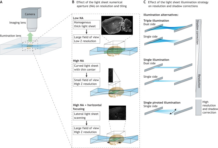
Fig. 3.
Optimization of light-sheet microscopy for embryology: maximizing the resolution. (A) Schematic of light-sheet illumination and light collection (example of the ultramicroscope). (B) Importance of the numerical aperture (NA) for the light-sheet generation and its effects on the homogeneity of the optical plane and the size of the field of view. Examples are given with vascular labeling: at low axial resolution, the vessels appear continuous, whereas at high resolution they are shown with their cross-sections as points. (C) Types of illumination. Ultramicroscopes incorporate multiple angles or dual-side illumination to reduce the shadows and improve the illumination width in large samples. The MesoSPIM uses dual-sided illumination and horizontal scanning to speed up the system efficiently. Finally, the Zeiss systems use a pivoted illumination system to reduce the shadows and incorporate a mechanical arm to rotate the sample.

