Abstract
Background
Coronavirus disease 2019 may have neurological manifestations including meningitis, encephalitis, post-infectious brainstem encephalitis and Guillain-Barre syndrome. Neuroinflammation has been claimed as a possible cause. Here, we present a child with multisystem inflammatory syndrome in children (MIS-C) who developed pseudotumor cerebri syndrome (PTCS) during the disease course.
Case
A 11-year-old girl presented with 5 days of fever, headache and developed disturbance of consciousness, respiratory distress, conjunctivitis and diffuse rash on her trunk. Immunoglobulin M and G antibodies against severe acute respiratory syndrome coronavirus 2 were positive in her serum. She was diagnosed with MIS-C. On day 10, she developed headache and diplopia. Left abducens paralysis and bilateral grade 3 papilledema were observed. Brain magnetic resonance imaging revealed optic nerve head protrusion, globe flattening. She was diagnosed with secondary PTCS. Papilledema and abducens paralysis improved under acetazolamide and topiramate. Neurological examination became normal after 2 months.
Conclusion
PTCS may emerge related to MIS-C.
Keywords: COVID-19, MIS-C, child, encephalitis, pseudotumor cerebri
Introduction
Severe acute respiratory syndrome coronavirus 2 (SARS CoV-2) infection has affected more than 194 000 000 people and caused more than 4 000 000 deaths worldwide, as of July 2021 [1]. Coronavirus disease 2019 (COVID-19), previously known to have a mild course among children, may cause severe respiratory distress or post-infectious features such as multisystem inflammatory syndrome in children (MIS-C) [2].
MIS-C is characterized by a severe illness with persisting fever (>38.0°C for ≥24 h), more than two organ system involvement (cardiac, gastrointestinal, renal, respiratory, hematologic, neurological, dermatologic) and laboratory evidence of inflammation. SARS-CoV-2 infection is verified either by reverse transcription-polymerase chain reaction (RT-PCR) or serology and an exposure to a suspected or confirmed COVID-19 case within the 4 weeks before the onset of symptoms, is declared [3].
Meningitis, encephalitis, post-infectious brainstem encephalitis, Guillain-Barre syndrome and non-specific symptoms such as headache, altered mental state, behavioral changes and movement disorders have been described as some of the neurological manifestations of the disease [4]. Neuroinflammation has been claimed as the possible cause but not clarified completely [5]. We, herein, aim to present a child with MIS-C, who developed pseudotumor cerebri syndrome (PTCS) during the disease course.
Case
A previously healthy, 11-year-old girl presented with 5 days of fever, headache and neck pain. She had a history of contact with COVID-19, 3 weeks before admission. She was conscious and oriented. Her heart rate was 94 beats/min, her blood pressure was 99/66 mmHg, SpO2 was 100%. Physical examination revealed neck stiffness, positive Kernig and Brudzinski signs. She was hospitalized on the ward, with a provisional diagnosis of meningoencephalitis. The laboratory findings on admission included leukocytosis (20 000/mm3), lymphopenia (1190/mm3) and increased procalcitonin (0.72 ng/mL, N ≤ 0.5).
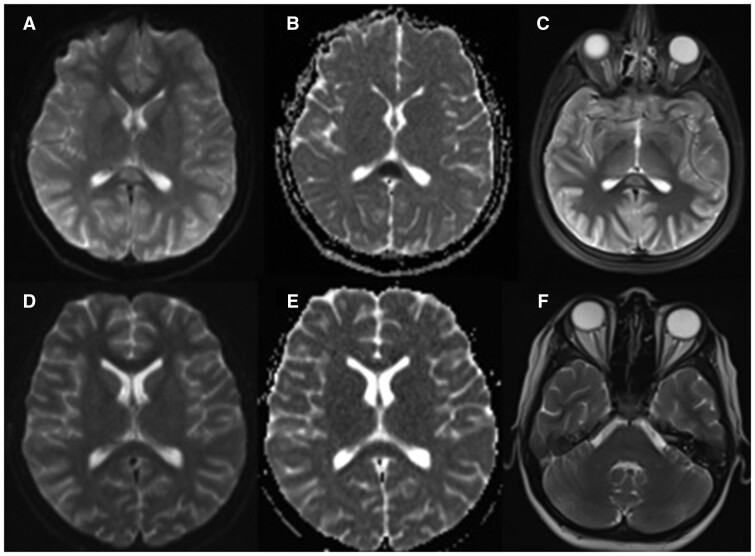
C-reactive protein (28.4 mg/dL, N ≤ 0.5), prohormone of brain natriuretic peptide (9730 ng/L, N ≤ 83) levels. Fibrinogen level was 295 mg/dL (N = 200–400). Cerebrospinal fluid (CSF) analysis was acellular. Protein and glucose levels (20 mg/dL and 90 mg/dL, respectively) were in normal range. Venous blood gas showed PCO2 level of 43 mmHg. Nasopharyngeal swab was negative, Immunoglobulin M and G antibodies were positive against SARS-CoV-2. Brain magnetic resonance imaging (MRI) pointed out hyperintensities on T2/FLAIR sequences and restricted diffusion in the splenium of the corpus callosum (Fig. 1). Electroencephalography showed diffuse slowing. Fundus examination could not be performed on admission. On day 3, she developed alteration of consciousness, respiratory distress, conjunctivitis and diffuse rash on her trunk. Her Glasgow Coma Scale was 12 and her blood pressure was 80/50 mmHg. She was diagnosed with MIS-C and was transferred to the pediatric intensive care unit. Thorax computed tomography showed bilateral ground glass opacity in her lungs. Non-invasive ventilation (NIV) was started. Milrinone (0.5 mcg/kg/min) and noradrenaline (0.05 mcg/kg/min) infusions were initiated due to mitral insufficiency and left ventricular dysfunction. She was treated with intravenous immunoglobulin (2 g/kg). Her fever persisted and therefore, methylprednisolone (2 mg/kg/day) treatment was added. On day 5, milrinone and noradrenaline infusions and NIV were discontinued. Methylprednisolone was continued at a lower dose (1 mg/kg/day). Her consciousness level returned back to normal. Brain MRI done on day 7 was normal.
Fig. 1.
Brain magnetic resonance imaging of the patient. T2-weighted axial (A), apparent diffusion coefficient (B) images of the patient on day 1 show hyperintensity and restricted diffusion in the splenium of the corpus callosum and T2-weighted axial (C) image shows normal optic nerve head. T2-weighted axial (D), apparent diffusion coefficient (E) images on day 7 show hyperintensity and restricted diffusion in the corpus callosum regressed and T2-weighted axial (F) image on day 10 shows optic nerve head protrusion and globe flattening.
On day 10, she developed headache and diplopia. Left abducens paralysis and bilateral grade 3 papilledema were observed. Arterial blood gas showed a PaCO2 level of 38 mmHg and PaO2 was 83 mmHg. Brain MRI revealed optic nerve head protrusion and globe flattening (Fig. 1). Brain magnetic resonance (MR) venography was normal. Lumbar puncture (LP) was not repeated since it was performed recently and CSF was acellular. She was diagnosed with a probable diagnosis of secondary PTCS associated with MIS-C based on the diagnostic criteria [6]. Acetazolamide (500 mg/day) and topiramate (25 mg/day) treatments were initiated. Papilledema and abducens paralysis improved gradually. Neurological examination became normal at 2-month follow-up.
Discussion
COVID-19 may lead to various neurological manifestations which have been declared in children and adult case series [4]. We, herein, describe a patient with MIS-C, who later developed PTCS during treatment.
PTCS was proclaimed in a 16-year-old, being the youngest of a study including 43 patients with COVID-19-related neurological disease. Having multiple congenital disorders and epilepsy, he developed non-convulsive status epilepticus. He had MIS-C components including cardiac, gastrointestinal and cutaneous involvements [7]. Vasogenic edema following status epilepticus could have been contributed to the neurological presentation of this case.
Corticosteroids used to treat the hyperinflammation in MIS-C might lead to PTCS. However, it seems unlikely for our patient since a distinguishably low dose was prescribed for a relatively short period of time.
A 14-year-old with PTCS secondary to MIS-C has recently been reported [8]. Similar to index case, our patient had intracranial hypertension signs at the end of the first week when shock signs and inotrope need regressed and mechanical ventilation support was weaned off. Therefore, regardless of the corticosteroid treatment, the underlying hyperinflammation may cause a resistance to CSF absorption and lead to PTCS. Pro-inflammatory cytokines have a role in developing PTCS and may have a relation with MIS-C [9].
Our patient also had hyperintensities on T2 and FLAIR sequences and diffusion restriction at the splenium of corpus callosum on her initial brain MRI, which later regressed. Reversible splenial lesion syndrome (RESLES) of corpus callosum has been reported as a distinct neurologic involvement of SARS CoV-2 infection. It has been described before COVID-19, in association with viral infections, metabolic disturbances, malnutrition, chemotherapeutic agents and anti-epileptic drug withdrawal. However, cytotoxic edema of the splenium of corpus callosum seems like the probable reason why it is more likely to occur in patients with MIS-C and why it resolves in one week with treatment that suppresses hyperinflammation [10, 11].
Singer et al. [12] discuss that microvascular infarcts in deep brain structures were more likely to occur in patients with MIS-C, which might lead to intracranial hypertension along with venous congestion and hypercoagulable state described by Silva et al. [13].
MR venography ruled out thrombosis in our patient which is one of the major complications of COVID-19. The viral infection and inflammation itself might have caused a decrease in CSF absorption and lead to secondary PTCS in our patient. LP was not repeated and CSF pressure could not be measured. However, PTCS seemed like the probable diagnosis since the other diagnostic criteria existed [6].
Our patient was not checked by an ophthalmologist on admission which was a limitation of our case report. Fundus examination holds a significance since MIS-C patients may have or develop a neurological manifestation at the beginning or during the disease course.
We emphasize that PTCS may emerge associated with MIS-C. It should be kept in mind when a sudden neurologic deterioration such as diplopia or severe headache occur during the treatment regime. Additional mechanisms leading to PTCS in MIS-C patients need to be enlightened.
Authors’ contribution
A.I.S., G.B., N.A. and E.S. treated the patient. A.I.S., G.B., N.A. and E.S. wrote and revised the manuscript. All authors approved the final manuscript.
Consent
The written inform consent to publication has been obtained from the parents.
Funding
This research did not receive any grant from the public, commercial, or not-for-profit sector funding agencies.
References
- 1. World Health Organization (WHO). Coronavirus Disease (COVID-19) Dashboard. https://covid19.who.int/table (27 July 2021, date last accessed).
- 2. Jiang L, Tang K, Levin M, et al. COVID-19 and multisystem inflammatory syndrome in children and adolescents. Lancet Infect Dis 2020;20:e276–88. [DOI] [PMC free article] [PubMed] [Google Scholar]
- 3. Centers for Disease Control and Prevention. Reporting Multisystem Inflammatory Syndrome in Children (MIS-C). https://www.cdc.gov/mis-c/hcp/index.html (21 December 2020, date last accessed).
- 4. Rimensberger PC, Kneyber MCJ, Deep A, et al. ; European Society of Pediatric and Neonatal Intensive Care (ESPNIC) Scientific Sections’ Collaborative Group. Caring for critically ill children with suspected or proven coronavirus disease 2019 infection: recommendations by the Scientific Sections' Collaborative of the European Society of Pediatric and Neonatal Intensive Care. Pediatr Crit Care Med 2021;22:56–67. [DOI] [PMC free article] [PubMed] [Google Scholar]
- 5. Chen TH. Neurological involvement associated with COVID-19 infection in children. J Neurol Sci 2020;418:117096. [DOI] [PMC free article] [PubMed] [Google Scholar]
- 6. Wall M, Corbett JJ.. Revised diagnostic criteria for the pseudotumor cerebri syndrome in adults and children. Neurology 2013;81:1159–65. [DOI] [PubMed] [Google Scholar]
- 7. Paterson RW, Brown RL, Benjamin L, et al. The emerging spectrum of COVID-19 neurology: clinical, radiological and laboratory findings. Brain 2020;143:3104–20. [DOI] [PMC free article] [PubMed] [Google Scholar]
- 8. Verkuil LD, Liu GT, Brahma VL, et al. Pseudotumor cerebri syndrome associated with MIS-C: a case report. Lancet 2020;396:532. [DOI] [PubMed] [Google Scholar]
- 9. Edwards LJ, Sharrack B, Ismail A, et al. Increased levels of interleukins 2 and 17 in the cerebrospinal fluid of patients with idiopathic intracranial hypertension. Am J Clin Exp Immunol 2013;2:234–44. [PMC free article] [PubMed] [Google Scholar]
- 10. Bektaş G, Akçay N, Boydağ K, et al. Reversible splenial lesion syndrome associated with SARS-CoV-2 infection in two children [published online ahead of print, 2020 Oct 13]. Brain Dev 2021;43:230–3. [DOI] [PMC free article] [PubMed] [Google Scholar]
- 11. Abdel-Mannan O, Eyre M, Löbel U, et al. Neurologic and radiographic findings associated with COVID-19 infection in children [published correction appears in JAMA Neurol. 2020 Dec 1;77(12):1582]. JAMA Neurol 2020;77:1440–5. [DOI] [PMC free article] [PubMed] [Google Scholar]
- 12. Singer TG, Evankovich KD, Fisher K, Demmler-Harrison GJ, et al. Coronavirus infections in the nervous system of children: scoping review making the case for long-term neurodevelopmental surveillance. Pediatr Neurol 2021;117:47–63. [DOI] [PMC free article] [PubMed] [Google Scholar]
- 13. Silva MTT, Lima MA, Torezani G, et al. Isolated intracranial hypertension associated with COVID-19. Cephalalgia 2020;40:1452–8. [DOI] [PMC free article] [PubMed] [Google Scholar]


