Abstract
Background
Members of the genus Planococcus have been revealed to utilize and degrade solvents such as aromatic hydrocarbons and alkanes, and likely to acquire tolerance to solvents. A yellow marine bacterium Planococcus maritimus strain iso-3 was isolated from an intertidal sediment that looked industrially polluted, from the Clyde estuary in the UK. This bacterium was found to produce a yellow acyclic carotenoid with a basic carbon 30 (C30) structure, which was determined to be methyl 5-glucosyl-5,6-dihydro-4,4′-diapolycopenoate. In the present study, we tried to isolate and identify genes involved in carotenoid biosynthesis from this marine bacterium, and to produce novel or rare C30-carotenoids with anti-oxidative activity in Escherichia coli by combinations of the isolated genes.
Results
A carotenoid biosynthesis gene cluster was found out through sequence analysis of the P. maritimus genomic DNA. This cluster consisted of seven carotenoid biosynthesis candidate genes (orf1–7). Then, we isolated the individual genes and analyzed the functions of these genes by expressing them in E. coli. The results indicated that orf2 and orf1 encoded 4,4′-diapophytoene synthase (CrtM) and 4,4′-diapophytoene desaturase (CrtNa), respectively. Furthermore, orf4 and orf5 were revealed to code for hydroxydiaponeurosporene desaturase (CrtNb) and glucosyltransferase (GT), respectively. By utilizing these carotenoid biosynthesis genes, we produced five intermediate C30-carotenoids. Their structural determination showed that two of them were novel compounds, 5-hydroxy-5,6-dihydro-4,4′-diaponeurosporene and 5-glucosyl-5,6-dihydro-4,4′-diapolycopene, and that one rare carotenoid 5-hydroxy-5,6-dihydro-4,4′-diapolycopene is included there. Moderate singlet oxygen-quenching activities were observed in the five C30-carotenoids including the two novel and one rare compounds.
Conclusions
The carotenoid biosynthesis genes from P. maritimus strain iso-3, were isolated and functionally identified. Furthermore, we were able to produce two novel and one rare C30-carotenoids in E. coli, followed by positive evaluations of their singlet oxygen-quenching activities.
Supplementary Information
The online version contains supplementary material available at 10.1186/s12934-021-01683-3.
Introduction
Carotenoids, essential pigments for photosynthesis, are known to protect cells from oxidative stress [1, 2]. In the domain bacteria, all of the photosynthetic bacteria, including cyanobacteria, as well as some of the non-photosynthetic bacteria, usually chemoheterotrophs, can produce carotenoids [3]. Among the non-photosynthetic bacteria, several species have been shown to produce acyclic carotenoids with a basic carbon 30 (C30) structure instead of the typical C40-carotenoids. Specifically, the carotenogenic-bacterial species of the phylum Firmicutes of low GC Gram-positive bacteria that have been reported to synthesize only C30-carotenoids include Streptococcus faecium [3], Staphylococcus aureus [4], Bacillus firmus [5], Halobacillus halophilus [6, 7], Planococcus maritimus [8, 9], Sporosarcina aquimarina [10], and Lactobacillus plantarum (recently reclassified to Lactiplantibacillus plantarum) [11]. C30-Carotenoids have also been found in bacterial species that belong to other phyla, such as Rubritalea squalenifaciens of the phylum Verrucomicrobia [12] and Methylomonas sp. strain 16a of the phylum Proteobacteria [13].
The biosynthesis of C30-carotenoids has mostly been studied in Staphylococcus aureus [4, 14–16]. Farnesyl diphosphate (FPP) is converted to 4,4′-diapophytoene (dehydrosqualene) by 4,4′-diapophytoene synthase (CrtM) and to 4,4′-diaponeurosporene by 4,4′-diapophytoene desaturase (CrtN) [16], which is likely to be the common biosynthetic route in all C30-carotenogenic bacteria. Lastly, staphyloxanthin [β-d-glucosyl 1-O-(4,4′-diaponeurosporen-4-oate)-6-O-(12-methyltetradecanoate)], which has shown to confer oxidative stress tolerance on S. aureus [17], is produced from 4,4′-diaponeurosporene as the final product with the mediation of four genes that encode oxidase (CrtP), dehydrogenase (AldH), glycosyltransferase (CrtQ), and acyltransferase (CrtO) [14, 15]. As for other C30-carotenogenic bacteria, a few studies have been published on their genes for metabolizing 4,4′-diaponeurosporene [13, 18–20].
Members of the genus Planococcus have been revealed to utilize and degrade solvents such as aromatic hydrocarbons and alkanes, and likely to acquire tolerance to solvents [21, 22]. A yellow marine bacterium Planococcus maritimus strain iso-3 was isolated from an intertidal sediment that looked industrially polluted, from the Clyde estuary in the UK [8]. Its yellow pigment was identified as methyl 5-glucosyl-5,6-dihydro-4,4′-diapolycopenoate (methyl 5-glucosyl-5,6-dihydro-apo-4,4′-lycopenoate), which showed potent antioxidative activity [8, 9]. Kim et al. [23] also reported that Planococcus faecalis AJ003T produced a similar C30-carotenoid glycosyl-4,4′-diaponeurosporen-4′-ol-4-oate.
In this study, we tried to isolate genes involved in the production of the glucosyl C30-carotenoid from P. maritimus strain iso-3, to identify the functions of the isolated genes using E. coli, and to produce novel and rare C30-carotenoids with anti-oxidative activity in E. coli by combinations of the obtained genes.
Materials and methods
Bacterial strains and growth conditions
Escherichia coli K12 DH5α was used for DNA manipulation. E. coli K12 JM101(DE3) was constructed from strain JM101 using the λDE3 lysogenization kit (Merk, Darmstadt, Germany) and used for expression of the carotenoid biosynthesis genes. These E. coli strains and their transformants were maintained as the frozen stocks including 20% glycerol at − 80 °C, and were grown in 2 × YT medium (16 g/L of tryptone, 10 g/L of yeast extract, 5 g/L of NaCl) at 37 °C as needed. Bacillus subtilis strain MI112 was kindly provided from Dr. Atsuhiko Shinmyo (NAIST, Japan). This bacterium was similarly maintained as the frozen stock and routinely grown in LB medium (10 g/L of tryptone, 5 g/L of yeast extract, 10 g/L of NaCl) at 37 °C.
DNA isolation of Planococcus maritimus strain iso-3
Genome DNA was prepared from Planococcus maritimus strain iso-3 according to a method described by Nishida et al. [24].
Functional cloning experiments
The genome DNA was digested with BamHI, ligated with vector pGETS103, and used to transform Bacillus subtilis strain MI112 as described previously [25]. In generated genome library, one yellow colony appeared, which was found to contain a 7344-bp insert.
Inverse PCR
To isolate the flanking region of the carotenoid biosynthesis gene cluster, inverse PCR was performed. One μg of genomic DNA from the Planococcus was digested with EcoRI. Digested DNA (0.1 μg/μL) was self-ligated with ligation solution (Takara Bio, Ohtsu, Japan). PCR was performed using 50 pg of these circular DNAs as the template in 25 μL of the reaction solution with the gene-specific primers which we designed. The amplified fragments were cloned into the pBC phagemid vector (Agilent, CA, USA), transformed into E. coli (DH5α) and sequenced. The primers used are summarized in Additional file 1: Table S1.
Sequence analysis
Homology search was performed by BLAST (http://blast.ncbi.nlm.nih.gov/Blast.cgi). Amino acid alignments and phylogenetic trees were constructed using MAFFT (http://www.mafft.ccbrc.jp/).
Cloning of the carotenoid biosynthesis genes from P. maritimus strain iso-3
Based on the sequences obtained, the primers containing the restriction sites were designed as shown in Additional file 1: Table S1 and the coding regions of each orf were amplified by PCR of the genomic DNAs.
Then PCR products were cloned into a plasmid vector and sequenced.
Expression of the Planococcus carotenoid biosynthesis genes in Escherichia coli
Plasmids used in this study are summarized in Additional file 1: Fig. S1. Firstly, we constructed the plasmid pAC-HI which contained the Haematococcus pluvialis IDI (isopentenyl diphosphate isomerase) gene between the tac promoter (Ptac) and the rrnB terminator (TrrnB) in the pACYC184 vector. The exogenous expression of the IDI gene in E. coli results in the increase of carotenoid production [26]. Then, the coding region of the Planococcus orf3 was inserted into the plasmid pAC-HI. The resultant plasmid was named as pAC-HIO3. The coding regions of the orf1, orf2, orf4 were cloned into the pAC-HIM which contained IDI and Leuconostoc mesenteroides crtM, independently. These plasmids were designated pAC-HIMO1, pAC-HIMO2, and pAC-HIMO4, respectively. The coding regions of the orf2, orf4 and orf7 were cloned into the pAC-HIMN which contained IDI and L. mesenteroides crtM and crtN, independently. The resultant plasmids were named as pAC-HIMNO2, pAC-HIMNO4, and pAC-HIMNO7, respectively. The orf2 and orf4 were also inserted into the pAC-HIMNF (same as pAC-HIMNO7) and the obtained plasmids were pAC-HIMNFO2 and pAC-HIMNFO4, respectively. The orf5 was further inserted into the pAC-HIMNFNb (same as pAC-HIMNFO4) and this plasmid was named pAC-HIMNFNbO5 (renamed pAC-HIMNFNbG). All plasmids were independently introduced into the wild type E. coli (JM101 (DE3)). Each transformed E. coli was grown in 2YT medium at 37 °C. Next day, this culture was inoculated in a new 2YT medium (100 mL medium in 500 mL Sakaguchi flask) and cultured at 21 °C for 2 days.
The accession no. of the sequences of the plasmids, pAC-HI, pAC-HIM, pAC-HIMN, pAC-HIMNF, pAC-HIMNFNb, and pAC-HIMNFNbG are LC635748, LC647532, LC647533, LC647534, LC647535 and LC647536, respectively.
Extraction and HPLC analysis of carotenoids from E. coli cells
Extraction of carotenoids from the recombinant E. coli was performed by the method described by Fraser et al. [27]. E. coli cultures were centrifuged and cell pellets were extracted with methanol (MeOH) using mixer for 5 min. Tris–HCl (50 mM, pH 7.5) and 1 M NaCl were added and mixed. Then chloroform was added to the mixture and mixed for 5 min. After centrifugation, the chloroform phase was collected and dried by centrifugal evaporation. Dried residues were re-suspended with ethyl acetate (EtOAc), and applied to HPLC with a Waters Alliance 2695–2996 (PDA) system (Waters, Milford, MA, USA). HPLC was carried out according to the method described [28] using TSKgel ODS-80Ts (4.6 × 150 mm, 5 μm; Tosoh, Tokyo, Japan). Briefly, the extract was eluted at a flow rate of 1.0 mL/min at 25 °C with solvent A [water (H2O)-MeOH, 5:95] for 5 min, followed by a linear gradient from solvent A to solvent B (tetrahydrofuran-MeOH, 3:7) for 5 min, and solvent B alone for 8 min. The produced carotenoids were identified by comparing both retention times and absorption spectra with those of our authentic standards. When the produced carotenoids are not compounds in our authentic standards as described in the following sections, we isolated the produced carotenoids and determined their structures using HRESI-MS (high resolution electrospray ionization-mass spectrometry) and NMR (nuclear magnetic resonance) analyses.
Isolation of 5-hydroxy-5,6-dihydro-4,4′-diaponeurosporene (3)
The transformed E. coli cells carrying crtM, crtNa, and cruF were collected by centrifugation from 4 L culture, and extracted with 50 mL acetone-MeOH (7:2) and dichloromethane (CH2Cl2) (×2) with sonication in a stepwise manner. The combined extract (150 mL) was concentrated to small volume in vacuo, and partitioned between EtOAc/H2O (each 100 mL). The n-hexane layer evaporated to dryness (54.2 mg) and subjected to silica gel chromatography (20 mm × 200 mm) using n-hexane–acetone (8:1), and fractionated by 10 mL. The fractions 15–30 containing a yellow compound [Rf 0.4 by silica gel TLC using developing solvent n-hexane–acetone (4:1)] were collected and concentrated to afford a yellow oil (27.1 mg). The yellow oil was applied on preparative HPLC (column: Develosil C-30-UG-5 (20 × 250 mm, Nomura Chemical, Co. Ltd, Aichi, Japan), solvent: CH2Cl2-CH3CN (4:6), flow rate: 8.0 mL/min, detect: PDA 250–700 nm), and a yellow peak eluted at 15.0 min was collected and concentrated to afford pure 5-hydroxy-5,6-dihydro-4,4′-diaponeurospore (3, 13.9 mg).
Isolation of 5-glucosyl-5,6-dihydro-4,4′-diapolycopene (5)
The transformed E. coli cells carrying crtM, crtNa, cruF, crtNb, and GT were collected by centrifugation from 4 L culture, and extracted with acetone-MeOH (7:2) and CH2Cl2-MeOH (1:1) (×2) with sonication in a stepwise manner. The extract (150 mL) was concentrated to small volume in vacuo, and partitioned between EtOAc/H2O (each 100 mL). The EtOAc layer evaporated to dryness (243.9 mg) and subjected to silica gel chromatography (25 mm × 190 mm) using CH2Cl2-MeOH (10:1) and fractionated by 10 mL. The fractions 13–15 containing an UV compound [Rf 0.36 by silica gel TLC using developing solvent CH2Cl2-MeOH (10:1)] to afford a yellow oil (11.2 mg). The yellow oil was applied on preparative HPLC [column: Develosil C-30-UG-5 (20 × 250 mm), solvent: CH2Cl2-MeOH (1:4), flow rate: 8.0 mL/min, detect: PDA 250–700 nm], and a yellow peak eluted at 17.0 min was collected and concentrated to afford pure 5-glucosyl-5,6-dihydro-4,4′-diapolycopene (5, 1.4 mg).
Singlet oxygen-quenching activity
For the measurement of singlet oxygen-quenching activity, 80 μL of 25 μM methylene blue and 100 μL of 0.24 M linoleic acid, with or without 40 μL of carotenoid (final concentration, 1–100 μM; each dissolved in ethanol), were added to 5 mL glass test tubes. The tubes were mixed well and were illuminated at 7000 lx at 22 °C for 3 h in a styrofoam box. Then, 50 μL of the reaction mixture was removed and diluted to 1.5 mL with ethanol, and OD235 was measured to estimate the formation of conjugated dienes [29]. The OD235 in the absence of carotenoids was measured as negative control [no singlet oxygen (1O2)-quenching activity], and the 1O2-quenching activity of carotenoids was calculated from OD235 in the presence of carotenoids relative to this reference value. The activity was indicated as the IC50 value, which represents the concentration at which 50% inhibition was observed.
Results
Isolation of the carotenoid biosynthesis gene cluster from P. maritimus strain iso-3
A yellow colony including a 7.3-kb genomic insert from P. maritimus strain iso-3 was obtained by functional cloning experiments using Bacillus subtilis as the host. Sequence analysis of this insert showed the presence of seven open reading frames (orfs), named orfs 2–8 (Fig. 1). Six orfs, orf2–7, were predicted to be involved in carotenoid biosynthesis, and the remaining orf, orf8, was unlikely to be involved in it. When the 7.3-kb (7344-bp) DNA fragment was expressed in E. coli, the recombinant cells did not become yellow. This DNA fragment contains the 4,4′-diapophytoene synthase (crtM) gene, but it does not include the 4,4′-diapophytoene desaturase (crtNa) gene, as shown later. It is thus likely that the recombinant E. coli produced only the colorless 4,4′-diapophytoene. On the other hand, the B. subtilis used as the host retained 4,4′-diapophytoene desaturase activity; therefore, the recombinant Bacillus cells turned yellow.
Fig. 1.

Gene organization of the DNA fragments obtained from the P. maritimus strain iso-3
The complete genome sequences of several Planococcus, such as P. plakortidis (NZ_CP016539.2), P. faecalis (NZ_CP019401.1), and P. halocrytophilus (NZ_CP016537.2), have been determined. Each of the sequences contains a similar carotenoid biosynthesis gene cluster that includes one more carotenoid biosynthesis gene other than the orfs2–8. Then, we performed inverse PCR to isolate the flanking region of orfs2–8. As a result, the 2.1-kb fragment adjacent to orf2 was isolated and the additional orf, named orf1, was found. In total, a 9.4-kb genomic DNA was analyzed to find out that eight orfs existed (Fig. 1). The accession no. of the sequences of this genomic DNA is LC620265.
Sequence analysis of the carotenoid biosynthesis gene candidates
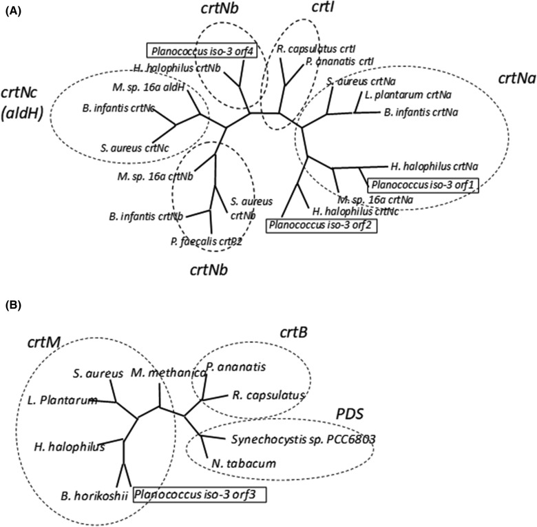
Next, we performed a sequence analysis of orf1–8. Homology searches suggested that seven orfs, orf1–7, were involved in carotenoid biosynthesis, but not orf8, which was homologous to the genes encoding the MurR/RpiR family of transcriptional regulators. The predicted amino acid sequences of Orf1, Orf2, and Orf4 were homologous to that of diapophytoene desaturase. Orf1 was most homologous, displaying approximately 61% identity to the H. halophilus CrtNa. Whereas Orf2 and Orf4 were homologous to the H. halophilus CrtNc and CrtNb, displaying about 50 and 52% identity, respectively (Fig. 2A). On the other hand, Orf3 showed homology to diapophytoene synthase (CrtM) (Fig. 2B). Meanwhile, Orf5 and Orf6 were similar to glycosyltransferase and acyltransferase, respectively. Lastly, Orf7 was homologous to the genes that were annotated as carotenoid biosynthesis genes but with unknown functions. However, we found that the Orf7 was slightly similar, at 22% identity, to CruF (1′-hydroxylase).
Fig. 2.
Phylogenetic trees of the crtN-related and crtM-related genes. Amino acid alignments and phylogenetic trees were constructed using MAFFT (http://www.mafft.ccbrc.jp/). A Phylogenetic tree of the crtN-related genes. The accession numbers of these sequences are shown in Additional file 1: Table S2. Orf1 and orf4 belonged to the crtNa and crtNb groups, respectively. B Phylogenetic tree of the crtM-related genes. The accession numbers of these sequences are shown in Additional file 1: Table S3. Orf3 belonged to the crtM group
Orf3 acts as a diapophytoene synthase
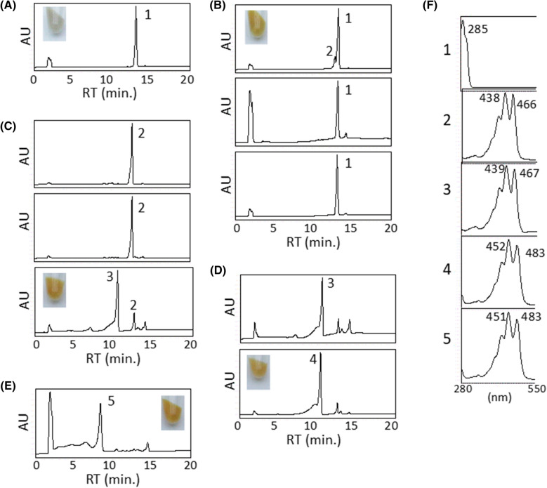
To investigate the function of orf3 which was homologous to diapophytoene synthase, we made the plasmid pAC-HIO3 which included H. pluvialis IDI and orf3, and introduced it into the E. coli (JM101(DE3)). Wild type E. coli cannot produce diapophytoene from farnesyl pyrophosphate (FPP). On the other hand, a new peak (named 1) was observed in the E. coli expressing pAC-HIO3 with IDI and orf3 (Fig. 3A). Then, the produced compound 1 was purified from the cells via acetone-MeOH extraction, n-hexane/90% MeOH partition, and silica gel column chromatography and analyzed by ESI–MS (+), 1H, and 13C NMR spectra. From these spectra, the produced compound was identified as 15-cis-4,4′-diapophytoene (carotenoid 1) (Fig. 4) [30]. Thus, orf3 was confirmed to encode a 15-cis-4,4′-diapophytoene synthase (CrtM).
Fig. 3.
Functional analysis of orfs. A HPLC chromatogram of cell extracts of the E. coli expressing pAC-HIO3. B HPLC chromatograms of the extracts of the E. coli expressing the plasmids pAC-HIMO1 (upper), pAC-HIMO2 (middle), and pAC-HIMO4 (lower). C HPLC chromatograms of the extracts of the E. coli expressing the plasmids pAC-HIMNO2 (upper), pAC-HIMNO4 (middle), and pAC-HIMNO7 (lower). D HPLC chromatograms of the extracts of the E. coli expressing the plasmids pAC-HIMNFO2 (upper) and pAC-HIMNFO4 (lower). E HPLC chromatogram of the extracts of the E. coli expressing the plasmid pAC-HIMNFNbO5. F The spectra correspond to the peak 1: 4,4′-diapophytoene, peak 2: 4,4′-diaponeurosporene, peak 3: 5-hydroxy-5,6-dihydro-4,4′-diaponeurosporene, peak 4: 5-hydroxy-5,6-dihydro-4,4′-diapolycopene, peak 5: 5-glucosyl-5,6-dihydro-4,4′-diapolycopene. Inset show the bacterial cell pelette of each recombinant E. coli
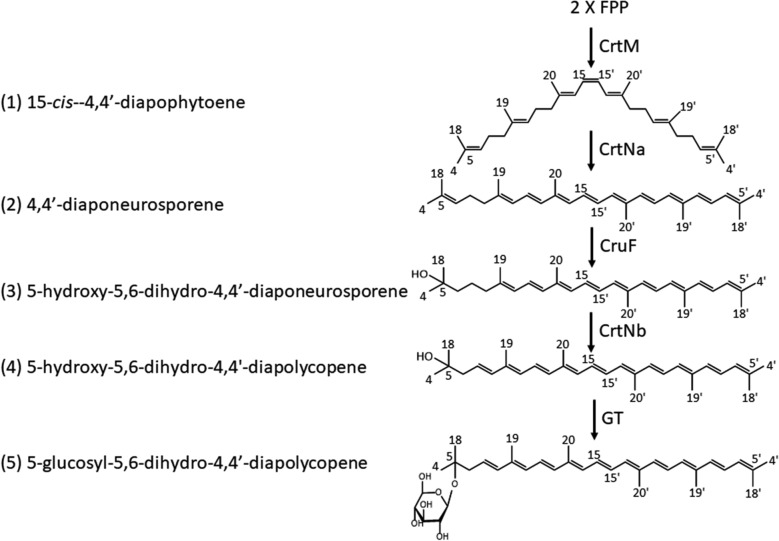
Fig. 4.
Carotenoid biosynthetic pathway in Planococcus strain iso-3
Orf1 acts as a diapophytoene desaturase
Because orf1, orf2, and orf4 were found to be homologous to phytoene desaturase, the catalytic activities of these Orfs were examined by constructing the plasmids pAC-HIMO1, pAC-HIMO2, and pAC-HIMO4 containing H. pluvialis IDI, Leuconostoc mesenteroides crtM, and each orf. The recombinant E. coli expressing pAC-HIMO1 generated a new carotenoid product, as indicated by the new peak (2) (Fig. 3B). On the other hand, the introductions of pAC-HIMO2 and pAC-HIOM4 did not affect the carotenoid profiles (Fig. 3B) We cultured the E. coli expressing pAC-HIMO1 and purified the carotenoid 2 using the same process described in the previous section. The ESI–MS (+), 1H, and 13C NMR spectral analyses indicated carotenoid 2 as 4,4′-diaponeurosporene (Fig. 4) [31]. These results suggest that the orf1, but not orf2 or orf4, acts as a diapophytoene desaturase (CrtNa). However, the activity of Orf1 was low in E. coli; therefore, we use L. mesenteroides crtN for further experiments.
Orf7 acts as a diaponeurosporene hydratase
The synthetic pathway from FPP to diaponeurosporene is thought to be general, but the reaction after diaponeurosporene varies. So, we investigated the activities of Orf2, Orf4, and Orf7 by constructing the plasmids, pAC-HIMNO2, pAC-HIMNO4, and pAC-HIMNO7, which contained H. pluvialis IDI, L. mesenteroides crtM, L. mesenteroides crtN and each orf. When pAC-HIMNO2 or pAC-HIMNO4 was introduced into the E. coli, it only produced 4,4′-diaponeurosporene (Fig. 3C). In the E. coli expressing pAC-HIMNO7 (orf7), a new peak (3) was detected, which could not be identified using our standards, in addition to 4,4′-diaponeurosporene (Fig. 3C). Thus, we cultured this recombinant E. coli and purified the new carotenoid 3 (13.9 mg) as orange powder as described in the Material and Methods section. HRESI-MS (+) analysis (C30H44ONa m/z 443.33015 (M+Na)+, calcd. for 443.32896) determined the molecular formula of peak 3 as C30H44O (4,4′-diaponeurosporene (2 + H2O). Detailed 1D NMR (1H and 13C) and 2D NMR [1H–1H COSY (correlation spectroscopy), HMQC (heteronuclear multiple quantum correlation), and HMBC (heteronuclear multiple bond correlation)] spectral analyses of 3 showed the 1H and 13C signals of carotenoids 2 and 3 to be quite similar except the 5,6Δ signals in carotenoid 2 and one methylene [H-6 (δH 1.42–1.49) and C-6 (δC 43.4)] and one sp3 quaternary carbon [C-5 (δC 80.0) signals in carotenoid 3 (Additional file 1: Fig. S2)]. Finally, the 1H–1H vicinal spin network of H-6–H-8 observed in 1H–1H COSY, and 1H–13C long-range couplings from H-4 and H-18 to C-5 and C-6 observed in HMBC indicated the structure of carotenoid 3 as 5-hydroxy-5,6-dihydro-4,4′-diaponeurosporene (Fig. 4). Carotenoid 3 was a new one according to the CAS database. The assigned 1H and 13C signals in 3 were listed in Table 1. These results indicate that orf7 encodes a diaponeurosporene hydratase; thus, we named orf7 as cruF, encoding a γ-carotene 1′ hydratase involved in myxoxanthophyll biosynthesis in Synechococcus. We also renamed the plasmid pAC-HIMNO7 to pAC-HIMNF.
Table 1.
1H and 13C NMR data for 5-hydroxy-5,6-dihydro-4,4′-diaponeurosporene (3) in CDCl3 and 5-glucosyl-5,6-dihydro-4,4′-diapolycopene (5) in DMSO-d6
| Position | 3 | 5 | ||
|---|---|---|---|---|
| δH | δC | δH | δC | |
| 4 | 1.22 (3H, s) | 29.3 (q) | 1.14 (3H, s) | 26.4 (q) |
| 5 | 80.0 (s) | 76.8 (s) | ||
| 6 | 1.42–1.49 (2H) | 43.4 (t) | 2.32 (2H) | 45.1 (t) |
| 7 | 1.46–1.52 (2H) | 22.6 (q) | 5.88 (1H, m) | 126.9 (d) |
| 8 | 2.11 (1H, dd, 7.0, 7.3) | 40.5 (t) | 6.14 (1H, d, 16.4) | 137.0 (d) |
| 9 | 139.5 (s) | 135.6 (s) | ||
| 10 | 5.96 (1H, d, 8.9) | 126.0 (d) | 6.13 (1H, d, 10.0) | 130.3 (d) |
| 11 | 6.49 (1H, dd, 8.9, 14.9) | 125.0 (d) | 6.62 (1H, dd, 10.0, 15.9) | 125.5 (d) |
| 12 | 6.24 (1H, d, 14.9) | 135.4 (d) | 6.36 (1H, d, 15.9) | 137.4 (d) |
| 13 | 136.0 (s) | 136.1 (s)b | ||
| 14 | 6.19 (1H, m) | 131.5 (d) | 6.32 (1H, m) | 132.8 (d)c |
| 15 | 6.60 (1H, m) | 130.1 (d) | 6.69 (1H, m) | 130.5 (d) |
| 18 | 1.22 (3H, s) | 29.3 (q) | 1.14 (3H, s) | 26.8 (q) |
| 19 | 1.82 (3H, s) | 16.8 (q) | 1.87 (3H, s) | 13.0 (q) |
| 20 | 1.96 (3H, s) | 12.8 (q)a | 1.92 (3H, s) | 12.7 (q)d |
| 4′ | 1.82 (3H, s) | 18.6 (q) | 1.78 (3H, s) | 26.2 (q) |
| 5′ | 136.0 (s) | 135.6 (s) | ||
| 6′ | 5.93 (1H, d, 9.6) | 126.1 (d) | 5.91 (1H, d, 10.0) | 126.9 (d) |
| 7′ | 6.47 (1H, dd, 9.6, 15.2) | 124.8 (d) | 6.46 (1H, dd, 10.0, 14.9) | 125.1 (d) |
| 8′ | 6.22 (1H, d, 15.2) | 135.0 (d) | 6.22 (1H, d, 14.9) | 169.1 (s) |
| 9′ | 136.4 (s) | 136.4 (s)b | ||
| 10′ | 6.25 (1H, d, 11.8) | 132.6 (d) | 6.21 (1H, d, 10.0) | 131.6 (d) |
| 11′ | 6.61 (1H, dd, 11.8, 14.9) | 124.9 (d) | 6.64 (1H, dd, 10.0, 15.2) | 125.4 (d) |
| 12′ | 6.35 (1H, d, 14.9) | 137.3 (d) | 6.37 (1H, d, 15.2) | 137.2 (d) |
| 13′ | 136.2 (s) | 136.4 (s)b | ||
| 14′ | 6.19 (1H, m) | 131.5 (d) | 6.32 (1H, m) | 132.6 (d)c |
| 15′ | 6.60 (1H, m) | 129.5 (d) | 6.69 (1H, m) | 130.5 (d) |
| 18′ | 1.82 (3H, s) | 26.3 (q) | 1.78 (3H, s) | 18.6 (q) |
| 19′ | 1.95 (3H, s) | 12.8 (q)a | 1.92 (3H, s) | 12.7 (q)d |
| 20′ | 1.96 (3H, s) | 12.9 (q)a | 1.92 (3H, s) | 13.0 (q)d |
| 1″ | 4.32 (1H, d, 7.9) | 97.4 (d) | ||
| 2″ | 2.89 (1H, dd, 7.9, 8.0) | 73.7 (d) | ||
| 3″ | 3.15 (1H, dd, 8.0, 8.8) | 76.8 (d) | ||
| 4″ | 3.05 (1H, m) | 70.4 (d) | ||
| 5″ | 3.05 (1H, m) | 71.6 (d) | ||
| 6″ | 3.40 (1H, m), 3.61 (1H, m) | 61.4 (t) | ||
a, b, c, dInterchangeable
Orf4 acts as a 5-hydroxy-5,6-dihydro-4,4′ diaponeurosporene desaturase
Since the subsequent reaction of carotenoid synthesis was expected to be desaturation, we constructed the plasmids pAC-HIMNFO2 and pAC-HIMNFO4. When pAC-HIMNFO2 was introduced into E. coli, only the production of 5-hydroxy-5,6-dihydro-4,4′-diaponeurosporene was detected (Fig. 3D). But in the E. coli expressing pAC-HIMNFO4, a new peak (4) was observed near the peak of 5-hydroxy-5,6-dihydro-4,4′-diaponeurosporene (Fig. 3D). The new compound 4 was identified as 5-hydroxy-5,6-dihydro-4,4′-diapolycopene, which was a rare compound in nature, using HPLC analysis with the standards (Fig. 4) [6, 7]. This data suggested that orf4 encodes a carotenoid desaturase; thus, we named orf4 as crtNb and changed the name of the plasmid pAC-HIMNFO4 to pAC-HIMNFNb.
Orf5 acts as a glycosyltransferase
A novel carotenoid previously isolated from Planococcus iso-3, methyl 5-glucosyl-5,6-dihydro-4,4′-diapolycopenate, was glycosylated. Because orf5 showed homology to glycosyltransferase, we investigated the activity of Orf5. The introduction of the plasmid pAC-HIMNFNbO5 into E. coli resulted in the detection of a new peak (5), which could not be identified using the standards (Fig. 3E). Thus, we cultured this recombinant E. coli and purified the new carotenoid (5, 13.9 mg) as orange powder. HRESI-MS (+) analysis [C36H52O6Na m/z 603.36794 (M+Na)+, calcd. for 603.36616] determined the molecular formula of carotenoid 5 as C36H52O6 (5-hydroxy-5,6-dihydro-4,4′-diapolycopene 4 + C6H12O6–H2O). Detailed 1D NMR (1H and 13C) and 2D NMR (1H–1H COSY, HSQC, and HMBC) spectral analyses of 5 demonstrated that the structure of 5-hydroxy-5,6-dihydro-4,4′-diapolycopene (carotenoid 4) was present also in carotenoid (5) and the attachment of a hexose at C-5 of carotenoid 4. The hexose in carotenoid 5 was shown to be β-glucose because all vicinal coupling constants among H-1″ to H-5″ were large (7.9–9.1 Hz) in the 1H NMR spectrum of acetylated carotenoid 5 in CDCl3 (Additional file 1: Fig. S3). From these observations, carotenoid 5 was determined to be 5-glucosyl-5,6-dihydro-4,4′-diapolycopene (Fig. 4). Carotenoid 5 was a new compound according to the CAS database. The assigned 1H and 13C signals in 5 were listed in Table 1. These results indicate that the orf5 encodes a glycosyltransferase and we renamed pAC-HIMNFNbO5 as pAC-HIMNFNbG.
Singlet oxygen-quenching activity of carotenoids 1 to 5
In the previous study, we reported 1O2-quenching activity of methyl 5-glucosyl-5,6-dihydro-4,4′-diapolycopenoate [8, 9]. Thus, we evaluated 1O2-quenching activities of the intermediates, carotenoids 1–5 isolated in this study. These five carotenoids showed milder 1O2-quenching activities than methyl 5-glucosyl-5,6-dihydro-4,4′-diapolycopenoate (Table 2).
Table 2.
Singlet oxygen quenching activity of the intermediate carotenoids
| Carotenoid | Singlet oxygen quenching IC50 (μM) |
|---|---|
| 15-cis-4,4′-Diapophytoene (1) | > 100 |
| 4,4′-Diaponeurosporene (2) | 45 |
| 5-Hydroxy-5,6-dihydro-4,4′-diaponeurosporene (3) | 56 |
| 5-Hydroxy-5,6-dihydro-4,4′-diapolycopene (4) | 30a |
| 5-Glucosyl-5,6-dihydro-4,4′-diapolycopene (5) | 30 |
| Methyl 5-glucosyl-5,6-dihydro-4,4′-diapolycopenoate | 5.1a |
| Astaxanthin | 3.7a |
aCited from the data of Shindo et al. [9]
Discussion
Until now, there are a few studies on the genes involved in the carotenoid biosynthesis in Planococcus. Recently, Lee et al. [19] reported the C30-carotenoid biosynthesis genes of Planococcus faecalis AJ003T, which mediated glycosyl-4,4′-diaponeurossporene-4′-ol-4-oic acid. Herein, we report the carotenoid biosynthesis genes in Planococcus maritimus strain iso-3, which produces the rare C30 carotenoid, methyl 5-glucosyl-5,6-dihydro-4,4′-diapolycopenoate.
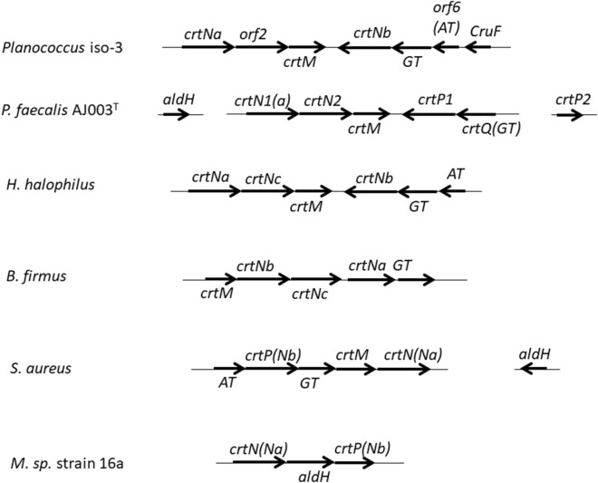
The whole-genome sequences of several kinds of Planococcus were available in the genome database (https://www.ncbi.nlm.nih.gov/genome/). All of them, including P. faecalis AJ003T, had identical carotenoid biosynthesis gene clusters to that found in this study [19, 23]. Similar gene clusters have also been reported in Halobacillus halophilus, Staphylococcus aureus, Bacillus indicus, and Bacillus firmus, which all belong to the order of Bacillales (Fig. 5) [14, 20]. In particular, H. halophilus has nearly the same gene organization as Planococcus regarding gene order and transcriptional direction, whereas they fall into distinct families, the Bacillaceae and Planococcaceae families, respectively [18]. On the other hand, the gene organization is much different between B. indicus and B. firmus [20]. These results suggested that the ancestor of Bacillales retained the same carotenoid biosynthesis gene cluster, and during evolution, the gene organizations have been rearranged independently.
Fig. 5.
Organization of the carotenoid biosynthesis genes from C30 carotenoid synthesizing bacteria
Functional analysis of the genes isolated from P. maritimus indicated that the crtM and crtNa genes encoded proteins with enzymatic activities as predicted from the sequence homology analysis. On the other hand, the enzyme encoded by crtNb was found to generate a product different from those by other crtNb gene products. For example, Steiger et al. [20] demonstrated that the proteins encoded by B. indicus and B. firmus crtNb functioned as aldehyde synthases for utilizing 4,4′-diapolycopene or 4,4′-diaponeurosporene. In contrast, the functions of the crtNb genes in H. halophilus and P. fecalis sp. Nov have not been revealed [18, 19, 32]. To discuss the evolution of crtNb gene, it will be required to characterize the function of various crtNb genes.
We also found a carotenoid 1,2-hydratase gene that showed a slight similarity to cruF, initially found in Deinococcus [33]. CruF catalyzes the reaction from γ-carotene to 1′-OH-γ-carotene and is required to produce deinoxanthin in Deinococcus. The homologous genes with this were present not only in the Planococcus genomes but also in the Halobacillus genomes at the same position, whereas their functions have not been elucidated. As for other-type hydratase genes, crtC has been reported in Rubivivax gelatinosus [34, 35]. However, the Orf7 did not show any similarity to the CrtC protein.
Here, the entire carotenoid biosynthetic pathway was almost elucidated using complementation analysis with E. coli (Fig. 4). After 15-cis-4,4′-diapophytoene (1) is synthesized, it is desaturated to 4,4′-diaponeurosporene (2), hydrated to produce 5-hydroxy-5,6-dihydro-4,4′-diaponeurosporene (3), and then desaturated to produce 5-hydroxy-5,6-dihydro-4,4′-diapolycopene (4). Finally, 5-glucosyl-5,6-dihydro-4,4′-diapolycopene (5) is produced by the glycosylation of a terminal group of 5-hydroxy-5,6-dihydro-4,4′-diapolycopene (4). However, according to previous reports, the final product is methyl 5-glucosyl-5,6-dihydro-apo-4,4′-lycopenoate, indicating that more reactions should occur. We found orf6, homologous to acyltransferase in the carotenoid gene cluster and speculated that it catalyzed the esterification. On the other hand, the genes involved in generating carotenoid carboxylic acid were not identified in the gene cluster. Indeed, the introduction of orf2 into the E. coli carrying the plasmid pAC-HIMNFNb or pAC-HIMNFNbO5 did not affect the carotenoid profile (Additional file 1: Fig. S4). Lee et al. [19] reported that crtP and aldH genes coded for 4,4-diaponeurosporene oxidase and aldehyde dehydrogenase, respectively; that these genes were located away from the carotenoid gene cluster in P. faecalis. Thus, it is necessary to analyze the genes outside of the carotenoid synthesis gene cluster in the Planococcus iso-3 genome.
We evaluated the 1O2-quenching activities of carotenoids 1–5, and compared their activities with 5-glucosyl-5,6-dihydro-4,4′-diapolycopenoate. It is very interesting that the compound produced through more advanced biosynthetic pathway shows more potent 1O2-quenching activity. P. maritimus strain iso-3 may have completed this biosynthetic pathway to obtain a strong singlet oxygen scavenger.
Conclusions
We isolated seven carotenoid biosynthesis gene candidates from P. maritimus strain iso-3, and functionally identified five of the seven candidates as the crtM, crtNa, crtNb, cruF and GT genes. By utilizing these genes, we produced two novel C30-carotenoids, 5-hydroxy-5,6-dihydro-4,4′-diaponeurospore (3) and 5-glucosyl-5,6-dihydro-4,4′-diapolycopene (5), in addition to one rare C30-carotenoid 5-hydroxy-5,6-dihydro-4,4′-diapolycopene (4), in E. coli. The yield of them were estimated to be 10.8 mg/L, 2.2 mg/L and 2.6 mg/L for 3, 4 and 5, respectively. These compounds were shown to exhibit moderate singlet oxygen-quenching activities. In the near future, by using new carotenogenic genes, other novel and rare C30-carotenoids will be produced in E. coli.
Supplementary Information
Additional file 1: Figure S1. The scheme of the constructed plasmids. Figure S2. NMR spectra of 5-hydroxy-5,6-dihydro-4,4′-diaponeurosporene (3) in CDCl3. Figure S3. NMR spectra of 5-glucosyl-5,6-dihydro-4,4′-diapolycopenene (5) in DMSO-d6. Figure S4. Functional analysis of orf2. Table S1. Primers used in this study. Table S2. Accession numbers of the genes used in Fig. 2A. Table S3. Accession numbers of the genes used in Fig. 2B.
Acknowledgements
We thank Chisako Fuchimoto and Miyuki Murakami for their technical assistances.
Authors’ contributions
MT, CT, KS and NM wrote the manuscript. NM, KS and MT designed this study and interpreted the data. SC, MI and MT cloned the genes and analyzed the sequences. MT constructed the genetic engineered E. coli. CT, MA, KA and KS purified and analyzed the carotenoids from E. coli. All authors read and approved the final manuscript.
Funding
Not applicable.
Availability of data and materials
The Planococcus genomic sequences decided during this study are available in GenBank with the accession no. LC620265. The sequences of the plasmids, pAC-HI, pAC-HIM, pAC-HIMN, pAC-HIMNF, pAC-HIMNFNb, and pAC-HIMNFNbG are available in GenBank with the accession no. LC635748, LC647532, LC647533, LC647534, LC647535 and LC647536, respectively. Data and materials used during this study are available from the corresponding author on reasonable request.
Declarations
Ethics approval and consent to participate
Not applicable.
Consent for publication
Not applicable.
Competing interests
The authors declare that they have no competing interests.
Footnotes
Publisher's Note
Springer Nature remains neutral with regard to jurisdictional claims in published maps and institutional affiliations.
Miho Takemura and Chiharu Takagi equally contributed to this work
References
- 1.Krinsky NI. Antioxidant functions of carotenoids. Free Radic Biol Med. 1989;7:617–635. doi: 10.1016/0891-5849(89)90143-3. [DOI] [PubMed] [Google Scholar]
- 2.Stahl W, Site H. Bioactivity and protective effects of natural carotenoids. Biochim Biophys Acta. 2004;1740:101–107. doi: 10.1016/j.bbadis.2004.12.006. [DOI] [PubMed] [Google Scholar]
- 3.Britton G, Liaaen-Jensen S, Pfander H. Carotenoid handbook. Basel: Birkhäuser Verlag; 2004. [Google Scholar]
- 4.Marshall JH, Wilmoth GJ. Proposed pathway of triterpenoid carotenoid biosynthesis in Staphylococcus aureus: evidence from a study of mutants. J Bacteriol. 1981;147:914–919. doi: 10.1128/jb.147.3.914-919.1981. [DOI] [PMC free article] [PubMed] [Google Scholar]
- 5.Steiger S, Perez-Fons L, Fraser PD, Sandmann G. Biosynthesis of a novel C30 carotenoid in Bacillus firmus isolates. J Appl Microbiol. 2012;113:888–895. doi: 10.1111/j.1365-2672.2012.05377.x. [DOI] [PubMed] [Google Scholar]
- 6.Osawa A, Ishii Y, Sasamura N, Morita M, Köcher S, Müller V, Sandmann G, Shindo K. Hydroxy-3,4-dehydro-apo-8′-lycopene and methyl hydroxy-3,4-dehydro-apo-8′-lycopenoate, novel C30-carotenoids produced by a mutant of marine bacterium Halobacillus halophilus. J Antibiot. 2010;63:291–295. doi: 10.1038/ja.2010.33. [DOI] [PubMed] [Google Scholar]
- 7.Osawa A, Ishii Y, Sasamura N, Morita M, Köcher S, Müller V, Sandmann G, Shindo K. 5-Hydroxy-5,6-dihydro-apo-4,4′-lycopene and methyl 5-hydroxy-5,6-dihydro-apo-4,4′-lycopenoate, novel C30-carotenoids produced by a mutant of marine bacterium Halobacillus halophilus. J Antibiot. 2014;67:733–735. doi: 10.1038/ja.2014.53. [DOI] [PubMed] [Google Scholar]
- 8.Shindo K, Endo M, Miyake Y, Wakasugi K, Morritt D, Bramley PM, Fraser PD, Kasai H, Misawa N. Methyl glucosyl-3,4-dehydro-apo-8′-lycopenoate, a novel antioxidative glyco-C30-carotenoic acid produced by a marine bacterium Planococcus maritimus. J Antibiot. 2008;61:729–735. doi: 10.1038/ja.2008.86. [DOI] [PubMed] [Google Scholar]
- 9.Shindo K, Endo M, Miyake Y, Wakasugi K, Morritt D, Bramley PM, Fraser PD, Kasai H, Misawa N. Methyl 5-glucosyl-5,6-dihydro-apo-4,4′-lycopenoate, a novel antioxidative glyco-C30-carotenoic acid produced by a marine bacterium Planococcus maritimus. J Antibiot. 2014;67:731–732. doi: 10.1038/ja.2014.52. [DOI] [PubMed] [Google Scholar]
- 10.Steiger S, Perez-Fons L, Fraser PD, Sandmann G. The biosynthetic pathway to a novel derivative of 4,4′-diapolycopene-4,4′-oate in a red strain of Sporosarcina aquimarina. Arch Micribiol. 2012;194:779–784. doi: 10.1007/s00203-012-0813-2. [DOI] [PubMed] [Google Scholar]
- 11.Garrido-Fernández J, Maldonado-Barragán A, Caballero-Guerrero B, Hornero-Méndez D, Ruiz-Barba JL. Carotenoid production in Lactobacillus plantarum. Int J Food Microbiol. 2010;140:34–39. doi: 10.1016/j.ijfoodmicro.2010.02.015. [DOI] [PubMed] [Google Scholar]
- 12.Shindo K, Asagi E, Sano A, Hotta E, Minemura N, Mikami K, Tamesada E, Misawa N, Maoka T. Diapolycopenedioic acid xylosyl esters A, B, and C, novel antioxidative glyco-C30-carotenoic acids produced by a new marine bacterium Rubritalea squalenifaciens. J Antibiot. 2008;61:185–191. doi: 10.1038/ja.2008.28. [DOI] [PubMed] [Google Scholar]
- 13.Tao L, Schenzle A, Odom JM, Cheng Q. Novel carotenoid oxidase involved in biosynthesis of 4,4′-diapophytoene dialdehyde. Appl Envion Microbiol. 2005;71:3294–3301. doi: 10.1128/AEM.71.6.3294-3301.2005. [DOI] [PMC free article] [PubMed] [Google Scholar]
- 14.Kim SH, Lee PC. Functional expression and extension of Staphylococcal staphyloxanthin biosynthesis pathway in Escherichia coli. J Biol Chem. 2012;287:21575–21583. doi: 10.1074/jbc.M112.343020. [DOI] [PMC free article] [PubMed] [Google Scholar]
- 15.Pelz A, Wieland K-P, Pitzbach K, Hentschel P, Alberl K, Götz F. Structure and biosynthesis of staphyloxanthin from Staphylococcus aureus. J Biol Chem. 2005;16:32493–32948. doi: 10.1074/jbc.M505070200. [DOI] [PubMed] [Google Scholar]
- 16.Wieland B, Feil C, Gloria-Maercker E, Thumm G, Lechner M, Bravo J-M, Poralla K, Gotz F. Genetic and biochemical analyses of the biosynthesis of the yellow carotenoid 4,4′-diaponeurosporene of Staphylococcus aureus. J Bacteriol. 1994;176:7719–7726. doi: 10.1128/jb.176.24.7719-7726.1994. [DOI] [PMC free article] [PubMed] [Google Scholar]
- 17.Clauditz A, Resch A, Wieland KP, Peschel A, Götz F. Staphyloxanthin plays a role in the fitness of Staphylococcus aureus and its ability to cope with oxidative stress. Infect Immun. 2006;74:4950–4953. doi: 10.1128/IAI.00204-06. [DOI] [PMC free article] [PubMed] [Google Scholar]
- 18.Köcher S, Breitenbach J, Müller V, Sandmann G. Structure, function and biosynthesis of carotenoids in the moderately halophilic bacterium Halobacillus halophilus. Arch Microbiol. 2009;191:95–104. doi: 10.1007/s00203-008-0431-1. [DOI] [PubMed] [Google Scholar]
- 19.Lee JH, Kim JW, Lee PC. Genome mining reveals two missing CrtP and AldH enzymes in the C30 carotenoid biosynthesis pathway in Planococcus faecalis AJ003T. Molecules. 2020;25:5892–5900. doi: 10.3390/molecules25245892. [DOI] [PMC free article] [PubMed] [Google Scholar]
- 20.Steiger S, Perez-Fons L, Cutting SM, Fraser PD, Sandmann G. Annotation and functional assignment of the genes for the C30 carotenoid pathways from the genomes of two bacteria: Bacillus indicus and Bacillus firmus. Microbiology. 2015;161:194–202. doi: 10.1099/mic.0.083519-0. [DOI] [PubMed] [Google Scholar]
- 21.Engelhardt MA, Daly K, Swannell RPJ, Head IM. Isolation and characterization of a novel hydrocarbon-degrading, Gram-positive bacterium, isolated from intertidal beach sediment, and description of Planococcus alkanoclasticus sp. Nov. J Appl Microbiol. 2001;90:237–247. doi: 10.1046/j.1365-2672.2001.01241.x. [DOI] [PubMed] [Google Scholar]
- 22.Li H, Liu YH, Luo N, Zhang XY, Luan TG, Hu JM, Wang ZY, Wu PC, Chen MJ, Lu JQ. Biodegradation of benzene and its derivatives by a psychrotolerant and moderately haloalkaliphilic Planococcus sp. strain ZD22. Res Microbiol. 2006;157:629–636. doi: 10.1016/j.resmic.2006.01.002. [DOI] [PubMed] [Google Scholar]
- 23.Kim JW, Choi BH, Kim JH, Kang H-J, Ryu H, Lee PC. Complete genome sequence of Planococcus faecalis AJ003T, the type species of the genus Planococcus and a microbial C30 carotenoid producer. J Biotechnol. 2018;266:72–76. doi: 10.1016/j.jbiotec.2017.12.005. [DOI] [PubMed] [Google Scholar]
- 24.Nishida Y, Adachi K, Kasai H, Shizuri Y, Shindo K, Sawabe A, Komemushi S, Miki W, Misawa N. Elucidation of a carotenoid biosynthesis gene cluster encoding a novel enzyme, 2,2′-β-hydroxylase, from Brevundimonas sp. strain SD212 and combinatorial biosynthesis of new or rare xanthophylls. Appl Environ Microbiol. 2005;71:4286–4296. doi: 10.1128/AEM.71.8.4286-4296.2005. [DOI] [PMC free article] [PubMed] [Google Scholar]
- 25.Tsuge K, Itaya M. Recombinational transfer of 100-kilobase genomic DNA to plasmid in Bacillus subtilis 168. J Bacteriol. 2001;183:5453–5458. doi: 10.1128/JB.183.18.5453-5458.2001. [DOI] [PMC free article] [PubMed] [Google Scholar]
- 26.Albrecht M, Misawa N, Sandmann G. Metabolic engineering of the terpenoid biosynthetic pathway of Escherichia coli for production of the carotenoids β-carotene and zeaxanthin. Biotechnol Lett. 1999;21:791–795. doi: 10.1023/A:1005547827380. [DOI] [Google Scholar]
- 27.Fraser PD, Elisabete M, Pinto S, Holloway DE, Bramley PM. Application of high-performance liquid chromatography with photodiode array detection to the metabolic profiling of plant isoprenoids. Plant J. 2000;24:551–558. doi: 10.1046/j.1365-313x.2000.00896.x. [DOI] [PubMed] [Google Scholar]
- 28.Yokoyama A, Miki W. Composition and presumed biosynthetic pathway of carotenoids in the astaxanthin-producing bacterium Agrobacterium aurantiacum. FEMS Microbiol Lett. 1995;128:139–144. doi: 10.1111/j.1574-6968.1995.tb07513.x. [DOI] [Google Scholar]
- 29.Hirayama O, Nakamura K, Hamada S, Kobayashi Y. Singlet oxygen quenching ability of naturally occurring carotenoids. Lipids. 1994;29:149–150. doi: 10.1007/BF02537155. [DOI] [PubMed] [Google Scholar]
- 30.Tiziani S, Schwartz SJ, Vodovotz Y. Profiling of carotenoids in tomato juice by one- and two-dimensional NMR. J Agric Food Chem. 2006;54:6094–6100. doi: 10.1021/jf061154m. [DOI] [PubMed] [Google Scholar]
- 31.Inomata M, Hirai N, Yoshida R, Ohigashi H. Biosynthesis of abscisic acid by the direct pathway via ionylideneethane in a fungus, Cercospora cruenta. Biosci Biotechnol Biochem. 2004;68:2571–2580. doi: 10.1271/bbb.68.2571. [DOI] [PubMed] [Google Scholar]
- 32.Kim JH, Kang HJ, Yu BJ, Kim SC, Lee PC. Planococcus faecalis sp. nov., a carotenoid-producing species isolated from stools of Antarctic penguins. Int J Syst Evol Microbiol. 2015;65:3373–3378. doi: 10.1099/ijsem.0.000423. [DOI] [PubMed] [Google Scholar]
- 33.Sun Z, Shen S, Wang C, Wang H, Hu Y, Jiao J, Ma T, Tian B, Hua Y. A novel carotenoid 1,2-hydratase (CruF) from two species of the non-photosynthetic bacterium Deinococcus. Microbiology. 2009;155:2775–2783. doi: 10.1099/mic.0.027623-0. [DOI] [PubMed] [Google Scholar]
- 34.Hiseni A, Arends IWCE, Otten LG. Biochemical characterization of the carotenoid 1,2-hydratases (CrtC) from Rubivivax gelatinosus and Thiocapsa roseopersicina. Appl Microbiol Biotechnol. 2011;91:1029–1036. doi: 10.1007/s00253-011-3324-1. [DOI] [PMC free article] [PubMed] [Google Scholar]
- 35.Steiger S, Mazet A, Sandmann G. Heterologous expression, purification, and enzymatic characterization of the acyclic carotenoid 1,2-hydratase from Rubivivax gelatinosus. Arch Biochem Biophys. 2003;414:51–58. doi: 10.1016/S0003-9861(03)00099-7. [DOI] [PubMed] [Google Scholar]
Associated Data
This section collects any data citations, data availability statements, or supplementary materials included in this article.
Supplementary Materials
Additional file 1: Figure S1. The scheme of the constructed plasmids. Figure S2. NMR spectra of 5-hydroxy-5,6-dihydro-4,4′-diaponeurosporene (3) in CDCl3. Figure S3. NMR spectra of 5-glucosyl-5,6-dihydro-4,4′-diapolycopenene (5) in DMSO-d6. Figure S4. Functional analysis of orf2. Table S1. Primers used in this study. Table S2. Accession numbers of the genes used in Fig. 2A. Table S3. Accession numbers of the genes used in Fig. 2B.
Data Availability Statement
The Planococcus genomic sequences decided during this study are available in GenBank with the accession no. LC620265. The sequences of the plasmids, pAC-HI, pAC-HIM, pAC-HIMN, pAC-HIMNF, pAC-HIMNFNb, and pAC-HIMNFNbG are available in GenBank with the accession no. LC635748, LC647532, LC647533, LC647534, LC647535 and LC647536, respectively. Data and materials used during this study are available from the corresponding author on reasonable request.


