PLOS ONE logoLink to PLOS ONE
. 2021 Oct 11;16(10):e0258226. doi: 10.1371/journal.pone.0258226

Alterations in children’s sub-dominant gut microbiota by HIV infection and anti-retroviral therapy

Quynh Thi Nguyen 1,#, Azumi Ishizaki 1,#, Xiuqiong Bi 1, Kazunori Matsuda 2, Lam Van Nguyen 3, Hung Viet Pham 3, Chung Thi Thu Phan 3, Thuy Thi Bich Phung 3, Tuyen Thi Thu Ngo 3, An Van Nguyen 3, Dung Thi Khanh Khu 3, Hiroshi Ichimura 1,*
Editor: Jennifer Manuzak4
PMCID: PMC8504761  PMID: 34634074

Abstract

Objective

We investigated the impact of human immunodeficiency virus (HIV) infection and anti-retroviral therapy (ART) on the gut microbiota of children.

Design

This cross-sectional study investigated the gut microbiota of children with and without HIV.

Methods

We collected fecal samples from 59 children with HIV (29 treated with ART [ART(+)] and 30 without ART [HIV(+)]) and 20 children without HIV [HIV(–)] in Vietnam. We performed quantitative RT-PCR to detect 14 representative intestinal bacteria targeting 16S/23S rRNA molecules. We also collected the blood samples for immunological analyses.

Results

In spearman’s correlation analyses, no significant correlation between the number of dominant bacteria and age was found among children in the HIV(−) group. However, the number of sub-dominant bacteria, including Streptococcus, Enterococcus, and Enterobacteriaceae, positively correlated with age in the HIV(−) group, but not in the HIV(+) group. In the HIV(+) group, Clostridium coccoides group positively associated with the CD4+ cell count and its subsets. In the ART(+) group, Staphylococcus and C. perfringens positively correlated with CD4+ cells and their subsets and negatively with activated CD8+ cells. C. coccoides group and Bacteroides fragilis group were associated with regulatory T-cell counts. In multiple linear regression analyses, ART duration was independently associated with the number of C. perfringens, and Th17 cell count with the number of Staphylococcus in the ART(+) group.

Conclusions

HIV infection and ART may influence sub-dominant gut bacteria, directly or indirectly, in association with immune status in children with HIV.

Introduction

The gut microbiota comprises approximately 100 trillion microbes from more than 1000 bacterial species [1, 2]. The gut microbiota plays a major role in nutrient absorption, food metabolism, intestinal barrier protection from pathogens, and the modulation of gut immune function [35]. Although the composition of the gut microbiota may be influenced by age, diet, genetics, and geography, four phyla (i.e., Firmicutes, Bacteroidetes, Actinobacteria, and Proteobacteria) are dominant and stable in healthy individuals [4, 6, 7]. CD4+ T cells and their subsets, such as type 1 helper T cells (Th1), Th2, Th17, and regulatory T (Treg) cells, have been associated with the gut microbiota, and their interactions are associated with various diseases, such as inflammatory bowel disease, rheumatoid arthritis, and cancer [8, 9].

Gut-associated lymphoid tissue (GALT) is the largest replication site, and it serves as a reservoir of human immunodeficiency virus type 1 (HIV) [1012]. Progressive HIV infections are characterized by a depletion of CD4+ T cells in the GALT, followed by microbial translocation, gut microbiota dysbiosis, and chronic immune activation [11, 1317]. Despite the sustained viral suppression and immune recovery provided by anti-retroviral therapy (ART), the imbalance in gut microbiota is, at best, only partially restored for a long time after initiating ART in adults [15, 16, 18].

In the gut microbiota of healthy children, the dominant phyla are Bacteroidetes and Actinobacteria, particularly the Bifidobacterium genus of Actinobacteria. These bacteria have a functional composition similar to that of healthy adults [7, 1922]. A few studies from Africa and India have shown reduced bacterial diversity in the gut microbiota of children with HIV and children treated with ART compared to the microbiota of children without HIV [2325]. However, no consensus exists on whether ART in children with HIV may restore the gut microbiota to the state observed in children without HIV [2325]. The impacts of HIV and ART on the gut microbiota in children remain poorly understood.

In Vietnam, no study has focused on understanding the gut microbiota in children with HIV. Therefore, the current study aimed to investigate the impact of HIV infection and ART on the gut microbiota among children in Vietnam.

Methods

Study population

This non-randomized, cross-sectional study was conducted at the Vietnam National Children’s Hospital (VNCH) in Hanoi, Vietnam, in 2012 [26, 27]. Children with HIV who did not start ART [HIV(+) group], children with HIV who received ART [ART(+) group], and children without HIV infection [HIV(−) group, control] were recruited.

The inclusion criteria for children with HIV were followed at the VNCH and ≥2 years old. Exclusion criteria were progression of HIV to acquired immunodeficiency syndrome (AIDS), treatment with anything that may influence the immune system, any antibiotics except cotrimoxazole, hospitalization in the prior 8 weeks, or symptoms of gastrointestinal infection, such as diarrhea, nausea, and vomiting, with fever, at the time of recruitment. The children in the ART(+) group resided at an orphanage near Hanoi. The children in the HIV(+) group resided at their own homes. The children in the HIV(−) group resided at a different orphanage in Hanoi [26, 27].

Collection and preparation of fecal samples

Immediately after defecation, fecal samples were collected in a plastic container (Sarstedt AG & Co., Nümbrecht, Germany), kept at 4°C, and transferred to the laboratory using containers maintained at 4°C. At the laboratory, the fecal samples were weighed, suspended in RNA-stabilizing solution (RNAlater; Ambion, Inc., Austin, TX, USA), and homogenized (20 mg of feces/mL). The fecal homogenate (200 μL) was added to 1 mL of Dulbecco’s Phospahte Buffered Saline (Nissui Pharmaceutical Co., Ltd., Tokyo, Japan). After centrifuging the mixture at 12,000 × g for 5 min, the pellet was stored at −80°C until used for RNA extraction. The whole process was completed within 24 hours after defecation [28].

Quantification of bacteria in human feces by RT-qPCR

Total RNA extraction and subsequent reverse-transcription and quantitative polymerase chain reaction (RT-qPCR) assays were performed using the methods described by Matsuda et al. [29, 30]. Briefly, 4 mg of feces were subjected to total RNA extraction, and each extracted RNA sample was serially diluted 10-fold. Three serial dilutions of the extracted RNA samples (corresponding to 1/400, 1/4,000, and 1/40,000 of the extracted RNA) were subjected to RT-qPCR with specific primer sets that targeted the 16S or 23S rRNA of the 14 representative intestinal bacteria in four main phyla, including: Firmicutes, such as Clostridium coccoides group, C. leptum subgroup, C. difficile, C. perfringens, Lactobacillus spp., Streptococcus, Enterococcus, and Staphylococcus; Actinobacteria, such as Bifidobacterium and Atopobium cluster; Bacteroidetes, such as Bacteroides fragilis group and Prevotella; and Proteobacteria, such as Enterobacteriaceae and Pseudomonas [2932]. The counts of Lactobacillus spp. obtained with RT-qPCR were expressed as the sum of six Lactobacillus subgroups and two species. In the same experiment, a standard curve was generated with the RT-qPCR data (by threshold cycle: CT value) and the cell counts (by DAPI staining) of the dilution series of total RNA from the standard strain for each bacterial target. The CT values from fecal samples were normalized to the standard curve to obtain the bacterial count per gram wet weight of feces.

In addition, the individual bacteria in the fecal microbiota are present at different microbial cell counts. Previous reports revealed that the average total bacterial count is approximately 1011 cells/g of feces [29, 31]. We regarded the threshold for dominance in abundance at 1.0% of the total bacterial count, and the threshold in counts was set at 109 cells/g [6, 3335].

Laboratory methods

White blood cell (WBC) counts, WBC differentiation, hemoglobin level (Beckman Coulter, Lh 780, USA), total cholesterol level, and fasting blood sugar (Olympus AU640, Japan) were measured at the clinical laboratory of VNCH. Plasma HIV viral loads were measured as described previously [26].

Immunological analysis

Immunological investigations were performed with blood samples as described previously [26]. Briefly, whole blood samples were stained with a combination of monoclonal antibodies to detect cell surface molecules within 6 hours after collection and analyzed using a JSAN flow cytometer (Bay Bioscience, Kobe, Japan). The obtained data were analyzed by Flowjo V.7.5.5 (FLOWJO, OR, USA). We defined CD38+HLA-DR+CD8+ cells as activated CD8+ cells, CXCR3+CCR6CD25lowCD4+ cells as Th1, CXCR3CCR6CD25lowCD4+ cells as Th2, CXCR3CCR6+CD25lowCD4+ cells as Th17 cells, and CD25highCD4+ cells as Treg cells [3638]. The gating strategy for cell staining analysis by flow cytometry is shown as S1 Fig. The absolute cell count was calculated as WBC count × percentage of lymphocytes × percentage of target cells obtained by flow cytometry.

Statistical analysis

Statistical analyses were performed using SPSS version 25 (IBM SPSS Statistics, USA) and R version 3.6.2 [39]. The chi-squared test or Fisher’s exact test was performed to assess the differences in bacterial detection rates. The gut microbiota patterns were presented as biplots with the principal component analysis (PCA) using the prcomp function from the ggbiplot package in R. The number of bacteria was compared between the groups using the Mann-Whitney U test. Spearman’s rank test was used to analyze the pairwise correlations between bacteria and possibly related factors, such as age, ART duration, CD4+ cells and their subsets, CD8+ cells, the proportion of activated CD8+ cells, and the use of cotrimoxazole. The correlations were visualized as a heatmap using the corrplot function in R. Simple linear regression was used to assess the linear relationship of the significantly correlated pairs. The significant relationship was confirmed in multiple linear stepwise regression analysis. CD4+ cells were not included in the multiple linear regression analysis due to the multicollinearity with their subsets. P < 0.05 was considered significant.

Study approval

This study was carried out according to the World Medical Association’s Declaration of Helsinki, the Japanese Ethics Guidelines for Human Genome/Gene Analysis Research, and the Vietnamese Ethics Guidelines. The protocol was approved by the Ethics Committee of Kanazawa University [2011–080 (5775)] and the Ethics Committee of the VNCH (09/2012/BVNTWW-HD3), Hanoi, Vietnam. Each child’s parents or guardians were informed, and written consent was obtained for all participants. This study is registered at UMIN-CTR: UMIN000015044.

Results

Recruitment and characteristics of the study population

Approximately 500 children with HIV were followed at the VNCH in Hanoi, Vietnam, in 2012, and 40 of them did not start ART according to the Guidelines of the Ministry of Health in Vietnam (No. 3003/QĐ-BYT dated 19/08/2009) [40]. We invited all 40 of the children who did not receive ART, 30 of whom consented to participate in this study [13 females and 17 males; median age 5.9 years, range 2.0–8.8 years; HIV(+) group]. We tried to recruit age- and gender-matched children with HIV who were on ART [n = 29: 12 females and 17 males; median age 6.1 years, range 3.6–8.6 years; median duration of ART: 3.5 years, range 0.8–5.8 years; ART(+) group] and children without HIV as a control [n = 20, 8 females and 12 males; median age 4.1, range 2.0–8.3 years; HIV(–) group], though we could only recruit a smaller number of children without HIV who were 2 years younger than the HIV(+) and ART(+) groups (P = 0.048 and P = 0.009, respectively). Their detailed demographic characteristics and immune status are provided in Table 1 and elsewhere [26, 27].

Table 1. Characteristics and immune status of each study group [25].

Characteristic HIV(−) (n = 20) HIV(+) (n = 30) ART(+) (n = 29) P-values
HIV(+) vs. HIV(−) ART(+) vs. HIV(−) HIV(+) vs. ART(+)
Age (years) 4.1 (2.0–8.3) 5.9 (2.0–8.8) 6.1 (3.6–8.6) 0.048 0.009 0.44
Gender, F/M 8/12 13/17 12/17 0.82 0.92 0.89
Height (cm) 110 (80–130) 107.5 (77–129.5) 110 (90–130) 0.88 0.42 0.41
Body weight (kg) 16 (9–35) 17.3 (10–27) 19.8 (12.0–32.8) 0.47 0.14 0.14
Hemoglobin (g/L) 130.5 (114–141) 121.5 (83–139) 129 (104–157) 0.001 0.74 0.001
Total cholesterol (mmol/L) 3.9 (3.2–5.0) 2.8 (1.8–4.3) 3.8 (2.8–5.3) <0.001 0.57 <0.001
Fasting blood sugar (mmol/L) 4.9 (4.2–5.3) 3.8 (2.6–8.3) 5.1 (3.5–6.0) < 0.001 0.03 <0.001
WHO clinical stage, 2/1 1/29 5/24 0.10
ART duration (years) 3.5 (0.8–5.8)
Age of ART initiation (years) 2.7 (0.4–6.9)
Viral load (log10 copies/μL) 5.0 (3.2–6.5) 3.6 (2.4–4.4)* <0.001
CD4+ cell count (cells/μL) 1050 (693–2688) 691 (97–1784) 894 (244–1711) 0.003 0.018 0.43
Th1 count (cells/μL) 136 (74–220) 78 (25–227) 147 (49–211) 0.004 0.61 0.003
Th2 count (cells/μL) 822 (413–2196) 532 (63–1375) 553 (119–1369) 0.017 0.009 0.98
Th17 count (cells/μL) 109 (51–192) 45 (6–116) 58 (23–144) <0.001 <0.001 0.02
Treg count (cells/μL) 48 (16–94) 14 (0–133) 30 (9–71) <0.001 0.004 <0.001
CD8+ cell count (cells/μL) 1101 (634–2874) 1417 (470–3127) 1212 (769–2064) 0.24 0.29 0.64
Activated CD8+ cells (%) 12.9 (5.8–38.6) 28.3 (12.2–53.3) 10.2 (5.0–27.7) <0.001 0.33 <0.001
CD4+/CD8+ ratio 1.03 (0.45–2.34) 0.49 (0.06–1.18) 0.66 (0.20–1.42) <0.001 0.001 0.19

Values are given as the median (range) or the number of patients. F: female: M: male; HIV(+): children with HIV and not treated with ART; ART(+): children with HIV and treated with ART; HIV(−): children not infected with HIV. P-values are based on the Mann-Whitney U test, except for the gender and WHO clinical stage comparison, which is based on the chi-squared test or Fisher’s exact test.

*Viral load was undetectable in 22 children in the ART(+) group.

The 29 children in the ART(+) group were all treated with two nucleoside reverse transcriptase inhibitors (NRTIs); 25 were also treated with one non-nucleoside reverse transcriptase inhibitor and the remaining 4 with one protease inhibitor (PI): 8 received zidovudine/lamivudine/nevirapine; 7 received stavudine/lamivudine/nevirapine; 6 received zidovudine/lamivudine/efavirenz; 4 received stavudine/lamivudine/efavirenz; 2 received zidovudine/lamivudine/lopinavir boosted with ritonavir; 1 received abacavir/lamivudine/lopinavir boosted with ritonavir; and 1 received abacavir/didanosine/lopinavir boosted with ritonavir. Fifteen children in the HIV(+) group and nine children in the ART(+) group received cotrimoxazole according to the Guidelines of the Ministry of Health in Vietnam (No. 3003/QĐ-BYT dated 19/08/2009) [40].

Gut microbiota profiles

The dominant bacteria in the gut microbiota (≥109 cells/g of feces) included C. coccoides group, C. leptum subgroup, Bifidobacterium, Atopobium cluster, B. fragilis group, and Prevotella. The sub-dominant gut microbiota (<109 cells/g) included C. difficile, C. perfringens, Streptococcus, Enterobacteriaceae, Lactobacillus spp., Enterococcus, Staphylococcus, and Pseudomonas (S1 Table). Due to the low detection frequencies of C. difficile and Pseudomonas (3.4% to 20% in all groups), these two bacteria were not included in further analyses (S2 Table).

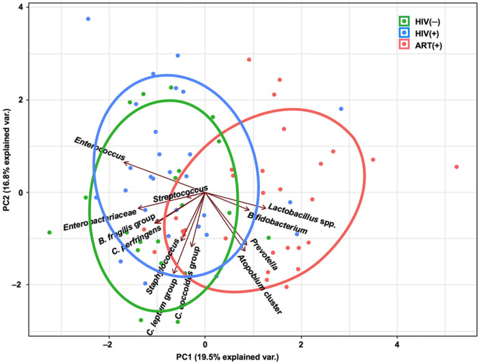
PCA revealed that the HIV(−) and HIV(+) groups had similar gut microbiota structures. The gut microbiota structure of the ART(+) group was different from the other groups and characterized by the abundance of Atopobium cluster, Bifidobacterium, Prevotella, and Lactobacillus spp. (Fig 1).

Fig 1. Principal component analysis based on the overall structure of the gut microbiota in three groups of children.

Fig 1

Each data point represents an individual sample. Ellipses represent the 95% confidence level. Treatment groups are color-coded: green, HIV(−); blue, HIV(+); and red, ART(+). Arrows indicate characteristic vectors of the 12 bacterial factors.

The number of bacteria in each group is shown in Fig 2. In the HIV(+) group, the numbers of C. perfringens and Atopobium cluster were significantly lower (P = 0.02 and P = 0.048, respectively) and the number of Lactobacillus spp. significantly higher (P = 0.02) than in the HIV(–) group. In the ART(+) group, the numbers of Enterococcus, B. fragilis group, and Enterobacteriaceae were significantly lower (P < 0.001, P = 0.04, P = 0.002, respectively) and the numbers of Bifidobacterium and Atopobium cluster significantly higher (both P < 0.001) than in the HIV(+) group (Fig 2 and S1 Table).

Fig 2. Box plots showing the abundance of bacteria in the gut microbiota of the three study groups.
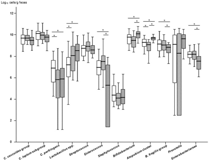
Fig 2

Phylum Firmicutes: Clostridium coccoides group, C. leptum subgroup, C. perfringens, Lactobacillus spp., Streptococcus, Enterococcus, and Staphylococcus; phylum Actinobacteria: Bifidobacterium and Atopobium cluster; phylum Bacteroidetes: Bacteroides fragilis group and Prevotella; and phylum Proteobacteria: Enterobacteriaceae. The lines and error bars correspond to the medians ± 95% confidence intervals. White box, HIV(−) group; oblique line box, HIV(+) group; gray box, ART(+) group. *P < 0.05, Mann-Whitney U test. C. difficile and Pseudomonas were not included due to the low detection frequencies (3.4% to 20% in all three groups; C. difficile, median = 1.15 log10 cells/g feces, and Pseudomonas, median = 1.45 log10 cells/g feces, S2 Table).

Association between age and gut microbiota

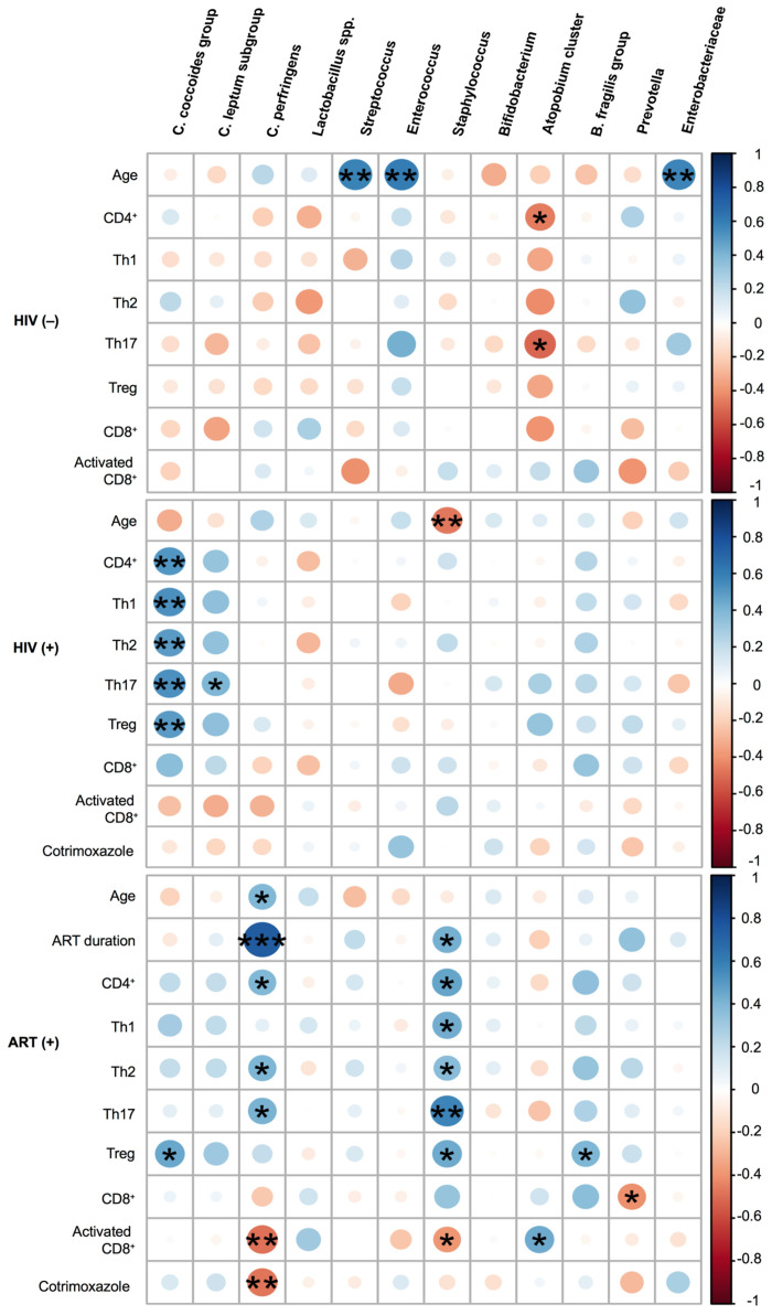
In the HIV(−) group, but not in the HIV(+) group, the numbers of Streptococcus, Enterococcus, and Enterobacteriaceae positively correlated with age (Rho = 0.59, P = 0.006; Rho = 0.61, P = 0.005; and Rho = 0.57, P = 0.008, respectively; Fig 3 and S3 Table). The number of Staphylococcus inversely correlated with age in the HIV(+) group (Rho = −0.47, P = 0.009).

Fig 3. Heatmap representing the correlation of gut microbiota with age, anti-retroviral therapy (ART) duration, immune status, and use of cotrimoxazole.

Fig 3

Blue shading indicates a positive association and red shading a negative association. The scale indicates the strengths of associations. C. difficile and Pseudomonas were not included due to the low detection frequencies (3.4% to 20% in all three groups: C. difficile, median = log10 1.15 cells/g feces, and Pseudomonas, median = log10 1.45 cells/g feces, S2 Table). The color intensity and size of the circles are proportional to the correlation coefficients. *P < 0.05, **P < 0.01, ***P < 0.001, based on Spearman’s rank-test.

Association between gut microbiota and immune status

In the HIV(−) group, the number of Atopobium cluster inversely correlated with CD4+ cell and Th17 counts (Rho = −0.46, P = 0.04 and Rho = −0.51, P = 0.02, respectively). In the HIV(+) group, the number of C. coccoides group significantly correlated with the CD4+ cell count and its subsets (CD4+ cells: Rho = 0.51, P = 0.004; Th1: Rho = 0.54, P = 0.002; Th2: Rho = 0.50, P = 0.005; Th17: Rho = 0.54, P = 0.002; and Treg: Rho = 0.49, P = 0.006).

In the ART(+) group, the number of Staphylococcus significantly correlated with the CD4+ cell count and its subsets (CD4+ cells: Rho = 0.46, P = 0.01; Th1: Rho = 0.44, P = 0.02; Th2: Rho = 0.37, P = 0.047; Th17: Rho = 0.58, P = 0.001; Treg: Rho = 0.45, P = 0.02), the percentage of activated CD8+ cells (Rho = −0.39, P = 0.04), and the ART duration (Rho = 0.42, P = 0.02). The number of C. perfringens significantly correlated with age (Rho = 0.39, P = 0.03), CD4+ cell count (Rho = 0.39, P = 0.04), Th2 count (Rho = 0.40, P = 0.03), Th17 count (Rho = 0.42, P = 0.03), and percentage of activated CD8+ cells (Rho = −0.49, P = 0.01), and most strongly with the ART duration (Rho = 0.75, P < 0.001). The C. coccoides group and B. fragilis group were associated with Treg cell count (Rho = 0.45, P = 0.01 and Rho = 0.40, P = 0.03, respectively). Prevotella was negatively associated with the CD8+ cell count (Rho = −0.41, P = 0.03; Fig 3 and S3 Table).

Impact of cotrimoxazole on the gut microbiota of children with HIV

Fifteen (50.0%) of the 30 children in the HIV(+) group and 9 (31%) of the 29 children in the ART(+) group received cotrimoxazole. Cotrimoxazole treatment did not significantly affect the gut microbiota profile in the HIV(+) group. However, in the ART(+) group, the number of C. perfringens was significantly lower among children treated with cotrimoxazole than those not treated with cotrimoxazole [with cotrimoxazole: 3.4 (2.2−5.4) vs. without cotrimoxazole: 6.2 (4.8−7.5), P = 0.01; S4 Table].

Independent predictors of the gut microbiota in children with HIV

The multiple linear regression analyses including age, ART duration, immune status, and use of cotrimoxazole showed that the ART duration was independently associated with the number of C. perfringens (Beta coefficient = 0.726, P < 0.001), and Th17 count with the number of Staphylococcus (Beta coefficient = 0.428, P = 0.02) in the ART(+) group (Tables 2 and 3). The linear regression analysis for C. coccoides group in the HIV(+) group showed no significant association (S5 Table).

Table 2. Linear regression analysis of Clostridium perfringens with age, ART duration, immune status, and use of cotrimoxazole in the ART(+) group.

Unadjusted linear regression Adjusted linear regression
Variable Beta SE P-value Beta SE P-value
Age (years) 0.323 0.261 0.09
ART duration (years) 0.726 0.166 <0.001 0.726 0.166 <0.001
Th2 count 0.418 0.001 0.024
Th17 count 0.371 0.015 0.048
Activated CD8+ cells (%) -0.484 0.068 0.008
Cotrimoxazole use (yes vs. no) -0.446 0.809 0.015

ART: anti-retroviral therapy; Beta: regression coefficient; SE: standard error.

The factors with P < 0.05 in the simple linear regression analysis were included in the stepwise multiple linear regression analysis. P-values in bold are significant.

Table 3. Linear regression analysis of Staphylococcus with ART duration and immune status in the ART(+) group.

Unadjusted linear regression Adjusted linear regression
Variable Beta SE P-value Beta SE P-value
ART duration (years) 0.370 0.109 0.048
Th1 count 0.309 0.005 0.103
Th2 count 0.279 0.001 0.142
Th17 count 0.428 0.007 0.020 0.428 0.007 0.020
Treg count 0.244 0.012 0.202
Activated CD8+ cells (%) -0.305 0.036 0.108

ART: anti-retroviral therapy; Treg: regulatory T cells; Beta: regression coefficient; SE: standard error. The factors with P < 0.05 in the simple linear regression analysis were included in the stepwise multiple linear regression analysis. P-values in bold are significant.

Discussion

In the present study, we investigated the impact of HIV infection and ART on the gut microbiota of Vietnamese children using RT-qPCR. We found that several sub-dominant gut bacteria were positively associated with age in children without HIV, but this was not observed in the children with HIV. In addition, Staphylococcus negatively correlated with age, i.e. the duration of HIV infection, in the children vertically infected with HIV, and ART duration had an independent positive association with C. perfringens and Th17 count with Staphylococcus in the HIV-infected children on ART. These findings indicate an impact of HIV infection and ART on the sub-dominant gut microbiota, such as C. perfringens and Staphylococcus, in children. Our findings highlight the importance of investigating the role of the sub-dominant gut microbiota in the pathogenesis of HIV infection.

To the best of our knowledge, this study is the first to apply RT-qPCR techniques to evaluate the gut microbiota, particularly sub-dominant bacteria, in children with and without HIV. The sum of the six dominant bacterial groups in fecal samples detected by RT-qPCR was previously reported to cover 71.3% of the total intestinal bacterial count estimated by hybridization with a universal probe [29, 41]. This finding ensures the validity of using RT-qPCR methods to identify the main gut microbiota profile in this study, though our results may not be comparable directly to the results of the other studies using NGS, since the RT-qPCR method is not appropriate to calculate the diversity indices and the relative abundance of the selected bacteria. In addition, the RT-qPCR approach can detect and enumerate the gut bacteria at the population level between 102 and 1011 cells/g of feces, whereas the lower detection limit of next generation sequencing (NGS) methods is 107 to 108 cells/g [33]. The counts of the sub-dominant bacteria, including Lactobacilli and potential pathogens, such as C. perfringens, were near the detection limit of NGS or lower [33]; thus, we took advantage of RT-qPCR to estimate the counts of these less abundant, but clinically significant, targets.

In this study, the numbers of dominant gut bacteria, including C. coccoides group, C. leptum subgroup, Bifidobacterium, and Atopobium cluster, did not correlate with age in the HIV(−) group, i.e., children aged 2 to 8 years. This finding is consistent with previous findings that the gut microbiota of healthy children stabilizes and becomes similar to that of adults at around 2 or 3 years of age [7, 21, 22, 33]. In contrast, several sub-dominant gut bacteria, including Streptococcus, Enterococcus, and Enterobacteriaceae, positively correlated with age in the HIV(−) group. This finding is also consistent with previous findings in Japanese children evaluated using the same RT-qPCR methods [33]. These results suggest that the dominant gut microbiota may reach stable levels by 2 or 3 years of age, whereas the sub-dominant bacteria may still be in transition in children aged 2 to 8 years.

We found that C. coccoides group positively correlated with the CD4+ cell count and its subsets in the HIV(+) group, as well as the Treg cell count in the ART(+) group. The C. coccoides spp. are known to promote the expansion and differentiation of Treg cells, which play a central role in regulating gut inflammation through the production of butyrate, a short-chain fatty acid (SCFA), in mice [42, 43]. In the ART(+) group, the B. fragilis group positively correlated with the Treg cell count. This finding is consistent with a previous study that found that B. fragilis promotes Treg cell function by producing polysaccharide A [44]. Thus, HIV infection and ART may also influence the immune status by changing the levels of gut bacterial metabolites, such as SCFAs [45, 46]. Further studies on bacterial metabolites and their networks in the gut microbiota of children with HIV treated with and without ART may elucidate the underlying mechanisms of immune modulation in HIV infection and ART interventions.

Multiple regression analysis showed a positive association between gut Staphylococcus and Th17 count in the ART(+) group, which was shown for the first time. Th17 cells produce interleukin-17, which is important for promoting neutrophil recruitment to clear bacteria and has a specific role in the host defense against Staphylococcus aureus skin infection [47]. Thus, it would be interesting to investigate the interaction between Th17 and gut Staphylococcus in order to understand the pathophysiology of HIV infection in children who are on ART.

The use of cotrimoxazole reportedly influences some gut bacteria and reduces gut inflammation in children with HIV [4850]. In the current study, the use of cotrimoxazole was associated only with C. perfringens in the ART(+) group. However, in multiple regression analysis, we found that ART duration, but not the use of cotrimoxazole, was independently associated with C. perfringens, which is a potentially harmful bacterium [51]. These findings suggest that a novel therapeutic approach, such as ingesting probiotics and/or prebiotics, may be necessary to restore gut microbiota homeostasis in children with HIV who are on ART [52].

In this study, all of the children in the ART(+) group were treated with NRTIs as backbone drugs and only four also received a PI. Therefore, the positive correlation between the number of C. perfringens and ART duration may be due to the use of NRTIs, but not PIs, even though PIs are known to lower the diversity of the gut microbiota [53, 54]. Our study is consistent with the previous study that ART, especially NRTI-including regimen, had more suppressive impacts on the composition and the variability of the gut microbiota [55]. Further study is needed to investigate whether NRTIs influence the gut microbiota, directly or indirectly, through the restoration of immune status in children with HIV who are on ART.

This study has some limitations. First, the children in the HIV(−) group were 2 years younger than the other groups. The diets were not controlled among the groups, though the children in the HIV(−) and ART(+) groups who resided at orphanages were provided the same diets. The children in the HIV(+) group who resided in their own homes appeared to have poorer nutritional status than the children in other groups, which could be due to the uncontrolled diet and/or HIV infection [26]. Considering the influence of age and diet factors on the gut microbiota [7, 21, 22, 33, 5658], we did not focus on comparing the gut microbiota between the groups, but highlighted the factors associated with the gut microbiota in each group. Second, the number of patients recruited in the present study was relatively small, which may limit the significance of our findings. Third, we have not mentioned the HIV-exposure history of the children in the HIV(−) group because we could not confirm the history via documents, though they were reportedly born to mothers with HIV. Machiavelli et al. reported that the gut microbiota of HIV-exposed but uninfected children is similar to that of HIV-unexposed and uninfected children at the age of 18 months except in several taxa [59]. Therefore, the impact of HIV-exposure history on the gut microbiota in the children without HIV over 2 years of age would be limited. These findings require confirmation in longitudinal studies that compare the gut microbiome between age-matched children with and without HIV, with and without HIV-exposure history, and/or before and after initiating ART to assess the effect of ART on the composition of the gut microbiota in children with HIV.

This study provided new insights into the alterations in the gut microbiota, particularly the sub-dominant groups of bacteria, among children with HIV. Our results suggest that HIV infection and ART influence the sub-dominant gut microbiota, directly or indirectly, in association with the immune status of children with HIV.

Supporting information

S1 Fig. The gating strategy for cell staining analysis in flow cytometry.

CD8+ cell activations was defined as the CD38+HLA-DR+ population. Regulatory T (Treg) cells were defined as CD25highCD4+ cells, Th1 as CXCR3+CCR6CD25lowCD4+ cells, Th2 as CXCR3CCR6CD25lowCD4+ cells, and Th17 as CXCR3CCR6+CD25lowCD4+ cells. Unstained cells were used for gating controls.

(TIF)

S1 Table. Number of bacteria in fecal samples from each study group.

Values are the median counts (IQR), based on RT-qPCR, expressed in units of log10 cells/g feces. The Lactobacillus spp. counts were obtained with RT-qPCR and are expressed as the sum of the six subgroups and two species; ART: anti-retroviral therapy; C.: Clostridium; L.: Lactobacillus; B.: Bacteroides. P-values in bold are statistically significant, based on the Man-Whitney U test.

(DOCX)

S2 Table. Detection frequency of bacteria in fecal samples from each study group.

Values are the detection frequency, defined as the % of samples that harbored detectable microbiota, among all samples in a given group. The Lactobacillus spp. counts were obtained with RT-qPCR and are expressed as the sum of the six subgroups and two species; ART: anti-retroviral therapy; C.: Clostridium; L.: Lactobacillus; B.: Bacteroides; P-values in bold are statistically significant, based on the Chi-square test or Fisher’s exact probability test.

(DOCX)

S3 Table. Association between gut microbiota and age, ART duration, immune status, and use of cotrimoxazole in children.

ART: anti-retroviral therapy; Treg: regulatory T cell; C.: Clostridium; B.: Bacteroides; P-values in bold are statistically significant, based on Spearman’s rank correlation analysis.

(DOCX)

S4 Table. Number of bacteria in fecal samples from the HIV(+) and ART(+) groups stratified by the use of cotrimoxazole.

Values are the median counts (IQR), based on RT-qPCR, expressed in units of log10 cells/g feces; ART: anti-retroviral therapy; C.: Clostridium; B.: Bacteroides; P-values in bold are statistically significant, based on the Mann−Whitney U test.

(DOCX)

S5 Table. Linear regression analysis of Clostridium coccoides group with immune status in the HIV(+) group.

ART: anti-retroviral therapy; Treg: regulatory T cells; Beta: regression coefficient; SE: standard error. Stepwise multiple linear regression analysis was not done, since no factor with P < 0.05 was found in the simple linear regression analysis.

(DOCX)

Acknowledgments

We would like to thank the children who participated in this study for their invaluable support through the use of their samples. We would also like to thank Ms. Thuy Thi Thanh Giang and other staff at the Department of Infectious Diseases and Molecular Laboratory of VNCH, who contributed enormously to this work.

Data Availability

All relevant data are within the manuscript and its Supporting information files.

Funding Statement

This study was supported, in part, by the Ministry of Education, Culture, Sports, Science, and Technology (MEXT) in Japan (the Program of Founding Research Centers for Emerging and Reemerging Infectious Disease; grant number 23406013, https://www.mext.go.jp/en/publication/whitepaper/title03/detail03/sdetail03/sdetail03/1372928.htm) and the Kanazawa University President Strategic Research Fund (https://www.kanazawa-u.ac.jp/research_bulletin/contact.html). The funders had no role in study design, data collection and analysis, decision to publish, or preparation of the manuscript.

References

  • 1.Qin J, Li R, Raes J, Arumugam M, Burgdorf KS, Manichanh C, et al. A human gut microbial gene catalogue established by metagenomic sequencing. Nature. 2010;464(7285):59–65. doi: 10.1038/nature08821 [DOI] [PMC free article] [PubMed] [Google Scholar]
  • 2.Ley RE, Peterson DA, Gordon JI. Ecological and evolutionary forces shaping microbial diversity in the human intestine. Cell. 2006;124(4):837–48. doi: 10.1016/j.cell.2006.02.017 [DOI] [PubMed] [Google Scholar]
  • 3.Kamada N, Nunez G. Regulation of the immune system by the resident intestinal bacteria. Gastroenterology. 2014;146(6):1477–88. doi: 10.1053/j.gastro.2014.01.060 [DOI] [PMC free article] [PubMed] [Google Scholar]
  • 4.Flint HJ, Scott KP, Louis P, Duncan SH. The role of the gut microbiota in nutrition and health. Nat Rev Gastroenterol Hepatol. 2012;9(10):577–89. doi: 10.1038/nrgastro.2012.156 [DOI] [PubMed] [Google Scholar]
  • 5.Maslowski KM, Mackay CR. Diet, gut microbiota and immune responses. Nat Immunol. 2011;12(1):5–9. doi: 10.1038/ni0111-5 [DOI] [PubMed] [Google Scholar]
  • 6.Mariat D, Firmesse O, Levenez F, Guimaraes V, Sokol H, Dore J, et al. The Firmicutes/Bacteroidetes ratio of the human microbiota changes with age. BMC Microbiol. 2009;9:123. doi: 10.1186/1471-2180-9-123 [DOI] [PMC free article] [PubMed] [Google Scholar]
  • 7.Yatsunenko T, Rey FE, Manary MJ, Trehan I, Dominguez-Bello MG, Contreras M, et al. Human gut microbiome viewed across age and geography. Nature. 2012;486(7402):222–7 doi: 10.1038/nature11053 [DOI] [PMC free article] [PubMed] [Google Scholar]
  • 8.Zheng D, Liwinski T, Elinav E. Interaction between microbiota and immunity in health and disease. Cell Res. 2020;30(6):492–506. doi: 10.1038/s41422-020-0332-7 [DOI] [PMC free article] [PubMed] [Google Scholar]
  • 9.Kamada N, Seo SU, Chen GY, Núñez G. Role of the gut microbiota in immunity and inflammatory disease. Nat Rev Immunol. 2013;13(5):321–35. doi: 10.1038/nri3430 [DOI] [PubMed] [Google Scholar]
  • 10.Sengupta S, Siliciano RF. Targeting the Latent Reservoir for HIV-1. Immunity. 2018;48(5):872–95. doi: 10.1016/j.immuni.2018.04.030 [DOI] [PMC free article] [PubMed] [Google Scholar]
  • 11.Mehandru S, Poles MA, Tenner-Racz K, Horowitz A, Hurley A, Hogan C, et al. Primary HIV-1 infection is associated with preferential depletion of CD4+ T lymphocytes from effector sites in the gastrointestinal tract. J Exp Med. 2004;200(6):761–70. doi: 10.1084/jem.20041196 [DOI] [PMC free article] [PubMed] [Google Scholar]
  • 12.Veazey RS, DeMaria M, Chalifoux LV, Shvetz DE, Pauley DR, Knight HL, et al. Gastrointestinal tract as a major site of CD4+ T cell depletion and viral replication in SIV infection. Science. 1998;280(5362):427–31. doi: 10.1126/science.280.5362.427 [DOI] [PubMed] [Google Scholar]
  • 13.Brenchley JM, Paiardini M, Knox KS, Asher AI, Cervasi B, Asher TE, et al. Differential Th17 CD4 T-cell depletion in pathogenic and nonpathogenic lentiviral infections. Blood. 2008;112(7):2826–35. doi: 10.1182/blood-2008-05-159301 [DOI] [PMC free article] [PubMed] [Google Scholar]
  • 14.Hunt PW. Th17, gut, and HIV: therapeutic implications. Curr Opin HIV AIDS. 2010;5(2):189–93. doi: 10.1097/COH.0b013e32833647d9 [DOI] [PMC free article] [PubMed] [Google Scholar]
  • 15.Zhou Y, Ou Z, Tang X, Zhou Y, Xu H, Wang X, et al. Alterations in the gut microbiota of patients with acquired immune deficiency syndrome. J Cell Mol Med. 2018;22(4):2263–71. doi: 10.1111/jcmm.13508 [DOI] [PMC free article] [PubMed] [Google Scholar]
  • 16.Lozupone CA, Li M, Campbell TB, Flores SC, Linderman D, Gebert MJ, et al. Alterations in the gut microbiota associated with HIV-1 infection. Cell Host Microbe. 2013;14(3):329–39. doi: 10.1016/j.chom.2013.08.006 [DOI] [PMC free article] [PubMed] [Google Scholar]
  • 17.Dillon SM, Lee EJ, Kotter CV, Austin GL, Dong Z, Hecht DK, et al. An altered intestinal mucosal microbiome in HIV-1 infection is associated with mucosal and systemic immune activation and endotoxemia. Mucosal Immunol. 2014;7(4):983–94. doi: 10.1038/mi.2013.116 [DOI] [PMC free article] [PubMed] [Google Scholar]
  • 18.McHardy IH, Li X, Tong M, Ruegger P, Jacobs J, Borneman J, et al. HIV Infection is associated with compositional and functional shifts in the rectal mucosal microbiota. Microbiome. 2013;1(1):26. doi: 10.1186/2049-2618-1-26 [DOI] [PMC free article] [PubMed] [Google Scholar]
  • 19.Ringel-Kulka T, Cheng J, Ringel Y, Salojarvi J, Carroll I, Palva A, et al. Intestinal microbiota in healthy U.S. young children and adults—a high throughput microarray analysis. PLoS One. 2013;8(5):e64315. doi: 10.1371/journal.pone.0064315 [DOI] [PMC free article] [PubMed] [Google Scholar]
  • 20.Derrien M, Alvarez AS, de Vos WM. The Gut Microbiota in the First Decade of Life. Trends Microbiol. 2019;27(12):997–1010. doi: 10.1016/j.tim.2019.08.001 [DOI] [PubMed] [Google Scholar]
  • 21.Stewart CJ, Ajami NJ, O’Brien JL, Hutchinson DS, Smith DP, Wong MC, et al. Temporal development of the gut microbiome in early childhood from the TEDDY study. Nature. 2018;562(7728):583–8. doi: 10.1038/s41586-018-0617-x [DOI] [PMC free article] [PubMed] [Google Scholar]
  • 22.Koenig JE, Spor A, Scalfone N, Fricker AD, Stombaugh J, Knight R, et al. Succession of microbial consortia in the developing infant gut microbiome. Proc Natl Acad Sci U S A. 2011;108 Suppl 1:4578–85. doi: 10.1073/pnas.1000081107 [DOI] [PMC free article] [PubMed] [Google Scholar]
  • 23.Kaur US, Shet A, Rajnala N, Gopalan BP, Moar P, D H, et al. High Abundance of genus Prevotella in the gut of perinatally HIV-infected children is associated with IP-10 levels despite therapy. Sci Rep. 2018;8(1):17679. doi: 10.1038/s41598-018-35877-4 [DOI] [PMC free article] [PubMed] [Google Scholar]
  • 24.Flygel TT, Sovershaeva E, Classen-Weitz S, Hjerde E, Mwaikono KS, Odland JO, et al. Composition of gut microbiota of children and adolescents with perinatal HIV infection taking antiretroviral therapy in Zimbabwe. J Infect Dis. 2019. [DOI] [PMC free article] [PubMed] [Google Scholar]
  • 25.Abange WB, Martin C, Nanfack AJ, Yatchou LG, Nusbacher N, Nguedia CA, et al. Alteration of the gut fecal microbiome in children living with HIV on antiretroviral therapy in Yaounde, Cameroon. Sci Rep. 2021;11(1):7666 doi: 10.1038/s41598-021-87368-8 [DOI] [PMC free article] [PubMed] [Google Scholar]
  • 26.Bi X, Ishizaki A, Nguyen LV, Matsuda K, Pham HV, Phan CT, et al. Impact of HIV Infection and Anti-Retroviral Therapy on the Immune Profile of and Microbial Translocation in HIV-Infected Children in Vietnam. Int J Mol Sci. 2016;17(8). [DOI] [PMC free article] [PubMed] [Google Scholar]
  • 27.Ishizaki A, Bi X, Nguyen LV, Matsuda K, Pham HV, Phan CTT, et al. Effects of Short-Term Probiotic Ingestion on Immune Profiles and Microbial Translocation among HIV-1-Infected Vietnamese Children. Int J Mol Sci. 2017;18(10). doi: 10.3390/ijms18102185 [DOI] [PMC free article] [PubMed] [Google Scholar]
  • 28.Kurakawa T, Kubota H, Tsuji H, Matsuda K, Asahara T, Takahashi T, et al. Development of a sensitive rRNA-targeted reverse transcription-quantitative polymerase chain reaction for detection of Vibrio cholerae/mimicus, V. parahaemolyticus/alginolyticus and Campylobacter jejuni/coli. Microbiol Immunol. 2012;56(1):10–20. doi: 10.1111/j.1348-0421.2011.00405.x [DOI] [PubMed] [Google Scholar]
  • 29.Matsuda K, Tsuji H, Asahara T, Matsumoto K, Takada T, Nomoto K. Establishment of an analytical system for the human fecal microbiota, based on reverse transcription-quantitative PCR targeting of multicopy rRNA molecules. Appl Environ Microbiol. 2009;75(7):1961–9. doi: 10.1128/AEM.01843-08 [DOI] [PMC free article] [PubMed] [Google Scholar]
  • 30.Matsuda K, Tsuji H, Asahara T, Takahashi T, Kubota H, Nagata S, et al. Sensitive quantification of Clostridium difficile cells by reverse transcription-quantitative PCR targeting rRNA molecules. Appl Environ Microbiol. 2012;78(15):5111–8. doi: 10.1128/AEM.07990-11 [DOI] [PMC free article] [PubMed] [Google Scholar]
  • 31.Matsuki T, Watanabe K, Fujimoto J, Takada T, Tanaka R. Use of 16S rRNA gene-targeted group-specific primers for real-time PCR analysis of predominant bacteria in human feces. Appl Environ Microbiol. 2004;70(12):7220–8. doi: 10.1128/AEM.70.12.7220-7228.2004 [DOI] [PMC free article] [PubMed] [Google Scholar]
  • 32.Sakaguchi S, Saito M, Tsuji H, Asahara T, Takata O, Fujimura J, et al. Bacterial rRNA-targeted reverse transcription-PCR used to identify pathogens responsible for fever with neutropenia. J Clin Microbiol. 2010;48(5):1624–8. doi: 10.1128/JCM.01724-09 [DOI] [PMC free article] [PubMed] [Google Scholar]
  • 33.Tsuji H, Matsuda K, Nomoto K. Counting the Countless: Bacterial Quantification by Targeting rRNA Molecules to Explore the Human Gut Microbiota in Health and Disease. Front Microbiol. 2018;9:1417. doi: 10.3389/fmicb.2018.01417 [DOI] [PMC free article] [PubMed] [Google Scholar]
  • 34.Harmsen HJ, Raangs GC, He T, Degener JE, Welling GW. Extensive set of 16S rRNA-based probes for detection of bacteria in human feces. Appl Environ Microbiol. 2002;68(6):2982–90. doi: 10.1128/AEM.68.6.2982-2990.2002 [DOI] [PMC free article] [PubMed] [Google Scholar]
  • 35.González-Hernández LA, Ruiz-Briseño MDR, Sánchez-Reyes K, Alvarez-Zavala M, Vega-Magaña N, López-Iñiguez A, et al. Alterations in bacterial communities, SCFA and biomarkers in an elderly HIV-positive and HIV-negative population in western Mexico. BMC Infect Dis. 2019;19(1):234. doi: 10.1186/s12879-019-3867-9 [DOI] [PMC free article] [PubMed] [Google Scholar]
  • 36.Hunt PW, Martin JN, Sinclair E, Bredt B, Hagos E, Lampiris H, et al. T cell activation is associated with lower CD4+ T cell gains in human immunodeficiency virus-infected patients with sustained viral suppression during antiretroviral therapy. J Infect Dis. 2003;187(10):1534–43. doi: 10.1086/374786 [DOI] [PubMed] [Google Scholar]
  • 37.Maecker HT, McCoy JP, Nussenblatt R. Standardizing immunophenotyping for the Human Immunology Project. Nat Rev Immunol. 2012;12(3):191–200. doi: 10.1038/nri3158 [DOI] [PMC free article] [PubMed] [Google Scholar]
  • 38.Kinter A, McNally J, Riggin L, Jackson R, Roby G, Fauci AS. Suppression of HIV-specific T cell activity by lymph node CD25+ regulatory T cells from HIV-infected individuals. Proc Natl Acad Sci U S A. 2007;104(9):3390–5. doi: 10.1073/pnas.0611423104 [DOI] [PMC free article] [PubMed] [Google Scholar]
  • 39.Team R. R:A language and environment for statistical computing. R Foundation for Statistical Computing Vienna, Austria; 2018. https://www.r-project.org.
  • 40.Ministry of Health, Vietnam. Guidelines for HIV/AIDS Diagnosis and Treatment; 2009. https://www.who.int/hiv/pub/guidelines/vietnam_art.pdf?ua=1.
  • 41.Hasegawa S, Goto S, Tsuji H, Okuno T, Asahara T, Nomoto K, et al. Intestinal Dysbiosis and Lowered Serum Lipopolysaccharide-Binding Protein in Parkinson’s Disease. PLoS One. 2015;10(11):e0142164. doi: 10.1371/journal.pone.0142164 [DOI] [PMC free article] [PubMed] [Google Scholar]
  • 42.Furusawa Y, Obata Y, Fukuda S, Endo TA, Nakato G, Takahashi D, et al. Commensal microbe-derived butyrate induces the differentiation of colonic regulatory T cells. Nature. 2013;504(7480):446–50. doi: 10.1038/nature12721 [DOI] [PubMed] [Google Scholar]
  • 43.Atarashi K, Tanoue T, Oshima K, Suda W, Nagano Y, Nishikawa H, et al. Treg induction by a rationally selected mixture of Clostridia strains from the human microbiota. Nature. 2013;500(7461):232–6. doi: 10.1038/nature12331 [DOI] [PubMed] [Google Scholar]
  • 44.Telesford KM, Yan W, Ochoa-Reparaz J, Pant A, Kircher C, Christy MA, et al. A commensal symbiotic factor derived from Bacteroides fragilis promotes human CD39(+)Foxp3(+) T cells and Treg function. Gut Microbes. 2015;6(4):234–42. doi: 10.1080/19490976.2015.1056973 [DOI] [PMC free article] [PubMed] [Google Scholar]
  • 45.Serrano-Villar S, Rojo D, Martínez-Martínez M, Deusch S, Vázquez-Castellanos JF, Bargiela R, et al. Gut Bacteria Metabolism Impacts Immune Recovery in HIV-infected Individuals. EBioMedicine. 2016;8:203–16. doi: 10.1016/j.ebiom.2016.04.033 [DOI] [PMC free article] [PubMed] [Google Scholar]
  • 46.Vázquez-Castellanos JF, Serrano-Villar S, Latorre A, Artacho A, Ferrús ML, Madrid N, et al. Altered metabolism of gut microbiota contributes to chronic immune activation in HIV-infected individuals. Mucosal Immunol. 2015;8(4):760–72. doi: 10.1038/mi.2014.107 [DOI] [PubMed] [Google Scholar]
  • 47.Miller LS, Cho JS. Immunity against Staphylococcus aureus cutaneous infections. Nature Reviews Immunology. 2011;11(8):505–518. doi: 10.1038/nri3010 [DOI] [PMC free article] [PubMed] [Google Scholar]
  • 48.Bourke CD, Gough EK, Pimundu G, Shonhai A, Berejena C, Terry L, et al. Cotrimoxazole reduces systemic inflammation in HIV infection by altering the gut microbiome and immune activation. Sci Transl Med. 2019;11(486). doi: 10.1126/scitranslmed.aav0537 [DOI] [PMC free article] [PubMed] [Google Scholar]
  • 49.Gough EK, Bourke CD, Berejena C, Shonhai A, Bwakura-Dangarembizi M, Prendergast AJ, et al. Strain-level analysis of gut-resident pro-inflammatory viridans group Streptococci suppressed by long-term cotrimoxazole prophylaxis among HIV-positive children in Zimbabwe. Gut Microbes. 2020;11(4):1104–15. doi: 10.1080/19490976.2020.1717299 [DOI] [PMC free article] [PubMed] [Google Scholar]
  • 50.Kyosiimire-Lugemwa J, Anywaine Z, Abaasa A, Levin J, Gombe B, Musinguzi K, et al. Effect of Stopping Cotrimoxazole Preventive Therapy on Microbial Translocation and Inflammatory Markers Among Human Immunodeficiency Virus-Infected Ugandan Adults on Antiretroviral Therapy: The COSTOP Trial Immunology Substudy. J Infect Dis. 2020;222(3):381–90. doi: 10.1093/infdis/jiz494 [DOI] [PMC free article] [PubMed] [Google Scholar]
  • 51.Kiu R, Hall LJ. An update on the human and animal enteric pathogen Clostridium perfringens. Emerg Microbes Infect. 2018;7(1):141. doi: 10.1038/s41426-018-0144-8 [DOI] [PMC free article] [PubMed] [Google Scholar]
  • 52.Monteiro CRAV, do Carmo MS, Melo BO, Alves MS, Dos Santos CI, Monteiro SG, et al. In Vitro Antimicrobial Activity and Probiotic Potential of. Nutrients. 2019;11(2). doi: 10.3390/nu11020448 [DOI] [PMC free article] [PubMed] [Google Scholar]
  • 53.Pinto-Cardoso S, Klatt NR, Reyes-Terán G. Impact of antiretroviral drugs on the microbiome: unknown answers to important questions. Curr Opin HIV AIDS. 2018;13(1):53–60. doi: 10.1097/COH.0000000000000428 [DOI] [PMC free article] [PubMed] [Google Scholar]
  • 54.Villanueva-Millan MJ, Perez-Matute P, Recio-Fernandez E, Lezana Rosales JM, Oteo JA. Differential effects of antiretrovirals on microbial translocation and gut microbiota composition of HIV-infected patients. J Int AIDS Soc. 2017;20(1):21526. doi: 10.7448/IAS.20.1.21526 [DOI] [PMC free article] [PubMed] [Google Scholar]
  • 55.Imahashi M, Ode H, Kobayashi A, Nemoto M, Matsuda M, Hashiba C, et al. Impact of long-term antiretroviral therapy on gut and oral microbiotas in HIV-1-infected patients. Sci Rep. 2021;11(1):960. doi: 10.1038/s41598-020-80247-8 [DOI] [PMC free article] [PubMed] [Google Scholar]
  • 56.Singh RK, Chang HW, Yan D, Lee KM, Ucmak D, Wong K, et al. Influence of diet on the gut microbiome and implications for human health. J Transl Med. 2017;15(1):73. doi: 10.1186/s12967-017-1175-y [DOI] [PMC free article] [PubMed] [Google Scholar]
  • 57.Nguyen DT, Van Hoorde K, Cnockaert M, De Brandt E, Aerts M, Binh Thanh L, et al. A description of the lactic acid bacteria microbiota associated with the production of traditional fermented vegetables in Vietnam. Int J Food Microbiol. 2013;163(1):19–27. doi: 10.1016/j.ijfoodmicro.2013.01.024 [DOI] [PubMed] [Google Scholar]
  • 58.Surono IS, Widiyanti D, Kusumo PD, Venema K. Gut microbiota profile of Indonesian stunted children and children with normal nutritional status. PLoS One. 2021;16(1):e0245399. doi: 10.1371/journal.pone.0245399 [DOI] [PMC free article] [PubMed] [Google Scholar]
  • 59.Machiavelli A, Duarte RTD, Pires MMS, Zárate-Bladés CR, Pinto AR. The impact of in utero HIV exposure on gut microbiota, inflammation, and microbial translocation. Gut Microbes. 2019;10(5):599–614. doi: 10.1080/19490976.2018.1560768 [DOI] [PMC free article] [PubMed] [Google Scholar]

Decision Letter 0

Jennifer Manuzak

13 Jul 2021

PONE-D-21-16737

Alterations in children's sub-dominant gut microbiota by HIV infection and anti-retroviral therapy

PLOS ONE

Dear Dr. Ichimura,

Thank you for submitting your manuscript to PLOS ONE. After careful consideration, we feel that it has merit but does not fully meet PLOS ONE’s publication criteria as it currently stands. Therefore, we invite you to submit a revised version of the manuscript that addresses the points raised during the review process.

Please be sure to pay particular attention to the following reviewer concerns: 

  • Ensure that all relevant literature is cited and the findings in the current study are put into context with the additional references 

  • Provide additional justification for the selection of flow cytometry markers and update gating strategy figure 

  • Provide justification for the experimental methods selected for the analysis of bacterial communities and include additional analyses on diversity measures and relative abundance analysis 

  • Address questions with regards to the impacts of age and ART duration and incorporate into manuscript

  • Address all other major and minor reviewer comments/concerns 

Please submit your revised manuscript by Aug 27 2021 11:59PM. If you will need more time than this to complete your revisions, please reply to this message or contact the journal office at plosone@plos.org. When you're ready to submit your revision, log on to https://www.editorialmanager.com/pone/ and select the 'Submissions Needing Revision' folder to locate your manuscript file. Do not submit a revised manuscript as a new submission. 

Please include the following items when submitting your revised manuscript:

  • A rebuttal letter that responds to each point raised by the academic editor and reviewer(s). You should upload this letter as a separate file labeled 'Response to Reviewers'.

  • A marked-up copy of your manuscript that highlights changes made to the original version. You should upload this as a separate file labeled 'Revised Manuscript with Track Changes'.

  • An unmarked version of your revised paper without tracked changes. You should upload this as a separate file labeled 'Manuscript'.

If you would like to make changes to your financial disclosure, please include your updated statement in your cover letter. Guidelines for resubmitting your figure files are available below the reviewer comments at the end of this letter.

If applicable, we recommend that you deposit your laboratory protocols in protocols.io to enhance the reproducibility of your results. Protocols.io assigns your protocol its own identifier (DOI) so that it can be cited independently in the future. For instructions see: http://journals.plos.org/plosone/s/submission-guidelines#loc-laboratory-protocols. Additionally, PLOS ONE offers an option for publishing peer-reviewed Lab Protocol articles, which describe protocols hosted on protocols.io. Read more information on sharing protocols at https://plos.org/protocols?utm_medium=editorial-email&utm_source=authorletters&utm_campaign=protocols.

If you choose not to submit a revision, please notify us. 

We look forward to receiving your revised manuscript.

Kind regards,

Dr. Jennifer Manuzak

Academic Editor

PLOS ONE

Journal requirements:

When submitting your revision, we need you to address these additional requirements.

1. Please ensure that your manuscript meets PLOS ONE's style requirements, including those for file naming. The PLOS ONE style templates can be found at

https://journals.plos.org/plosone/s/file?id=wjVg/PLOSOne_formatting_sample_main_body.pdf and

https://journals.plos.org/plosone/s/file?id=ba62/PLOSOne_formatting_sample_title_authors_affiliations.pdf

2. In your Data Availability statement, you have not specified where the minimal data set underlying the results described in your manuscript can be found. PLOS defines a study's minimal data set as the underlying data used to reach the conclusions drawn in the manuscript and any additional data required to replicate the reported study findings in their entirety. All PLOS journals require that the minimal data set be made fully available. For more information about our data policy, please see http://journals.plos.org/plosone/s/data-availability.

Upon re-submitting your revised manuscript, please upload your study’s minimal underlying data set as either Supporting Information files or to a stable, public repository and include the relevant URLs, DOIs, or accession numbers within your revised cover letter. For a list of acceptable repositories, please see http://journals.plos.org/plosone/s/data-availability#loc-recommended-repositories. Any potentially identifying patient information must be fully anonymized.

Important: If there are ethical or legal restrictions to sharing your data publicly, please explain these restrictions in detail. Please see our guidelines for more information on what we consider unacceptable restrictions to publicly sharing data: http://journals.plos.org/plosone/s/data-availability#loc-unacceptable-data-access-restrictions. Note that it is not acceptable for the authors to be the sole named individuals responsible for ensuring data access.

We will update your Data Availability statement to reflect the information you provide in your cover letter.

3. We note that you have included the phrase “data not shown” in your manuscript. Unfortunately, this does not meet our data sharing requirements. PLOS does not permit references to inaccessible data. We require that authors provide all relevant data within the paper, Supporting Information files, or in an acceptable, public repository. Please add a citation to support this phrase or upload the data that corresponds with these findings to a stable repository (such as Figshare or Dryad) and provide and URLs, DOIs, or accession numbers that may be used to access these data. Or, if the data are not a core part of the research being presented in your study, we ask that you remove the phrase that refers to these data.

[Note: HTML markup is below. Please do not edit.]

Reviewers' comments:

Reviewer's Responses to Questions

Comments to the Author

1. Is the manuscript technically sound, and do the data support the conclusions?

The manuscript must describe a technically sound piece of scientific research with data that supports the conclusions. Experiments must have been conducted rigorously, with appropriate controls, replication, and sample sizes. The conclusions must be drawn appropriately based on the data presented.

Reviewer #1: Partly

Reviewer #2: Partly

**********

2. Has the statistical analysis been performed appropriately and rigorously?

Reviewer #1: Yes

Reviewer #2: Yes

**********

3. Have the authors made all data underlying the findings in their manuscript fully available?

The PLOS Data policy requires authors to make all data underlying the findings described in their manuscript fully available without restriction, with rare exception (please refer to the Data Availability Statement in the manuscript PDF file). The data should be provided as part of the manuscript or its supporting information, or deposited to a public repository. For example, in addition to summary statistics, the data points behind means, medians and variance measures should be available. If there are restrictions on publicly sharing data—e.g. participant privacy or use of data from a third party—those must be specified.

Reviewer #1: Yes

Reviewer #2: Yes

**********

4. Is the manuscript presented in an intelligible fashion and written in standard English?

PLOS ONE does not copyedit accepted manuscripts, so the language in submitted articles must be clear, correct, and unambiguous. Any typographical or grammatical errors should be corrected at revision, so please note any specific errors here.

Reviewer #1: Yes

Reviewer #2: Yes

**********

5. Review Comments to the Author

Please use the space provided to explain your answers to the questions above. You may also include additional comments for the author, including concerns about dual publication, research ethics, or publication ethics. (Please upload your review as an attachment if it exceeds 20,000 characters)

Reviewer #1: This study examined select bacteria via RT-PCR in the feces of children from Vietnam who were HIV- or HIV+ (ART+/-). The authors then looked for associations between the absolute number of bacteria and a variety of other parameters (Age of child, length of time on ART, T cell counts, activation of CD8+ T cells and use of cotrimoxazole) antibiotic.

Major

1. This study is similar to previous work including Flygel et al 2020 Journ Infec Dis and Kaur et al 2018 Sci Rep, which the authors did cite in the introduction. This work is also similar to the studies by Dirajlal-Fargo et al 2020 AIDS (Brazil) and Abange et al 2021 Sci Repo (Cameroon) which was not cited by the authors.

A). Since the current study is so similar to other studies, the authors must provide additional justification for the current study in the introduction.

B). The authors should reference the additional two studies listed above.

C). There is no mention of these comparable studies in the discussion. The current study’s findings must be discussed in the context of existing literature (i.e. what findings were similar between studies, what findings were different and speculation on why there are differences between studies). For example, in the Flygel et al study, children were on ART for at least 6 months at the time of sampling. How does this differ since it is known that the length of time on ART impacts gut microbiome (Imahashi et al 2021)? How did T cell activation differ between the appropriate studies? Does geographical region impact differences between studies?

2. The gating strategy in S1 Figure needs some modification.

A). For the CD8 vs CD4 flow plot, most of the events appear to be out of view on the axis. This flow plot needs to be adjusted so that all events are brought into view.

B). For the CD38 vs HLA-DR flow plot, please provide and isotype control flow plot or FMO controls. The authors may be missing some of the activated CD8 cells based on where the quadrant gate currently sits.

C). It is confusing why the authors have an arrow pointing downwards from the lymphocyte gate towards CD4 when they could have gated from the CD4+ cells found in the CD8 vs CD4 flow plot.

4. The authors need to provide justification for why CD3 was not included in the panel since monocytes and NK cells in the blood can express CD4 and CD8.

Minor

1. Provide some explanation in the discussion as to why the antibiotic cotrimoxazole only effected C. perfringens.

2. Please provide some commentary on whether the use of RT-PCR instead of sequencing could overlook changes since not all bacteria were examined.

Reviewer #2: The manuscript by Nguyen Q, et al, entitled “Alterations in children’s sub-dominant gut microbiota by HIV infection and anti-retroviral therapy” seeks to investigate the gut microbiota of children with HIV in Vietnam and the impact of ART. The paper has important implications as there is no clear consensus on the microbiome in children with HIV, both on and off ART. While the authors find that HIV and ART may influence sub-dominant gut bacteria, there is surprisingly not a significant difference between the microbiome of children uninfected and infected with HIV, opposing current research in the field that has shown reduced bacterial diversity with HIV. Overall, the paper is thought-provoking and provides broad impact; however, there are many key points that the authors need to address:

Major points:

1. A major claim within the paper is that HIV and ART influence the sub-dominant gut microbiota; however, support for this claim rely upon various associations based on the number of bacteria. This case would be strengthened by looking at alpha and beta diversity and the relative abundance of bacterial species.

2. The methods of the RT-qPCR are unclear. The primers appear to be specific for the 14 listed bacteria. If this is true, this eliminates the analysis of a large variety of other intestinal bacteria. Could the authors explain their selection criteria for these 14 selected bacteria? Are these the most abundant bacteria in children? Could the authors also explain their rationale for not sequencing with a universal 16S rRNA primer to identify the most abundant bacteria?

3. FoxP3 and IL-17 are expressed by Tregs and Th17, respectively. Have the authors considered performing a functional analysis of the T cell populations to better classify these T cells?

4. The authors state that they “regarded the threshold for dominance in abundance at 1.0% of the total bacterial count, and the threshold in counts was set at 109 cells/g”. What percentage of the bacterial counts passed this threshold? How was the threshold for sub-dominant bacteria determined at <109 cells/g?

5. Could the authors explain whether the bacterial counts were normalized across groups?

6. It appears that there were many comparisons examined to produce the associations highlighted in this manuscript. Was the data corrected for multiple comparisons using a false discovery rate?

7. Table S2 provides the detection frequency of bacteria in fecal samples from each study group. However, this only accounts for the percentage of samples that harbored the detectable bacteria. Providing the relative abundance of bacterial frequencies within the entire group would strengthen the data and provide a better comparison of the distinct patterns across the groups.

8. The association between number of bacteria and age represent a correlation, not a causation. Can the authors expand upon the impact of the data especially since the groups differed in the average age? Could the positive association be due to the development of the microbiome and the lower age of the HIV(-) group?

9. Can the authors speculate on the impact of the ART duration? Was the duration long enough to see an impact on and restoration of the microbiome?

10. Studies have shown that an altered gut microbiota is associated with elevated circulating inflammatory markers. Since blood was collected in this study, did the authors consider performing an ELISA on the plasma to check for elevated markers of inflammation and microbial translocation?

11. HIV(-) and HIV(+) groups had similar gut microbiota structures both of which differed from the ART(+) group. Could the authors speculate as to whether this was due to ART itself, irrespective of HIV infection?

Minor points:

1. Could the authors include the age at which the children in the ART(+) group started ART?

2. Could the authors provide the specific antiretroviral used in the study, including the specific nucleoside reverse transcriptase inhibitors, non-nucleoside reverse transcriptase inhibitor, and protein inhibitor?

**********

6. PLOS authors have the option to publish the peer review history of their article (what does this mean?). If published, this will include your full peer review and any attached files.

If you choose “no”, your identity will remain anonymous but your review may still be made public.

Do you want your identity to be public for this peer review? For information about this choice, including consent withdrawal, please see our Privacy Policy.

Reviewer #1: Yes: Moriah J. Castleman

Reviewer #2: No

[NOTE: If reviewer comments were submitted as an attachment file, they will be attached to this email and accessible via the submission site. Please log into your account, locate the manuscript record, and check for the action link "View Attachments". If this link does not appear, there are no attachment files.]

While revising your submission, please upload your figure files to the Preflight Analysis and Conversion Engine (PACE) digital diagnostic tool, https://pacev2.apexcovantage.com/. PACE helps ensure that figures meet PLOS requirements. To use PACE, you must first register as a user. Registration is free. Then, login and navigate to the UPLOAD tab, where you will find detailed instructions on how to use the tool. If you encounter any issues or have any questions when using PACE, please email PLOS at figures@plos.org. Please note that Supporting Information files do not need this step.

PLoS One. 2021 Oct 11;16(10):e0258226. doi: 10.1371/journal.pone.0258226.r002

Author response to Decision Letter 0


3 Sep 2021

Responses to the Reviewers' comments:

The authors would like to thank all the reviewers for their valuable suggestions and precise comments to clarify the major contribution of the work. Moreover, we sincerely appreciate the reviewers’ great efforts in pointing out the existing inconsistencies and errors for the improvement. To our best, the manuscript has been carefully revised and clarified according to the reviewers' comments.

Reviewer #1:

This study examined select bacteria via RT-PCR in the feces of children from Vietnam who were HIV- or HIV+ (ART+/-). The authors then looked for associations between the absolute number of bacteria and a variety of other parameters (Age of child, length of time on ART, T cell counts, activation of CD8+ T cells and use of cotrimoxazole) antibiotic.

Major

Comment 1. This study is similar to previous work including Flygel et al 2020 Journ Infec Dis and Kaur et al 2018 Sci Rep, which the authors did cite in the introduction. This work is also similar to the studies by Dirajlal-Fargo et al 2020 AIDS (Brazil) and Abange et al 2021 Sci Repo (Cameroon) which was not cited by the authors.

A) Since the current study is so similar to other studies, the authors must provide additional justification for the current study in the introduction.

Response:

Following the reviewer's comment, we have revised the sentences in the introduction as follows: “Although the composition of the gut microbiota may be influenced by age, diet, genetics, and geography, four phyla (i.e., Firmicutes, Bacteroidetes, Actinobacteria, and Proteobacteria) are dominant and stable in healthy individuals [4,6,7]” (lines 56–58); and “A few studies from Africa and India have shown reduced bacterial diversity in the gut microbiota of children with HIV and children treated with ART compared to the microbiota of children without HIV [23¬¬−25]. However, no consensus exists on whether ART in children with HIV may restore the gut microbiota to the state observed in children without HIV [23¬¬−25].” (lines 74–78)

The composition of gut microbiota is known to be influenced by geography [ref 7]. Poor understanding of gut microbiota in children with HIV living in different geography is one of the rationales for conducting the current study. Kaur et al. investigated the gut microbiota of HIV-infected and -uninfected children in India, Flygel et al. in Zimbabwe, and Abange et al. in Cameroon. In Vietnam, no study has focused on understanding the gut microbiota in children with HIV so far.

B) The authors should reference the additional two studies listed above.

Response:

Following the reviewer's comment, we have included the paper by Abange et al. 2021 Sci Repo as a reference in our revised manuscript as mentioned above. However, we have not included the paper by Dirajlal-Fargo et al. (AIDS, 2020) in the revised manuscript because of the reason as mentioned below:

Dirajlal-Fargo et al. (AIDS, 2020) investigated the correlation between fungal translocation and immune status in the adolescents with and without HIV infection, whose median age was 13 years (IQR 11-15), in Uganda. In contrast, we investigated the composition of gut microbiota and the correlation between gut microbiota and immune status in the children with and without HIV infection, whose age was between 2 and 8 years old, in Vietnam. Thus, their report does not match the context of our current study. Therefore, we have not included the paper in the reference list of this revised manuscript.

Dirajlal-Fargo et al. (AIDS 2019) also reported that HIV-exposed-uninfected infants had higher levels of inflammation and monocyte activation compared to HIV-unexposed infants at birth, and that the elevated markers of inflammation were associated with a lower weight, in Brazil. As mentioned above, we consider that their report does not match the context of our current study, and have not included their paper in the reference list of the revised manuscript.

C) There is no mention of these comparable studies in the discussion. The current study’s findings must be discussed in the context of existing literature (i.e. what findings were similar between studies, what findings were different and speculation on why there are differences between studies). For example, in the Flygel et al study, children were on ART for at least 6 months at the time of sampling. How does this differ since it is known that the length of time on ART impacts gut microbiome (Imahashi et al 2021)? How did T cell activation differ between the appropriate studies? Does geographical region impact differences between studies?

Response:

We believe that we have tried to discuss about most of our important findings in the context of existing literatures in each paragraph of the discussion section. However, as we have mentioned in the second paragraph of the discussion section that "our results may not be comparable directly to the results of the other studies using NGS, since the RT-qPCR method is not appropriate to calculate the diversity indices and the relative abundance of the selected bacteria (lines 336−338), we would think that direct comparison of our results with those of other studies conducted in different countries may not be appropriate.

In addition, we have already mentioned the limitation of our study in the second last paragraph in the discussion section that "First, the children in the HIV(−) group were 2 years younger than the other groups. The diets were not controlled among the groups, though the children in the HIV(−) and ART(+) groups who resided at orphanages were provided the same diets. The children in the HIV(+) group who resided in their own homes appeared to have poorer nutritional status than the children in other groups, which could be due to the uncontrolled diet and/or HIV infection [26]. Considering the influence of age and diet factors on the gut microbiota [7,21,22,33,56–58], we did not focus on comparing the gut microbiota between the groups, but highlighted the factors associated with the gut microbiota in each group." (lines 398-406)

As for the impacts of the length of time on ART on gut microbiome, we found that ART duration had an independent positive association only with C. perfringens, a sub-dominant gut bacterium, in the HIV-infected children on ART with a median duration of 3.5 years (range: 0.8–5.8 years). This is the most important finding in our study using RT-qPCR, the authors think. The changes of gut microbiota by ART have been reported in HIV-infected children on ART with a minimum duration of 6 months (Flygel, 2020; Imahishi, 2021). On the other hand, Flygel et al. reported that the gut microbiota of children on ART longer than 10 years was similar to that of the HIV-uninfected children. Considering these previous studies, we would think that the duration of ART in our study was long enough to investigate the impact of ART on the gut microbiota, but not long enough to observe the restoration of the gut microbiota by ART.

As for the correlation between gut bacteria and T-cell activation, we could not find any significant correlation between gut bacteria and CD8+-cell activation in the HIV(+) group nor ART(+) group in our study.

Comment 2. The gating strategy in S1 Figure needs some modification.

A) For the CD8 vs CD4 flow plot, most of the events appear to be out of view on the axis. This flow plot needs to be adjusted so that all events are brought into view.

Response:

We have adjusted the gating of CD4+ and CD8+ cells as density plots in Logicle scale to show the events much more clearly.

B) For the CD38 vs HLA-DR flow plot, please provide and isotype control flow plot or FMO controls. The authors may be missing some of the activated CD8 cells based on where the quadrant gate currently sits.

Response:

We did not use isotype control or FMO controls, but used the unstained cells as gating controls. We have added two mini-figures, including no-staining, Per-CP vs. PE, PE-Cy7 vs. FITC, in the S1 Figure. Also, one sentence has been added in the S1 Figure legend as follows: "Unstained cells were used as gating controls.” (line 608)

C) It is confusing why the authors have an arrow pointing downwards from the lymphocyte gate towards CD4 when they could have gated from the CD4+ cells found in the CD8 vs CD4 flow plot.

Response:

Following the reviewer's comment, we have revised the S1 Figure.

Comment 3. The authors need to provide justification for why CD3 was not included in the panel since monocytes and NK cells in the blood can express CD4 and CD8.

Response:

The data were retrieved from our previous study (ref 26: Bi, et al., 2016). The stained immune cells were analyzed by JSAN flow cytometer (Bay Bioscience, Kobe, Japan) that could detect only 4 colors, which limited us including CD3 in the panel. Before conducting the study, we stained the cells in three colors: CD3, CD4 and CD8, and analyzed them with the flow cytometer to confirm if our cell-gating strategy would be appropriate. We found that CD4 and CD8 molecules were expressed more strongly on the CD3-positive CD4+ and CD8+ cells than CD3-negative CD4+ and CD8+ cells, respectively (below figures). We, therefore, gated the cells with highly-expressed CD4 and CD8 molecules as CD4+ and CD8+ cells, respectively, as shown in the below figure C and S1 Figure. The cells with highly-expressed CD4 molecule (46.17% in the figure C) were almost equivalent to the CD3+CD4+ cells (45.58% in the figure A), and the cells with highly-expressed CD8 molecule (17.51% in the figure C) to the CD3+CD8+ cells (17.86% in the figure B). In addition, as monocytes are larger than lymphocytes, most of the monocytes could be gated out by our lymphocyte gating strategy, especially when we used fresh whole blood for staining (S1 Figure). Thus, we thought that our gating strategy could be used for this study.

Minor

Comment 1. Provide some explanation in the discussion as to why the antibiotic cotrimoxazole only effected C. perfringens.

Response:

We have already discussed some about cotrimoxaole in the 6th paragraph in the discussion section as follows: "The use of cotrimoxazole reportedly influences some gut bacteria and reduces gut inflammation in children with HIV [48−50]. In the current study, the use of cotrimoxazole was associated only with C. perfringens in the ART(+) group. However, in multiple regression analysis, we found that ART duration, but not the use of cotrimoxazole, was independently associated with C. perfringens, which is a potentially harmful bacterium [51]."(lines 379−384). Thus, we determined that the association of the cotrimoxazole with C. perfringens in the ART(+) group was a spurious correlation.

Comment 2. Please provide some commentary on whether the use of RT-PCR instead of sequencing could overlook changes since not all bacteria were examined.

Response:

We have already discussed our choice of an RT-qPCR approach rather than NGS in the second paragraph in the discussion section in the original manuscript. Following the reviewer's comment, we have added some limitation of the RT-qPCR approach in the second paragraph in the discussion section (lines 336–338).

Reviewer #2: The manuscript by Nguyen Q, et al, entitled “Alterations in children’s sub-dominant gut microbiota by HIV infection and anti-retroviral therapy” seeks to investigate the gut microbiota of children with HIV in Vietnam and the impact of ART. The paper has important implications as there is no clear consensus on the microbiome in children with HIV, both on and off ART. While the authors find that HIV and ART may influence sub-dominant gut bacteria, there is surprisingly not a significant difference between the microbiome of children uninfected and infected with HIV, opposing current research in the field that has shown reduced bacterial diversity with HIV. Overall, the paper is thought-provoking and provides broad impact; however, there are many key points that the authors need to address:

Major points:

Comment 1. A major claim within the paper is that HIV and ART influence the sub-dominant gut microbiota; however, support for this claim rely upon various associations based on the number of bacteria. This case would be strengthened by looking at alpha and beta diversity and the relative abundance of bacterial species.

Response:

In this study, we did not employ the sequencing approach, but used RT-qPCR approach for analyzing specific bacteria. Because of large range of the bacteria (10^2-10^10 cells/g feces), we thought it would not be appropriate to calculate the diversity indices and the relative abundance from the microbiota data of the selected bacteria. This discussion has been added in the second paragraph of the discussion section as follows: "though our results may not be comparable directly to the results of the other studies using NGS, since the RT-qPCR method is not appropriate to calculate the diversity indices and the relative abundance of the selected bacteria. (lines 336–338)

Comment 2. The methods of the RT-qPCR are unclear. The primers appear to be specific for the 14 listed bacteria. If this is true, this eliminates the analysis of a large variety of other intestinal bacteria. Could the authors explain their selection criteria for these 14 selected bacteria? Are these the most abundant bacteria in children? Could the authors also explain their rationale for not sequencing with a universal 16S rRNA primer to identify the most abundant bacteria?

Response:

As the reviewer pointed out, we used the specific primer sets for the 14 listed bacteria. We selected Clostridium coccoides group, Clostridium leptum subgroup, Bacteroides fragilis group, Bifidobacterium, Atopobium cluster, and Prevotella, since more than 70% of total intestinal bacteria were covered by these groups [ref 29]. The other 8 bacterial groups including lactobacilli and potential pathogens were selected from the perspectives of their associations with health and diseases. We employed the RT-qPCR approach since it is proven to be an efficient and valuable tool for an exhaustive analysis of gut microbiota over a wide dynamic range. The RT-qPCR approach can detect and enumerate the gut bacteria at the population level between 102 and 1011 cells/g of stool, while the lower detection limit of the sequencing approach is 107 to 108 cells/g of feces. The counts of the subdominant bacteria were around the detection limit of the sequencing approach or lower [ref 33], we thus took advantage of RT-qPCR to estimate the counts of these less abundant but clinically significant targets.

Comment 3. FoxP3 and IL-17 are expressed by Tregs and Th17, respectively. Have the authors considered performing a functional analysis of the T cell populations to better classify these T cells?

Response:

The immunological data were retrieved from our previous study (ref 26: Bi, et al, 2016). It is obvious that the cytokine production is the best marker for classifying CD4 cell subsets such as Th17. Unfortunately, we could not carry out cell stimulation experiment at that time mainly due to the limited amount of the blood samples collected from the children with HIV infection (2-3ml of whole blood per child). Thus, we used cell surface markers to define the cells as an alternative method (ref 26: Bi, et al, 2016).

Comment 4. The authors state that they “regarded the threshold for dominance in abundance at 1.0% of the total bacterial count, and the threshold in counts was set at 109 cells/g”. What percentage of the bacterial counts passed this threshold? How was the threshold for sub-dominant bacteria determined at <109 cells/g?

Response:

The previous reports revealed that the average of total bacterial counts was around 1011 cells/g of feces [ref 29,31]. We regarded the threshold for dominance in abundance at 1.0% of total bacterial counts, and thus the threshold in counts was set at 109 cells/g [ref 6,33–35]. We have already mentioned quantification of bacteria in human stool with RT-qPCR paragraph in the methodology section: “Previous reports revealed that the average total bacterial count is approximately 1011 cells/g of feces [29,31]. We regarded the threshold for dominance in abundance at 1.0% of the total bacterial count, and the threshold in counts was set at 109 cells/g [6,33−35].” (lines 133–136). In detail, the dominant bacteria in the gut microbiota (≥109 cells/g of feces) included C. coccoides group, C. leptum subgroup, Bifidobacterium, Atopobium cluster, B. fragilis group, and Prevotella. The sub-dominant gut microbiota (<109 cells/g) included C. difficile, C. perfringens, Streptococcus, Enterobacteriaceae, Lactobacillus spp., Enterococcus, Staphylococcus, and Pseudomonas. In this study, the sum of the dominant gut microbiota accounted for 90.14% ± 14.66% (mean ± SD) of the total gut microbiota, and the percentage of the sub-dominant gut microbiota was 9.86%.

Comment 5. Could the authors explain whether the bacterial counts were normalized across groups?

Response:

We have already mentioned in the methods parts: “In the same experiment, a standard curve was generated with the RT-qPCR data (by threshold cycle: CT value) and the cell counts (by DAPI staining) of the dilution series of total RNA from the standard strain for each bacterial target. The CT values from fecal samples were normalized to the standard curve to obtain the bacterial count per gram wet weight of feces.” (lines 126–130)

The processing of stool samples was standardized by those wet weights. Fecal samples were weighed, and their portion of 4 mg was subjected to RNA extraction as written in the method part (lines 114–130). In RT-qPCR assay, a standard curve was generated from dilution series of total RNA extracted from the standard strain for each bacterial target based on the cell counts, and the RT-qPCR data of fecal samples were normalized to the standard curve to obtain the bacterial count per gram wet weight of feces of all the children in the same batch.

Comment 6. It appears that there were many comparisons examined to produce the associations highlighted in this manuscript. Was the data corrected for multiple comparisons using a false discovery rate?

Response:

As we have mentioned in the study limitation paragraph in the discussion section, "Considering the influence of age and diet factors on the gut microbiota [7,21,22,33,56–58], we did not focus on comparing the gut microbiota between the groups, but highlighted the factors associated with the gut microbiota in each group." (lines 403–406). Additionally, to avoid type 1 error (false positive), we conducted a simple correlation analysis and confirmed with multiple linear analysis.

Comment 7. Table S2 provides the detection frequency of bacteria in fecal samples from each study group. However, this only accounts for the percentage of samples that harbored the detectable bacteria. Providing the relative abundance of bacterial frequencies within the entire group would strengthen the data and provide a better comparison of the distinct patterns across the groups.

Response:

In the current study, we quantified the dominant bacteria in the gut microbiota (≥109 cells/g of feces), including C. coccoides group, C. leptum subgroup, Bifidobacterium, Atopobium cluster, B. fragilis group, and Prevotella. The sub-dominant gut microbiota (<109 cells/g) included C. difficile, C. perfringens, Streptococcus, Enterobacteriaceae, Lactobacillus spp., Enterococcus, Staphylococcus, and Pseudomonas. There was a big gap of the percentage of relative abundance between dominant groups and subdominant groups. Taking advantage of RT-PCR method, we could detect absolute number of bacteria, and focused on the analysis of the factors associated with the gut microbiota in each group.

C. difficile and Pseudomonas were not included in further analyses due to the low detection frequencies (3.4% to 20% in all three groups). The other bacteria groups with the detection frequency of greater than 50% were added dummy data for further analyses. Missing values were imputed using the half of dectected limitation values.

Comment 8. The association between number of bacteria and age represent a correlation, not a causation. Can the authors expand upon the impact of the data especially since the groups differed in the average age? Could the positive association be due to the development of the microbiome and the lower age of the HIV(-) group?

Response:

As the reviewer pointed out, the average age of the children in the HIV(–) group was 2 years younger than the other groups. This would limit our data comparison among the groups. Considering these, we have tried to make it clear that we did not focus on comparing the gut microbiota between the groups but highlighted the analysis of the factors associated with the gut microbiota in each group. Please refer to the second last paragraph of the discussion section (lines 398–406): “First, the children in the HIV(−) group were 2 years younger than the other groups. The diets were not controlled among the groups, though the children in the HIV(−) and ART(+) groups who resided at orphanages were provided the same diets. The children in the HIV(+) group who resided in their own homes appeared to have poorer nutritional status than the children in other groups, which could be due to the uncontrolled diet and/or HIV infection [26]. Considering the influence of age and diet factors on the gut microbiota [7,21,22,33,56–58], we did not focus on comparing the gut microbiota between the groups, but highlighted the factors associated with the gut microbiota in each group.”

Comment 9. Can the authors speculate on the impact of the ART duration? Was the duration long enough to see an impact on and restoration of the microbiome?

Response:

Our multiple linear regression analysis showed a significant association only between C. perfringens and ART duration. The HIV-infected children in our study were on ART with a median duration of 3.5 years [range: 0.8–5.8]. The changes of gut microbiota by ART have been reported in HIV-infected children on ART with a minimum duration of 6 months (Flygel, 2020; Imahishi, 2021). On the other hand, Flygel et al. reported that the gut microbiota of children on ART longer than 10 years was similar to that of the HIV-uninfected children. Considering these previous studies, we would think that the duration of ART in our study was long enough to investigate the impact of ART on the gut microbiota, but not long enough to observe the restoration of the gut microbiota by ART.

Comment 10. Studies have shown that an altered gut microbiota is associated with elevated circulating inflammatory markers. Since blood was collected in this study, did the authors consider performing an ELISA on the plasma to check for elevated markers of inflammation and microbial translocation?

Response:

In our previous study targeting same cohort (ref 26: Bi, at al, 2016), we could not detect any bacterial 16S/23S rRNA from all children, though a few of the targeted bacterial 16S/23S rRNA gene (rDNA) were detected in the children of HIV(+) and HIV (–) groups but not in the ART(+) group. The level of sCD14 was not significantly associated with the detection frequency of the bacterial rDNA in the serum. Therefore, we did not include sCD14 as an indicator of microbial translocation in the current analysis. We also measured IL-2, 4, 6, 10, 17, IFN-γ, and TNF-α in the serum using two different Multiplex cytokine detection kits, however, all of these cytokines but IFN-γ were under the detection limits. Therefore, we did not include these data in current study. Other makers for inflammation and microbial translocation such as sTNFR-I and II, sCD163, IP-10, D-Dimer, hsCRP and so on, could not be evaluated due to the limited amount of the blood samples collected from the children (2-3ml of whole blood per child).

Comment 11. HIV(-) and HIV(+) groups had similar gut microbiota structures both of which differed from the ART(+) group. Could the authors speculate as to whether this was due to ART itself, irrespective of HIV infection?

Response:

As we mentioned in the study limitation paragraph, the children in the HIV(−) group were 2 years younger than the other groups and the diets were not controlled among the groups, though the children in the HIV(−) and ART(+) groups who resided at orphanages were provided the same diets. Considering the influence of age and diet factors on the gut microbiota, we did not focus on comparing the gut microbiota between the groups but highlighted the factors associated with the gut microbiota in each group.

As we have mentioned in the first paragraph of the discussion section, "We found that several sub-dominant gut bacteria were positively associated with age in children without HIV, but this was not observed in the children with HIV. In addition, Staphylococcus negatively correlated with age, i.e. the duration of HIV infection, in the children vertically infected with HIV, and ART duration had an independent positive association with C. perfringens, a sub-dominant gut bacteria, in the HIV-infected children on ART. These findings indicate an impact of HIV infection and ART on the sub-dominant gut microbiota in children." (lines 319-326) Thus, we would think that HIV infection and ART influence the sub-dominant gut microbiota, directly or indirectly, in association with the immune status of children with HIV. (lines 45-46)

Minor points:

Comment 1. Could the authors include the age at which the children in the ART(+) group started ART?

Response:

Children in the ART(+) group started ART with the median age of 2.67 (0.42–6.92) years. This information has been added in the Table 1.

Comment 2. Could the authors provide the specific antiretroviral used in the study, including the specific nucleoside reverse transcriptase inhibitors, non-nucleoside reverse transcriptase inhibitor, and protein inhibitor?

Response:

Following the reviewer's comment, the following sentence has been added in the results section (lines 205–210): "8 received zidovudine/lamivudine/nevirapine; 7 received stavudine/lamivudine/nevirapine; 6 received zidovudine/lamivudine/efavirenz; 4 received stavudine/lamivudine/efavirenz; 2 received zidovudine/lamivudine/lopinavir boosted with ritonavir; 1 received abacavir/lamivudine/lopinavir boosted with ritonavir; and 1 received abacavir/didanosine/lopinavir boosted with ritonavir."

Editorial comment 3. We note that you have included the phrase “data not shown” in your manuscript. Unfortunately, this does not meet our data sharing requirements. PLOS does not permit references to inaccessible data. We require that authors provide all relevant data within the paper, Supporting Information files, or in an acceptable, public repository. Please add a citation to support this phrase or upload the data that corresponds with these findings to a stable repository (such as Figshare or Dryad) and provide and URLs, DOIs, or accession numbers that may be used to access these data. Or, if the data are not a core part of the research being presented in your study, we ask that you remove the phrase that refers to these data.

Response:

We have re-conducted the multiple linear regression analysis for Staphylococcus and found that the Th17 count was independently associated with the number of Staphylococcus in the ART(+) group. We sincerely apology our mistake.

However, fortunately the change did not affect our conclusion: “HIV infection and ART may influence sub-dominant gut bacteria, directly or indirectly, in association with immune status in children with HIV”, since Staphylococcus belongs to the sub-dominant gut microbiota.

The sentences have been added or revised in the abstract, results, and discussion section as follows:

In the abstract:

“In multiple linear regression analyses, ART duration was independently associated with C. perfringens, and and Th17 cell count with the number of Staphylococcus in the ART(+) group.” (lines 42–44)

In the result section:

”The multiple linear regression analyses including age, ART duration, immune status, and use of cotrimoxazole showed that the ART duration was independently associated with the number of C. perfringens (Beta coefficient = 0.726, P < 0.001) and the Th17 count with the number of Staphylococcus (Beta coefficient = 0.428, P = 0.02) in the ART(+) group (Tables 2 and 3). The linear regression analysis for C. coccoides group in the HIV(+) group showed no significant association (S5 Table).” (lines 299–304)

In the discussion part:

“We found that several sub-dominant gut bacteria were positively associated with age in children without HIV, but this was not observed in the children with HIV. In addition, Staphylococcus negatively correlated with age, i.e. the duration of HIV infection, in the children vertically infected with HIV, and ART duration had an independent positive association with C. perfringens and Th17 count with Staphylococcus in the HIV-infected children on ART. These findings indicate an impact of HIV infection and ART on the sub-dominant gut microbiota, including C. perfringens and Staphylococcus, in children.” (lines 311–318)

Fifth paragraph has been newly added as follows:

“Multiple regression analysis showed a positive association between gut Staphylococcus and Th17 counts in the ART(+) group, which was shown for the first time. Th17 cells produce interleukin-17, which is important for promoting neutrophil recruitment to clear bacteria and has a specific role in the host defense against Staphylococcus aureus skin infection [47]. Thus, it would be interesting to investigate the interaction between Th17 and gut Staphylococcus in order to understand the pathophysiology of HIV infection in children who are on ART.” (lines 361–367)

The title of the table 2 has been modified as follows: Table 2. Linear regression analysis of Clostridium perfringens with age, ART duration, immune status, and use of cotrimoxazole in the ART(+) group

Table 3 and S5 Table have been newly added in the revised manuscript.

Attachment

Submitted filename: Response to Reviewers.docx

Decision Letter 1

Jennifer Manuzak

22 Sep 2021

Alterations in children's sub-dominant gut microbiota by HIV infection and anti-retroviral therapy

PONE-D-21-16737R1

Dear Dr. Ichimura,

We’re pleased to inform you that your manuscript has been judged scientifically suitable for publication and will be formally accepted for publication once it meets all outstanding technical requirements.

Within one week, you’ll receive an e-mail detailing the required amendments. When these have been addressed, you’ll receive a formal acceptance letter and your manuscript will be scheduled for publication.

An invoice for payment will follow shortly after the formal acceptance. To ensure an efficient process, please log into Editorial Manager at http://www.editorialmanager.com/pone/, click the 'Update My Information' link at the top of the page, and double check that your user information is up-to-date. If you have any billing related questions, please contact our Author Billing department directly at authorbilling@plos.org.

If your institution or institutions have a press office, please notify them about your upcoming paper to help maximize its impact. If they’ll be preparing press materials, please inform our press team as soon as possible -- no later than 48 hours after receiving the formal acceptance. Your manuscript will remain under strict press embargo until 2 pm Eastern Time on the date of publication. For more information, please contact onepress@plos.org.

Kind regards,

Jennifer Manuzak

Academic Editor

PLOS ONE

Additional Editor Comments (optional):

Reviewers' comments:

Reviewer's Responses to Questions

Comments to the Author

1. If the authors have adequately addressed your comments raised in a previous round of review and you feel that this manuscript is now acceptable for publication, you may indicate that here to bypass the “Comments to the Author” section, enter your conflict of interest statement in the “Confidential to Editor” section, and submit your "Accept" recommendation.

Reviewer #1: All comments have been addressed

Reviewer #2: All comments have been addressed

**********

2. Is the manuscript technically sound, and do the data support the conclusions?

The manuscript must describe a technically sound piece of scientific research with data that supports the conclusions. Experiments must have been conducted rigorously, with appropriate controls, replication, and sample sizes. The conclusions must be drawn appropriately based on the data presented.

Reviewer #1: Partly

Reviewer #2: (No Response)

**********

3. Has the statistical analysis been performed appropriately and rigorously?

Reviewer #1: Yes

Reviewer #2: (No Response)

**********

4. Have the authors made all data underlying the findings in their manuscript fully available?

The PLOS Data policy requires authors to make all data underlying the findings described in their manuscript fully available without restriction, with rare exception (please refer to the Data Availability Statement in the manuscript PDF file). The data should be provided as part of the manuscript or its supporting information, or deposited to a public repository. For example, in addition to summary statistics, the data points behind means, medians and variance measures should be available. If there are restrictions on publicly sharing data—e.g. participant privacy or use of data from a third party—those must be specified.

Reviewer #1: Yes

Reviewer #2: (No Response)

**********

5. Is the manuscript presented in an intelligible fashion and written in standard English?

PLOS ONE does not copyedit accepted manuscripts, so the language in submitted articles must be clear, correct, and unambiguous. Any typographical or grammatical errors should be corrected at revision, so please note any specific errors here.

Reviewer #1: Yes

Reviewer #2: (No Response)

**********

6. Review Comments to the Author

Please use the space provided to explain your answers to the questions above. You may also include additional comments for the author, including concerns about dual publication, research ethics, or publication ethics. (Please upload your review as an attachment if it exceeds 20,000 characters)

Reviewer #1: (No Response)

Reviewer #2: (No Response)

**********

7. PLOS authors have the option to publish the peer review history of their article (what does this mean?). If published, this will include your full peer review and any attached files.

If you choose “no”, your identity will remain anonymous but your review may still be made public.

Do you want your identity to be public for this peer review? For information about this choice, including consent withdrawal, please see our Privacy Policy.

Reviewer #1: No

Reviewer #2: No

Acceptance letter

Jennifer Manuzak

30 Sep 2021

PONE-D-21-16737R1

Alterations in children's sub-dominant gut microbiota by HIV infection and anti-retroviral therapy

Dear Dr. Ichimura:

I'm pleased to inform you that your manuscript has been deemed suitable for publication in PLOS ONE. Congratulations! Your manuscript is now with our production department.

If your institution or institutions have a press office, please let them know about your upcoming paper now to help maximize its impact. If they'll be preparing press materials, please inform our press team within the next 48 hours. Your manuscript will remain under strict press embargo until 2 pm Eastern Time on the date of publication. For more information please contact onepress@plos.org.

If we can help with anything else, please email us at plosone@plos.org.

Thank you for submitting your work to PLOS ONE and supporting open access.

Kind regards,

PLOS ONE Editorial Office Staff

on behalf of

Dr. Jennifer Manuzak

Academic Editor

PLOS ONE

Associated Data

    This section collects any data citations, data availability statements, or supplementary materials included in this article.

    Supplementary Materials

    S1 Fig. The gating strategy for cell staining analysis in flow cytometry.

    CD8+ cell activations was defined as the CD38+HLA-DR+ population. Regulatory T (Treg) cells were defined as CD25highCD4+ cells, Th1 as CXCR3+CCR6CD25lowCD4+ cells, Th2 as CXCR3CCR6CD25lowCD4+ cells, and Th17 as CXCR3CCR6+CD25lowCD4+ cells. Unstained cells were used for gating controls.

    (TIF)

    S1 Table. Number of bacteria in fecal samples from each study group.

    Values are the median counts (IQR), based on RT-qPCR, expressed in units of log10 cells/g feces. The Lactobacillus spp. counts were obtained with RT-qPCR and are expressed as the sum of the six subgroups and two species; ART: anti-retroviral therapy; C.: Clostridium; L.: Lactobacillus; B.: Bacteroides. P-values in bold are statistically significant, based on the Man-Whitney U test.

    (DOCX)

    S2 Table. Detection frequency of bacteria in fecal samples from each study group.

    Values are the detection frequency, defined as the % of samples that harbored detectable microbiota, among all samples in a given group. The Lactobacillus spp. counts were obtained with RT-qPCR and are expressed as the sum of the six subgroups and two species; ART: anti-retroviral therapy; C.: Clostridium; L.: Lactobacillus; B.: Bacteroides; P-values in bold are statistically significant, based on the Chi-square test or Fisher’s exact probability test.

    (DOCX)

    S3 Table. Association between gut microbiota and age, ART duration, immune status, and use of cotrimoxazole in children.

    ART: anti-retroviral therapy; Treg: regulatory T cell; C.: Clostridium; B.: Bacteroides; P-values in bold are statistically significant, based on Spearman’s rank correlation analysis.

    (DOCX)

    S4 Table. Number of bacteria in fecal samples from the HIV(+) and ART(+) groups stratified by the use of cotrimoxazole.

    Values are the median counts (IQR), based on RT-qPCR, expressed in units of log10 cells/g feces; ART: anti-retroviral therapy; C.: Clostridium; B.: Bacteroides; P-values in bold are statistically significant, based on the Mann−Whitney U test.

    (DOCX)

    S5 Table. Linear regression analysis of Clostridium coccoides group with immune status in the HIV(+) group.

    ART: anti-retroviral therapy; Treg: regulatory T cells; Beta: regression coefficient; SE: standard error. Stepwise multiple linear regression analysis was not done, since no factor with P < 0.05 was found in the simple linear regression analysis.

    (DOCX)

    Attachment

    Submitted filename: Response to Reviewers.docx

    Data Availability Statement

    All relevant data are within the manuscript and its Supporting information files.


    Articles from PLoS ONE are provided here courtesy of PLOS

    RESOURCES