Abstract
Scopulariopsis chartarum was reported as the agent of a multisystemic infection in a dog. The clinical syndromes in this dog with a fulminating mycotic disease mimicked those observed in dogs infected with canine distemper virus.
CASE REPORT
A 2-year-old, mixed-breed, male dog that had been captured by animal control officials was presented to the Oklahoma Animal Disease Diagnostic Laboratory for euthanasia and diagnostic tests. The dog was initially found to be dehydrated, and the animal had diarrhea over the first 10 days of laboratory confinement. The course worsened to episodes of myoclonia and convulsions. The dog displayed “chewing gum fits” and other nervous disorders and ultimately advanced to a comatose state which caused the kennel personnel to request that the animal be euthanized. The clinical diagnosis was infectious canine distemper.
At necropsy, the predominant lesions discovered were enlarged kidneys with a purulent exudate in the renal pelvis and scattered, 3- to 4-mm yellow foci in the renal parenchyma and raised 1- to 3-cm splenic plaques and white foci in the interventricular septum of the heart. The meninges had 2- to 3-mm yellow foci, and there was an area of liquefaction necrosis in the rostral area of the left cerebral hemisphere. The gross postmortem conclusion was that the animal displayed ascending pyelonephritis and septicemia which is not consistent with infectious canine distemper.
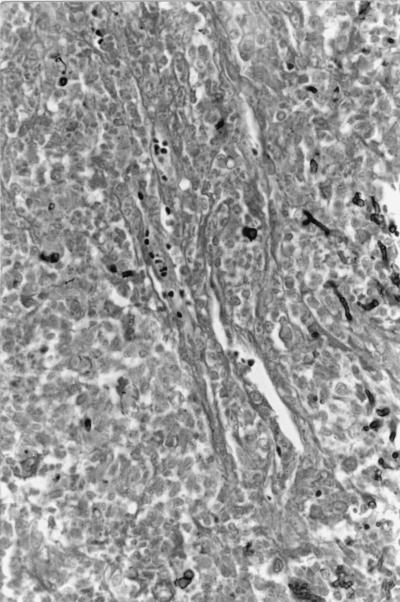
Histopathologic examination revealed an area of necrosis and hemorrhage surrounded by granulomatous inflammation in the splenic parenchyma. There was marked centrilobular hepatocellular fatty change and midzonal cloudy swelling in the liver. Marked tubular and ductal dilations were observed in the renal cortex and medulla. Numerous pyogranulomatous foci were present in the renal cortical interstitium with several small red infarcts in the renal cortex. Pyogranulomatous and necrotizing foci were distributed throughout the renal parenchyma (Fig. 1).
FIG. 1.
Photomicrograph of a section of the kidney of the studied animal. Note the pyogranulomatous inflammation and branched septate hyphae. Gomori’s methenamine-silver stain was used. Magnification, ×486.
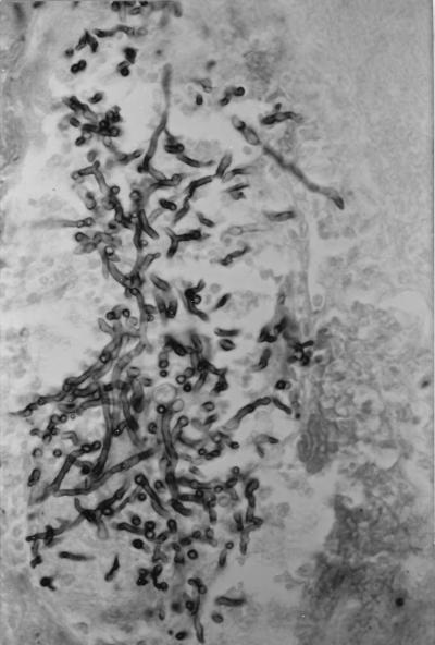
As determined microscopically, the meninges were infiltrated with neutrophils, lymphocytes, and macrophages. Multiple granulomas were centered on blood vessels in the neuropil of the brain. A large area of necrosis was observed rostral to the thalamus. Fibrinopurulent inflammation and edema were associated with the cerebral hemisphere, with several septate, branching hyphae observed in the areas of necrosis and granulomas (Fig. 2). The histopathologic conclusion was that the animal displayed mycotic granulomatous encephalitis, meningitis, myocarditis, nephritis, and splenitis.
FIG. 2.
Photomicrograph of a section of the brain of the studied animal. Note the branching septate hyphae. Gomori’s methenamine-silver stain was used. Magnification, ×486.
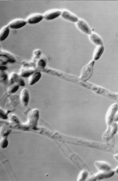
Frozen sections of the lung, brain, and urinary bladder tested negative for canine distemper virus by specific immunofluorescent staining (Veterinary Medical Research and Development, Pullman, Wash.). Routine cultures of kidney, urinary bladder, lung, liver, spleen, and cerebral spinal fluid on blood agar at 37°C did not yield any bacteria. Cells from the cerebral hemisphere were cultured on Sabouraud dextrose agar and mycobiotic agar (Difco Laboratories, Detroit, Mich.) at 25°C, and a filamentous fungus was isolated. Young colonies were white and glabrous at 2 to 3 days and became brown and granular after 6 to 7 days of growth. The reverse was tan with a brownish center. A slide culture stained with lactophenol cotton blue showed branched penicillus-like structures with chains of echinulate conidia (Fig. 3). The final, taxonomic evaluation identified the organism as Scopulariopsis chartarum (cataloged as UTMB 3307 by Michael McGinnis, UTMB Mycology Identification Services, The University of Texas Medical Branch at Galveston).
FIG. 3.
Cylindric annellide and catenulate annellocondia of S. chartarum. Differential interference contrast microscopy was used. Magnification, ×1,000.
S. chartarum infection of dogs has not been previously reported in the literature. The pathogenesis of infection in this case is speculative due to the absence of clinical history. However, if the mechanism for infection is similar to other filamentous, nondimorphic mycoses in dogs, the portal of entry is the skin (5, 6).
Reports of brain abscesses in domestic animals due to fungal infections are conspicuously deficient in the literature. Although rare, most cases of mycotic brain infections are caused by Cladosporium spp., but the sources of infections were not determined in previous studies (2, 7). Likewise, in humans, mycotic brain abscesses are generally cerebral phaeohyphomycoses caused by Cladosporium spp. (1).
There is a paucity of information on Scopulariopsis sp. infections in humans and animals, but these fungi are known to infect the nails, and they are occasionally associated with infections of soft tissue, bone, and lungs in immunocompromised people (4). Since members of the genus Scopulariopsis are common soil fungi, it seems obvious that most infections of animals would be via the cutaneous route. A recent report regarding these infections in humans suggests an invasive cutaneous or pulmonary route of infection with the teleomorphic stage of Scopulariopsis spp. (3).
Acknowledgments
We thank Sam Crosby of Edmond, Okla., for referral of this case and Roger Nieman and Lester Pasarell for expert technical assistance.
REFERENCES
- 1.Chandler F W, Kaplan W, Ajello L. Color atlas & textbook of the histopathology of mycotic diseases. Chicago, Ill: Year Book Medical Publishers, Inc.; 1980. pp. 253–262. [Google Scholar]
- 2.Jang S S, Biberstein E L, Rinald M G, et al. Feline brain abscesses due to Cladosporium trichoides. Sabouraudia. 1977;15:115–123. [PubMed] [Google Scholar]
- 3.Krishner K K, Holdridge N B, Mustafa M M, Rinaldi M G, McGough D A. Disseminated Microascus cirrosus infection in pediatric bone marrow transplant recipient. J Clin Microbiol. 1995;33:735–737. doi: 10.1128/jcm.33.3.735-737.1995. [DOI] [PMC free article] [PubMed] [Google Scholar]
- 4.Larone D H. Medically important fungi: a guide to identification. 3rd ed. Washington, D.C: ASM Press; 1995. p. 195. [Google Scholar]
- 5.Migaki G, Casey H W, Bayles W B. Cerebral phaeohyphomycosis in a dog. J Am Vet Med Assoc. 1987;191:997–998. [PubMed] [Google Scholar]
- 6.Waurzyniak B J, Hoover J P, Clinkenbeard K D, et al. Dual systemic mycosis caused by Bipolaris spicifera and Torulopsis glabrata in a dog. Vet Pathol. 1992;29:566–569. doi: 10.1177/030098589202900620. [DOI] [PubMed] [Google Scholar]
- 7.Welsh R D. Cerebral cladosporiosis in the feline. Southwest Vet. 1984;36:12. [Google Scholar]


