Abstract
Purpose
The Korean Gastric Cancer Association (KGCA) has been conducting nationwide surveys on patients with surgically treated gastric cancer, every 5 years, since 1995. This study details the results of the survey conducted in 2019.
Materials and Methods
This survey was conducted from March to December 2020 using a standardized case report form, which was sent to every member of the KGCA via e-mail. We collected data on 54 items, including patient demographics, tumor characteristics, surgical procedures, and surgical outcomes. We compared the results of the 2019 survey with previous surveys.
Results
Data of 14,076 cases were collected from 68 institutions. The mean patient age was 62.9 years and the proportion of patients who were aged ≥71 years increased from 9.1% in 1995 to 28.8% in 2019. The proportion of upper-third tumors steadily increased from 11.2% in 1995 to 20.9% in 2019 and that of early gastric cancer increased from 57.7% in 2009 to 63.6% in 2019. Regarding operative procedures, a total laparoscopic approach was used in more than half of the cases (55.1%) in 2019. The most common anastomotic method was the Billroth II procedure (45.0%) after distal gastrectomy and double tract reconstruction (81.2%) after proximal gastrectomy in 2019. The postoperative mortality rate was 1.0%, and the overall postoperative complication rate was 14.5%.
Conclusions
The results of the 2019 nationwide survey demonstrate the current status of gastric cancer treatment in Korea. This information will provide a basis for gastric cancer research in the future.
Keywords: Stomach neoplasm, Health care survey, Korea
INTRODUCTION
Gastric cancer is the most common cancer and the fourth most common cause of cancer-related deaths in the Republic of Korea [1]. According to the Korea Central Cancer Registry report, 29,279 people were newly diagnosed with gastric cancer in 2018, accounting for 12.0% of the total cancer incidence in the Republic of Korea [1].
The Korea Central Cancer Registry has annually published nationwide cancer statistics, including the incidence, mortality, and prevalence of all types of cancer. However, these reports do not include detailed information, such as the clinicopathological characteristics and treatment methods of each cancer type. Thus, the Korean Gastric Cancer Association (KGCA) has been conducting nationwide surveys for gastric cancer, every 5 years, since 1995. The initial survey collected information on patient demographics (age and sex), pathological results (tumor size, location, gross type, number of resected lymph nodes, depth of invasion, lymph node metastasis, distant metastasis, and cancer stage), and operative methods [2,3,4,5]. Thereafter, information on the World Health Organization (WHO) classification of tumors and Lauren classification, surgical approaches (open or laparoscopic), curability, and reconstruction methods was added to the survey in 2004 and 2009, respectively [3,4]. Information on the type of stapler for anastomosis, estimated blood loss, and operating time was added to the survey to better understand the widespread trend of minimally invasive surgery in 2014 [5]. These nationwide surveys have been the basis for academic research on gastric cancer in the Republic of Korea.
In this report, we present the results of the nationwide survey on patients with surgically treated gastric cancer in 2019. Several items, such as the extent of lymph node dissection and surgery-related morbidity and mortality, were investigated in this survey.
MATERIALS AND METHODS
Data collection
The Information Committee of the KGCA created a case report form for the 2019 nationwide survey based on the data from previous surveys. All institutions, which were registered with the KGCA, were asked to participate in this survey via e-mail. Designated representatives were responsible for collecting and submitting data across all participating institutions. The case report form was sent to each institution and data were collected from March to December 2020.
The Information Committee of the KGCA was responsible for reviewing the collected data and filtering incorrect or missing data. Representatives in each institution were queried regarding any incorrect or missing data. Responses to all queries were provided by February 2021.
This study followed the ethical principles for medical research in accordance with the tenets of the Declaration of Helsinki and was approved by the Institutional Review Boards of each participating institution (Representative approval No. NCC2020-0206).
Survey data
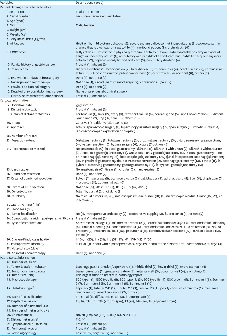
The survey consisted of 54 questions, including patient demographics, past medical history, pathological findings, operative methods and surgical outcomes (Appendix 1). Patient demographics included age, sex, height, weight, body mass index (BMI), American Society of Anesthesiologists physical status classification and Eastern Cooperative Oncology Group performance score. Some information, such as family history of gastric cancer, comorbidities, previous endoscopic treatment or chemotherapy, previous abdominal surgery, and other cancer treatment history were added to this survey.
Regarding pathological information, histological types were classified according to the 2010 WHO classification [6]. Pathological staging was determined according to the eighth edition of the American Joint Committee on Cancer tumor-node-metastasis (TNM) classification system [7]. The presence of lymphovascular invasion and perineural invasion by the cancer cells and washing cytology were newly added in this survey.
As for surgical information, data on surgical approach, operation type, and combined resection and reconstruction method were collected. Several data, such as the number of trocars used in laparoscopic/robotic surgery, extent of lymph node dissection, omentectomy, and tumor localization were newly included in this survey. In particular, postoperative morbidity and mortality rates were investigated for the first time. Postoperative complications were defined as any complications that occurred within 30 days of the surgery. They were categorized into 14 types (anastomotic leakage, anastomotic stricture, duodenal stump leakage, intra-abdominal bleeding, luminal bleeding, pancreatic fistula, intra-abdominal abscess, fluid collection, wound problem, mechanical ileus, pneumonia, cerebrovascular accident, cardiac disease, and others), and the severity of postoperative complications was determined using the Clavien-Dindo classification [8]. Postoperative mortality was defined as death within 30 days of surgery or during hospitalization.
Statistical analysis
Continuous variables were presented as averages and standard deviations, and nominal variables were presented as numbers and proportions. Descriptive analyses were conducted to compare the results of the 2019 survey with previous results since 1995.
Statistical analyses were performed using IBM SPSS version 26.0, for Windows (IBM Corp., Armonk, NY, USA).
Results
Participating institutions and patients
Sixty-eight institutions participated in this survey and collected data from 14,076 patients who underwent surgery for gastric adenocarcinoma in 2019. The number of surgeries performed per year was more than 1,000 in 3 institutions and 500–999 in 3 other institutions. These 6 institutions accounted for 44.9% (6,318/14,076) of all surgeries performed. Eleven institutions performed 200–499 surgeries, and 17 institutions performed 100–199 surgeries. Thirty-four institutions performed fewer than 100 surgeries.
Age, sex, and BMI distribution
Patients' age, sex, and preoperative BMI are shown in Table 1. The mean age was 62.9±11.9 years, which was slightly higher than that reported in the 2014 survey (60.9±12.1 years). The proportion of patients who were aged ≥71 years increased from 9.1% in 1995 to 28.8% in 2019, whereas the proportion of patients aged ≤40 years decreased from 13.3% in 1995 to 3.9% in 2019.
Table 1. Age and sex distribution according to the study period.
| Factor | Subgroup | 1995 (n=5,356) | 1999 (n=6,314) | 2004 (n=11,293) | 2009 (n=14,658) | 2014 (n=15,613) | 2019 (n=14,076) |
|---|---|---|---|---|---|---|---|
| Age (yr) | 59.2±11.9 | 60.9±12.1 | 62.9±11.9 | ||||
| Age distribution (yr) | ≤30 | 103 (1.9) | 115 (1.8) | 142 (1.3) | 134 (0.9) | 88 (0.6) | 59 (0.4) |
| 31–40 | 612 (11.4) | 622 (9.9) | 855 (7.6) | 972 (6.6) | 810 (5.2) | 491 (3.5) | |
| 41–50 | 910 (17.0) | 1,033 (16.4) | 2,106 (18.7) | 2,492 (17.0) | 2,313 (14.8) | 1,625 (11.5) | |
| 51–60 | 1,727 (32.2) | 1,848 (29.3) | 2,732 (24.2) | 3,762 (25.7) | 4,257 (27.3) | 3,688 (26.2) | |
| 61–70 | 1,516 (28.3) | 1,971 (31.2) | 3,866 (34.2) | 4,527 (30.9) | 4,195 (26.9) | 4,162 (29.6) | |
| ≥71 | 488 (9.1) | 725 (11.5) | 1,589 (14.1) | 2,768 (18.9) | 3,949 (25.3) | 4,051 (28.8) | |
| Sex | Male | 3,569 (66.5) | 3,949 (62.5) | 7,586 (67.2) | 9,816 (67.0) | 10,298 (66.0) | 9,228 (65.6) |
| Female | 1,799 (33.5) | 2,368 (37.5) | 3,705 (32.8) | 4,839 (33.0) | 5,315 (34.0) | 4,848 (34.4) | |
| Ratio | 1.98:1 | 1.67:1 | 2.05:1 | 2.03:1 | 1.94:1 | 1.90:1 | |
| BMI (kg/m2) | NA | NA | NA | NA | 23.4±3.3 | 23.9±3.4 | |
| BMI distribution (kg/m2) | <18.5 | 900 (5.9) | 667 (4.8) | ||||
| 18.5–24.9 | 10,228 (65.4) | 8,343 (59.9) | |||||
| 25.0–29.9 | 3,950 (25.7) | 4,320 (31.0) | |||||
| ≥30 | 462 (3.0) | 600 (4.3) |
Values are presented as mean±standard deviation or number (%). The sum of the percentages does not equal 100% because of rounding.
BMI = body mass index; NA = not available (items were not included in the survey).
The male-to-female ratio was 1.9:1, with little change since 1995., the mean BMI was 23.9±3.4 kg/m2 and 59.9% of the patients had normal BMI (<25 kg/m2) according to the WHO classification in the 2019 survey.
Histopathological characteristics of gastric cancer
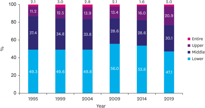
Most patients (n=13,185, 95.8%) had a single lesion, 516 patients (3.7%) had 2 lesions, and 60 patients (0.5%) had 3 or more lesions that were detected simultaneously. The proportions of lower-, middle-, and upper-third tumors were 47.1%, 30.1%, and 20.9%, respectively. The proportion of lower-third tumors was slightly lower than that in previous reports (Table 2). In contrast, the proportion of upper-third tumors constantly increased from 11.2% in 1995 to 20.9% in 2019 (Fig. 1). The tumor size was almost unchanged over time, and the most common tumor size was 2.0–3.9 cm (38.8%).
Table 2. Histopathological characteristics of gastric cancer according to the study period.
| Factor | Subgroup | 1995 | 1999 | 2004 | 2009 | 2014 | 2019 |
|---|---|---|---|---|---|---|---|
| Location | Lower | 2,374 (49.3) | 2,919 (49.6) | 5,347 (49.8) | 7,919 (56.0) | 7,959 (53.8) | 6,422 (47.1) |
| Middle | 1,798 (37.4) | 2,050 (34.8) | 3,635 (33.8) | 4,045 (28.6) | 4,233 (28.6) | 4,100 (30.1) | |
| Upper | 539 (11.2) | 738 (12.5) | 1,493 (13.9) | 1,895 (13.4) | 2,365 (16.0) | 2,844 (20.9) | |
| Entire | 100 (2.1) | 178 (3.0) | 299 (2.8) | 292 (2.1) | 244 (1.6) | 272 (2.0) | |
| Tumor size (cm) | <2.0 | 812 (19.5) | 1,164 (21.8) | 2,675 (24.8) | 3,063 (22.0) | 3,300 (22.3) | 3,146 (23.5) |
| 2.0–3.9 | 1,342 (32.3) | 1,650 (30.9) | 3,528 (32.7) | 5,212 (37.5) | 5,751 (38.8) | 5,187 (38.8) | |
| 4.0–5.9 | 972 (23.4) | 1,183 (22.1) | 2,235 (20.7) | 2,821 (20.3) | 2,990 (20.2) | 2,689 (20.1) | |
| 6.0–7.9 | 548 (13.2) | 598 (13.1) | 1,215 (11.3) | 1,437 (10.3) | 1,359 (9.2) | 1,180 (8.8) | |
| 8.0–9.9 | 270 (6.5) | 364 (6.8) | 626 (5.8) | 673 (4.8) | 670 (4.5) | 538 (4.0) | |
| ≥10.0 | 215 (5.2) | 286 (5.4) | 508 (4.7) | 690 (5.0) | 754 (5.1) | 624 (4.7) | |
| Macroscopic type | EGC type I | 106 (8.6) | 124 (8.0) | 253 (5.9) | 400 (5.1) | 401 (4.6) | 332 (4.0) |
| EGC type IIa | 138 (11.2) | 138 (8.9) | 435 (10.2) | 937 (12.0) | 1,222 (13.9) | 1,262 (15.2) | |
| EGC type IIb | 241 (19.6) | 293 (18.8) | 902 (21.1) | 1,578 (20.3) | 1,938 (22.1) | 2,126 (25.5) | |
| EGC type IIc | 695 (56.6) | 901 (57.9) | 2,346 (54.9) | 4,408 (56.6) | 4,757 (54.1) | 4,233 (50.9) | |
| EGC type III | 49 (4.0) | 99 (6.4) | 339 (7.9) | 462 (5.9) | 470 (5.3) | 370 (4.4) | |
| Borrmann 1 | 159 (4.9) | 137 (4.0) | 198 (3.6) | 270 (4.8) | 274 (5.0) | 192 (4.0) | |
| Borrmann 2 | 763 (23.6) | 825 (23.8) | 1,165 (21.3) | 1,235 (21.9) | 1,242 (22.5) | 1,030 (21.2) | |
| Borrmann 3 | 1,867 (57.7) | 1,980 (57.1) | 3,377 (61.8) | 3,464 (61.3) | 3,338 (60.4) | 2,983 (61.4) | |
| Borrmann 4 | 445 (13.8) | 523 (15.1) | 720 (13.2) | 679 (12.0) | 674 (12.2) | 581 (12.0) | |
| Borrmann 5 | NA | NA | NA | NA | NA | 73 (1.5) | |
| Histology | Papillary | 61 (0.6) | 168 (1.2) | 86 (0.6) | 90 (0.7) | ||
| Tubular WD | 1,517 (14.7) | 1,761 (12.5) | 1,733 (11.5) | 1,359 (10.0) | |||
| Tubular MD | 3,091 (29.9) | 4,283 (30.3) | 4,538 (30.2) | 4,428 (32.5) | |||
| Tubular PD | 3,721 (35.9) | 4,820 (34.1) | 4,288 (28.5) | 3,630 (26.6) | |||
| PCC (SRC)* | 1,597 (15.4) | 2,686 (19.0) | 2,715 (18.1) | 2,603 (19.1) | |||
| Mucinous | 249 (2.4) | 324 (2.3) | 380 (2.5) | 187 (1.4) | |||
| Mixed carcinoma* | NA | NA | 714 (4.7) | 930 (6.8) | |||
| Others | 118 (1.1) | 100 (0.7) | 573 (3.8) | 398 (3.0) |
Values are presented as number (%). The sum of the percentages does not equal 100% because of rounding.
EGC = early gastric cancer; WD = well differentiated; MD = moderately differentiated; PD = poorly differentiated; PCC = poorly cohesive carcinoma; SRC = signet ring cell carcinoma; NA = not available (items were not included in the survey).
*Mixed carcinomas display a mixture of discrete morphologically identifiable glandular (tubular/papillary) and poorly cohesive cellular histological components (signet ring cell).
Fig. 1. Distribution of tumor location over time.
Macroscopic and histological types did not significantly change over time. The most prevalent gross types were type IIc (50.9%) for early gastric cancer (EGC) and Borrmann type 3 (61.4%) for advanced gastric cancer. Borrmann type 5, classified as a category for the first time in this survey, accounted for 1.5%. Of the EGC types, proportions of types I, IIc, and III tended to decrease, whereas those of types IIa and IIb tended to increase Among the histological types, moderately differentiated tubular adenocarcinoma was the most common (32.5%), followed by poorly differentiated tubular adenocarcinoma (26.6%) in 2019. The proportion of poorly differentiated tubular adenocarcinoma gradually decreased from 35.9% in 2004 to 26.6% in 2019. In contrast, the proportions of moderately differentiated tubular adenocarcinoma and poorly cohesive carcinoma slightly increased from 29.9% and 15.4% in 2004 to 32.5% and 19.1% in 2019, respectively.
Tumor stage
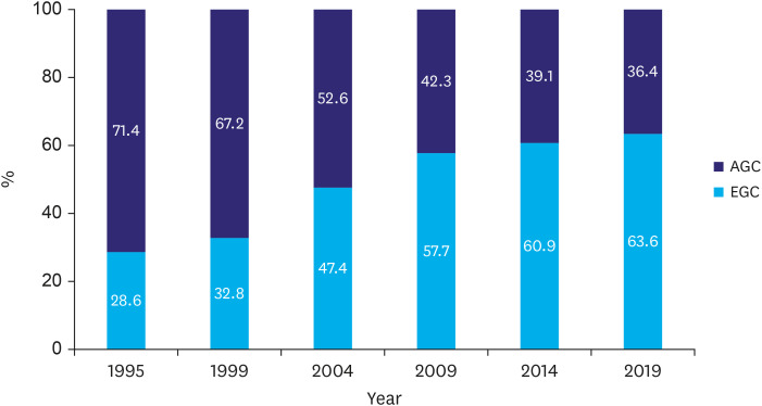
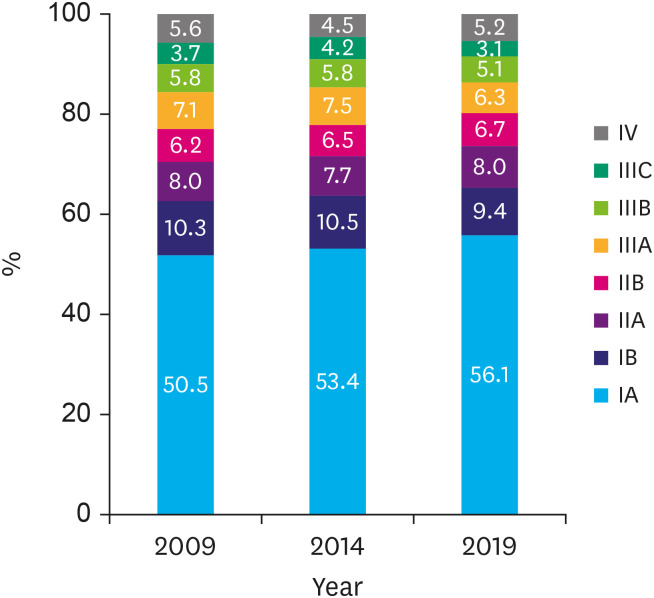
The pathological stages of the tumors are shown in Table 3. The proportion of EGC (pT1Nany) consistently increased from 57.7% in 2009 to 63.6% in 2019 (Fig. 2). The proportion of node-negative cancers also slightly increased from 65.5% to 71.3% during the same period. Regarding TNM stage, stage I accounted for 65.5%, increasing by 3.0% every 5 years since 2009 (Fig. 3). The mean number of harvested lymph nodes was 38.9±18.2, and 95.2% of the patients met the National Comprehensive Cancer Network recommendation for harvesting lymph nodes (≥16 lymph nodes) [9].
Table 3. The tumor-node-metastasis stages according to the American Joint Committee on Cancer classification, eighth edition.
| Factor | Subgroup | 2009 | 2014 | 2019 |
|---|---|---|---|---|
| Depth of invasion | T1a (mucosa) | 4,507 (32.0) | 5,145 (34.6) | 4,667 (34.5) |
| T1b (submucosa) | 3,618 (25.7) | 3,935 (26.4) | 3,945 (29.1) | |
| T2 (proper muscle) | 1,726 (12.3) | 1,668 (11.2) | 1,327 (9.8) | |
| T3 (subserosa) | 2,038 (14.5) | 1,822 (12.2) | 1,758 (13.0) | |
| T4a (serosa) | 1,799 (12.8) | 1,890 (12.7) | 1,611 (11.9) | |
| T4b (adjacent organ) | 388 (2.8) | 421 (2.8) | 226 (1.7) | |
| Lymph node metastasis | N0 | 9,176 (65.5) | 10,201 (68.1) | 9,536 (71.3) |
| N1 (1–2) | 1,516 (10.8) | 1,629 (10.9) | 1,370 (10.2) | |
| N2 (3–6) | 1,361 (9.7) | 1,276 (8.5) | 1,044 (7.8) | |
| N3a (7–15) | 1,165 (8.3) | 1,072 (7.2) | 866 (6.5) | |
| N3b (≥16) | 792 (5.7) | 793 (5.3) | 567 (4.2) | |
| Distant metastasis | M0 | 13,511 (94.5) | 14,404 (95.5) | 13,167 (94.9) |
| M1 | 788 (5.5) | 684 (4.5) | 711 (5.1) | |
| Stage | IA | 7,127 (50.5) | 8,051 (53.4) | 7,703 (56.1) |
| IB | 1,461 (10.3) | 1,582 (10.5) | 1,291 (9.4) | |
| IIA | 1,129 (8.0) | 1,160 (7.7) | 1,102 (8.0) | |
| IIB | 987 (6.2) | 975 (6.5) | 918 (6.7) | |
| IIIA | 1,118 (7.1) | 1,138 (7.5) | 863 (6.3) | |
| IIIB | 925 (5.8) | 869 (5.8) | 704 (5.1) | |
| IIIC | 582 (3.7) | 629 (4.2) | 430 (3.1) | |
| IV | 788 (5.6) | 684 (4.5) | 711 (5.2) | |
| Harvested lymph nodes | 38.3±17.8 | 41.6±20.0 | 38.9±18.2 | |
| <16 | 975 (7.0) | 514 (3.4) | 639 (4.8) | |
| ≥16 | 12,978 (93.0) | 14,435 (96.6) | 12,733 (95.2) |
Values are presented as number (%). The sum of the percentages does not equal 100% because of rounding.
Fig. 2. Proportions of early and advanced gastric cancers over time.
AGC = advanced gastric cancer; EGC = early gastric cancer.
Fig. 3. Proportions of pathological stage according to the American Joint Committee on Cancer, eighth edition.
Surgery-related factors
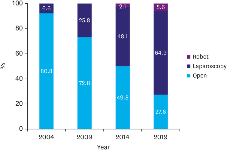
The operative approach (open vs. laparoscopic) has markedly changed over time, and the proportion of minimally invasive surgery (laparoscopic/robot) has increased from 6.6% in 2004 to 70.5% in 2019 (Fig. 4). The detailed method of the laparoscopic approach (laparoscopy-assisted vs. total laparoscopy) has been investigated since 2014, and more than half of the cases (55.1%) were performed via a total laparoscopic approach in 2019 (Table 4). Compared with the 2014 data, the proportion of laparoscopy-assisted approach cases decreased by approximately 8%, but that of the total laparoscopic approach cases increased by 25.0%. The proportion of robotic approaches increased slightly from 2.1% in 2014 to 5.6% in 2019.
Fig. 4. Proportions of operative approach over time.
Table 4. Operative methods and curability.
| Factor | Subgroup | 2004 | 2009 | 2014 | 2019 | |
|---|---|---|---|---|---|---|
| Approach | Open | 9,129 (80.8) | 10,672 (72.8) | 7,760 (49.8) | 3,853 (27.6) | |
| Laparoscopy | 740 (6.6) | 3,783 (25.8) | 7,493 (48.1) | 9,052 (64.9) | ||
| Assisted | NA | NA | 2,805 (18.0) | 1,369 (9.8) | ||
| Totally | NA | NA | 4,688 (30.1) | 7,683 (55.1) | ||
| Robot | NA | NA | 325 (2.1) | 787 (5.6) | ||
| Others* | NA | 176 (1.2) | 1 (<0.1) | 258 (1.8) | ||
| Operation type | DG | 7,959 (70.5) | 10,375 (70.8) | 10,808 (69.2) | 10,091 (71.7) | |
| TG | 2,645 (23.4) | 3,348 (23.3) | 3,659 (23.4) | 2,855 (20.3) | ||
| NTG | NA | 105 (0.7) | 119 (0.8) | NA | ||
| PG | 119 (1.1) | 141 (1.0) | 168 (1.1) | 365 (2.6) | ||
| PPG | 29 (0.3) | 86 (0.6) | 233 (1.5) | 242 (1.7) | ||
| Segmental resection | NA | NA | 10 (0.1) | NA | ||
| Wedge resection | 38 (0.3) | 51 (0.3) | 58 (0.4) | 37 (0.3) | ||
| Bypass | 170 (1.5) | 196 (1.3) | 163 (1.0) | 157 (1.1) | ||
| Biopsy or exploration only | 243 (2.2) | 251 (1.7) | 300 (1.9) | 239 (1.7) | ||
| Others | NA | 105 (0.7) | 75 (0.6) | 86 (0.6) | ||
| Combined resection† | No | 14,176 (92.6) | 12,495 (90.6) | |||
| Yes | 1,139 (7.4) | 1,299 (9.4) | ||||
| Lymph node dissection | Not done | 336 (2.4) | ||||
| <D1 | 83 (0.6) | |||||
| D1 | 210 (1.5) | |||||
| D1+ | 4,961 (36.1) | |||||
| D2 | 7,781 (56.7) | |||||
| >D2 | 357 (2.6) | |||||
| Curability | R0 | 10,068 (81.9) | 13,537 (92.4) | 14,043 (89.9) | 13,115 (93.2) | |
| R1 | 174 (1.4) | 291 (2.0) | 223 (1.4) | 127 (0.9) | ||
| R2 | 364 (3.0) | 257 (1.8) | 349 (2.2) | 171 (1.2) | ||
| No resection | 384 (3.1) | 513 (3.5) | 286 (1.8) | 455 (3.2) | ||
| NA | 303 (2.5) | 60 (0.4) | 712 (4.6) | 208 (1.5) | ||
Values are presented as number (%).
DG = distal gastrectomy; TG = total gastrectomy; NTG = near total gastrectomy; PG = proximal gastrectomy; PPG = pylorus-preserving gastrectomy; NA = not available (items were not included in the survey).
*Others refer to laparoscopic or open exploration without gastrectomy, such as open or closed; †Combined resection was initially evaluated in the 2014 survey.
The most common operation type was distal gastrectomy (71.7%), followed by total gastrectomy (20.3%). The proportion of patients who underwent proximal gastrectomy was 2.6% in 2019, which is more than double of that in 2014 (1.1%). The proportion of pylorus-preserving gastrectomy was 1.7%, which was similar to that in 2014 (1.5%). Most patients (≥95.0%) underwent D1+ or more lymph node dissection, and curative (R0) resection was performed in 93.2% of patients.
Reconstruction method and surgical outcomes according to the surgical approach
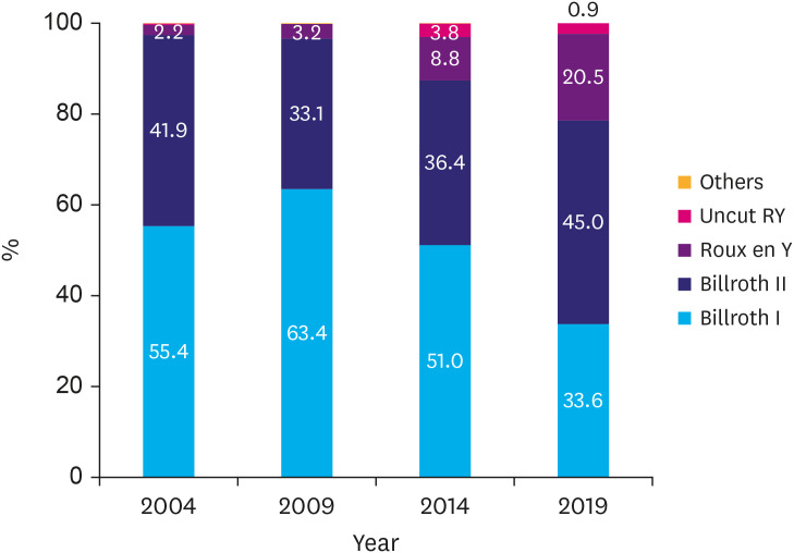
The reconstruction methods are presented in Table 5. After distal gastrectomy, Billroth II reconstruction was the most frequently performed (45.0%) method, followed by Billroth I (33.6%) and Roux-en-Y reconstruction (20.5%) (Fig. 5). The proportions of Billroth II and Roux-en-Y reconstruction increased compared with previous reports, while that of Billroth I gradually decreased. The most common reconstruction method after proximal gastrectomy was double tract reconstruction (286/352 cases, 81.3%), which was higher than the 2014 data (82/132 cases, 62.1%).
Table 5. Methods of anastomosis according to the types of gastrectomy.
| Resection type | Anastomosis | 2004 | 2009 | 2014 | 2019 |
|---|---|---|---|---|---|
| Distal gastrectomy | Billroth I | 4,340 (55.3) | 6,581 (63.4) | 5,426 (51.0) | 3,347 (33.6) |
| Billroth II | 3,285 (41.9) | 3,437 (33.1) | 3,869 (36.4) | 4,477 (45.0) | |
| Roux-en-Y | 175 (2.2) | 332 (3.2) | 933 (8.8) | 2,038 (20.5) | |
| Loop | 11 (0.1) | 0 (0) | NA | NA | |
| Jejunal interposition | 33 (0.4) | 23 (0.2) | 0 (0) | NA | |
| Uncut Roux-en-Y | NA | NA | 404 (3.8) | 90 (0.9) | |
| Others | 3 (<0.1) | 2 (<0.1) | 3 (<0.1) | 3 (<0.1) | |
| Near total gastrectomy* | Billroth II | 46 (67.6) | 59 (56.2) | 23 (21.5) | NA |
| Roux-en-Y | 22 (32.4) | 39 (37.1) | 81 (75.7) | NA | |
| Jejunal interposition | 0 (0) | 5 (4.8) | 0 (0) | NA | |
| Uncut Roux-en-Y | NA | NA | 3 (2.8) | NA | |
| Others | 0 (0) | 2 (1.9) | 0 (0) | NA | |
| Total gastrectomy | Roux-en-Y | 2,407 (91.1) | 3,308 (98.8) | 3,418 (97.8) | 2,874 (99.3) |
| Loop | 155 (5.9) | 18 (0.5) | 13 (0.4) | 12 (0.4) | |
| Jejunal interposition | 49 (1.9) | 10 (0.3) | 8 (0.2) | 5 (0.2) | |
| Uncut Roux-en-Y | NA | NA | 56 (1.6) | NA | |
| Others | 30 (1.1) | 12 (0.4) | 3 (<0.1) | 4 (0.1) | |
| Proximal gastrectomy | Esophagogastrostomy | NA | NA | 50 (37.9) | 66 (18.8) |
| Double tract | NA | NA | 82 (62.1) | 286 (81.2) |
Values are presented as number (%).
NA = not available (items were not included in the survey).
*Near total gastrectomy was not included as a choice in the 2019 survey.
Fig. 5. Proportions of anastomotic method following distal gastrectomy over time.
The reconstruction methods and surgical outcomes according to surgical approach are presented in Table 6. Billroth I (70.9%) was the most common reconstruction method in laparoscopy-assisted distal gastrectomy and Billroth II (51.4%) in totally laparoscopic distal gastrectomy. During open distal gastrectomy, the frequency of performing Billroth I and Billroth II reconstruction methods was similar (approximately 40%). As for the stapler type, circular staplers were frequently used for anastomosis in open or laparoscopy-assisted distal gastrectomy cases (approximately 60%); however, linear staplers were used in more than 95% of totally laparoscopic and robotic distal gastrectomy cases. In total gastrectomy, a circular stapler (94.4%) is commonly used in open surgery, whereas a linear stapler is commonly used in other approaches.
Table 6. Reconstruction methods, type of stapler, amount of blood loss, and operative time according to the surgical approaches in 2019.
| Operation type | Factors | Subgroup | Open | Laparoscopy-assisted | Total laparoscopic approach | Robot |
|---|---|---|---|---|---|---|
| Distal gastrectomy | Reconstruction method | Billroth I | 959 (44.9) | 676 (70.9) | 1,460 (23.3) | 252 (41.4) |
| Billroth II | 797 (37.3) | 225 (23.6) | 3,215 (51.4) | 240 (39.4) | ||
| Roux-en-Y | 373 (17.5) | 52 (5.5) | 1,503 (24.0) | 110 (18.1) | ||
| Uncut Roux-en-Y | 5 (0.2) | 0 (0) | 78 (1.2) | 7 (1.1) | ||
| Others | 1 (<0.1) | 0 (0) | 2 (<0.1) | 0 (0) | ||
| Stapler | Linear | 608 (28.5) | 323 (33.9) | 5,960 (95.3) | 595 (97.4) | |
| Circular | 1,321 (61.9) | 603 (63.3) | 285 (4.6) | 15 (2.4) | ||
| Others | 205 (9.6) | 27 (2.8) | 12 (0.2) | 1 (0.2) | ||
| Blood loss (mL) | 168.7±209.1 | 115.1±181.8 | 78.4±90.3 | 63.1±69.4 | ||
| Operation time (min) | 164.9±60.1 | 154.6±59.8 | 174.6±62.1 | 189.5±64.7 | ||
| Total gastrectomy | Stapler | Linear | 84 (5.5) | 148 (56.7) | 802 (84.4) | 60 (83.3) |
| Circular | 1,429 (94.0) | 112 (42.9) | 145 (15.3) | 11 (15.3) | ||
| Others | 8 (0.5) | 1 (0.4) | 3 (0.3) | 1 (1.4) | ||
| Blood loss (mL) | 253.0±302.2 | 135.7±123.9 | 128.3±172.3 | 107.5±102.3 | ||
| Operation time (min) | 197.5±69.4 | 239.7±74.2 | 218.9±89.0 | 247.3±79.3 | ||
Values are presented as mean±standard deviation or number (%).
The amount of blood loss was lower in the robotic approach than in the other approaches. The robotic surgery showed the longest operating time, and the open surgery had a relatively shorter operating time compared to laparoscopic or robotic surgery.
Postoperative morbidity and mortality
Postoperative mortality data were obtained from 13,420 (95.3%) patients, and 136 (1.0%) patients died within 30 days of surgery or during hospitalization (Table 7).
Table 7. Postoperative mortality and complications in 2019.
| Factors | Subgroup | Values | |
|---|---|---|---|
| Mortality | Survival | 13,284 (99.0) | |
| Died | 136 (1.0) | ||
| Complications within postoperative 30 days | Absent | 11,340 (85.5) | |
| Present | 1,930 (14.5) | ||
| Type of complications* | Local complications | 1,230 (9.3) | |
| Anastomotic leakage | 138 (1.2) | ||
| Duodenal stump leakage | 76 (0.7) | ||
| Anastomotic stricture | 92 (0.8) | ||
| Intra-abdominal bleeding | 82 (0.7) | ||
| Intra-luminal bleeding | 51 (0.4) | ||
| Pancreatic fistula | 36 (0.3) | ||
| Fluid collection | 250 (2.2) | ||
| Intra-abdominal abscess | 97 (0.9) | ||
| Wound problem | 207 (1.8) | ||
| Mechanical ileus | 174 (1.5) | ||
| Systemic complications | 375 (3.3) | ||
| Pulmonary | 304 (2.7) | ||
| Cardiac | 59 (0.5) | ||
| Cerebrovascular | 12 (0.1) | ||
| Others | 627 (5.5) | ||
Values are presented as number (%).
*Data on 13,420 patients for mortality and 13,270 patients for complications.
Postoperative morbidity data were obtained from 13,270 (94.3%) patients, and 1,930 (14.5%) patients experienced postoperative complications. The incidence of local and systemic complications was 9.3% and 3.3%, respectively. The most common local complication was intra-abdominal fluid collection (2.2%), followed by wound problems (1.8%), mechanical ileus (1.5%), and anastomotic leakage (1.2%). Pulmonary complications (2.7%) were the most common systemic complications.
Discussion
The KGCA has regularly performed nationwide surveys to investigate the clinicopathological characteristics of gastric cancer and its surgical treatment trends since 1995. The 2019 survey identified that the proportions of the elderly, proximal gastric cancer, and EGC have been increasing. Regarding the surgical method, the use of the laparoscopic approach, especially the intracorporeal anastomosis technique, has markedly increased. Furthermore, this survey firstly reported the morbidity and mortality rates, which were 14.5% and 1.0%, respectively.
In the current survey, the number of patients who underwent gastrectomy was 13,553 among the 14,076 patients surveyed. This number of gastrectomy cases is nearly equal to the number of cases in the 5th adequacy evaluation of stomach cancer by the Health Insurance Review and Assessment Service [10]. In the 5th adequacy evaluation, 13,597 gastrectomy cases from 182 institutions were evaluated, which were nearly all the cases undergoing gastrectomy for primary gastric cancer in Korea in 2019. Therefore, we believe that this nationwide survey by the KGCA may cover almost all patients who underwent gastric cancer surgery in 2019.
The proportion of patients who were aged ≥71 years increased by approximately 20% (from 9.1% in 1995 to 28.8% in 2019). However, the proportion of middle-aged group patients (31–50 years) decreased by more than 5%. These changes in age distribution indicate that Korea is becoming an ultra-aged society and the number of surgeries for older adult patients would increase in the future [11]. In addition, a change in the distribution of tumor locations was observed. The proportion of upper-third gastric cancer has increased by approximately 10%, which may be related to a westernized diet and increased body weight in Koreans [12,13]. This increase in upper-third gastric cancer can also be associated with an increase in proximal gastrectomy cases in this survey.
Another notable change was the consistent increase in EGCs. The proportion of EGC was 67.9% in 2019, which indicates a 10% increase over the last 10 years. The overall proportion of EGC in all gastric cancers is thought to be higher than that in our report, because this survey included only patients who underwent surgery and did not include those who underwent endoscopic resection. According to the 5th adequacy evaluation of stomach cancer by the Health Insurance Review and Assessment Service in 2019, a total of 9,185 patients underwent endoscopic treatment for gastric cancer, which accounts for 40.3% of all registered gastric cancer cases. Considering that endoscopic treatment is performed for EGC, we can expect that the proportion of EGC would be over 75% of all gastric cancer cases. This high incidence of EGC may be attributed to the widespread national cancer screening program in Korea. A recent survey showed that the screening rate for gastric cancer markedly increased from 39.2% in 2004 to 72.8% in 2018 [14].
This survey also noted a remarkable change in the surgical approach. The proportion of open surgery decreased from 80.8% in 2004 to 27.6% in 2019, while the proportion of laparoscopic surgery increased from 6.6% to 64.9% during the same period. In particular, the total laparoscopic approach with intracorporeal anastomosis has increased by 25% over the last 5 years. These changes indicate that laparoscopic techniques have rapidly evolved from extracorporeal to intracorporeal anastomosis.
Changes in the surgical approach affected the reconstruction method and stapler use. Billroth I reconstruction was more commonly performed during distal gastrectomy than Billroth II or Roux-en-Y reconstruction until 2014. However, this trend was reversed in 2019. Billroth II or Roux-en-Y reconstruction is less affected by the tumor location or assistants' proficiency. These methods are technically more feasible when performing a totally laparoscopic approach, and some surgeons suggest that Roux-en-Y reconstruction can be beneficial for long-term survivors because of less bile reflux and a low risk of remanent gastric cancer [15,16]. This technical feasibility and possible long-term clinical benefits might increase the preference for Billroth II and Roux-en-Y reconstruction.
We also found that the surgical stapler usage is different based on the surgical approach. A circular stapler was commonly used in open or laparoscopy-assisted approach. In contrast, a linear stapler was more commonly used in totally laparoscopic approach. It is probably more convenient because there is no need for anvil insertion, and placement of linear staplers is less affected by assistant abilities.
Postoperative morbidity and mortality rates were first reported in this survey, which were 14.5% and 1.0%, respectively. These results were comparable to those of clinical trials conducted by the Korean Laparoendoscopic Gastrointestinal Surgery Study group. In a multicenter randomized controlled trial that compared surgical outcomes between open and laparoscopic distal gastrectomy for EGC (KLASS-01 trial), the morbidity and mortality rates were 13.0% and 0.6%, respectively [17]. Another randomized controlled trial that compared open and laparoscopic distal gastrectomy for advanced gastric cancer (KLASS-02 trial) demonstrated morbidity and mortality rates of 16.6% and 0.4%, respectively [18]. In the KLASS-03 trial that investigated laparoscopic total gastrectomy, the morbidity and mortality rates were 20.6% and 0.3%, respectively [19].
This study had some limitations. First, there were some missing data despite our efforts to ensure data quality. However, more than 95% of the data were collected for most variables, but some variables, such as family history, blood loss, and tumor localization, had less than 90% of the data. Second, although we corrected the data with obvious errors via queries, it is possible that unidentified errors persist in the data. Finally, this survey included only patients who were surgically treated for gastric cancer. Patients who received other treatments, such as endoscopic resection or chemotherapy, were excluded.
In conclusion, this survey showed the clinicopathological characteristics of gastric cancer and the chronological changes in the surgical treatment of gastric cancer in Korea. The postoperative morbidity and mortality rates were first reported in this survey. We believe these results will be an important basis for understanding the current status of gastric cancer management in Korea.
Appendix 1
Survey data
ASA = American Society of Anesthesiologists; ECOG = Eastern Cooperative Oncology Group; ESD = endoscopic submucosal dissection; LN = lymph node; EGC = early gastric cancer; WD = well differentiated; MD = moderately differentiated; PD = poorly differentiated.
*World Health Organization classification of tumors of the stomach, 2010; †Based on the eighth edition of the American Joint Committee on Cancer classification.
Footnotes
Participating Institutions: The participating institutions are as follows: Gachon University Gil Medical Center; The Catholic University of Korea, Daejeon St. Mary's Hospital, The Catholic University of Korea, Bucheon St. Mary's Hospital; The Catholic University of Korea, Seoul St. Mary's Hospital; The Catholic University of Korea, St. Vincent's Hospital; The Catholic University of Korea, Yeouido St. Mary's Hospital; The Catholic University of Korea, Eunpyeong St. Mary's Hospital; The Catholic University of Korea, Uijeongbu St. Mary's Hospital; The Catholic University of Korea, Incheon St. Mary's Hospital; Kyung Hee University Hospital at Gangdong; Konkuk University Medical Center; Konyang University Hospital; Kyungpook National University Hospital; Kyungpook National University Chilgok Hospital; Gyeongsang National University Hospital; Gyeongsang National University Changwon Hospital; Keimyung University Dongsan Hospital; Korea University Guro Hospital; Korea University Ansan Hospital; Kosin University Gospel Hospital; National Cancer Center; National Medical Center; Daegu Catholic University Medical Center; Daejeon Sun Medical Center; Daejeon Eulji Medical Center; Dongguk University Ilsan Hospital; Dongnam Institution of Radiological and Medical Science; Dong-A University Hospital; Myonggi Hospital; Pusan National University Hospital; Pusan National University Yangsan Hospital; Busan Medical Center; Bundang Jesaeng Hospital; Bundang Cha Hospital; Samsung Medical Center; Seoul National University Hospital; Seoul National University Boramae Medical Center; Seoul National University Bundang Hospital; Asan Medical Center; Soonchunhyang University Bucheon Hospital; Soonchunhyang University Seoul Hospital; Soonchunhyang University Cheonan Hospital; Ajou University Medical Center; Yeonsei University Gangnam Severance Hospital; Yeonsei University Severance Hospital; Wonju Severance Christian Hospital; Yeungnam University Medical Center; Presbyterian Medical Center; Ulsan University Hospital; Wonkang University Hospital; Ewha Woman's University Seoul Hospital; Inje University Busan Paik Hospital; Inje University Haeundae Paik Hospital; Inha University Hospital; Seoul Red Cross Hospital; Chonnam National University Hospital; Chonnam National University Hwasun Hospital; Chonbuk National University Hospital; Jeju National University Hospital; Chungang University Hospital; Chinjujeil Hospital; Cheongju St. Mary's Hospital; Chungnam National University Hospital; Chungbuk National University Hospital; Paju Hospital; Hallym University Dongtan Sacred Heart Hospital; Hallym University Chuncheon Sacred Heart Hospital; Hanyang University Hospital.
Conflict of Interest: No potential conflict of interest relevant to this article was reported
References
- 1.Hong S, Won YJ, Lee JJ, Jung KW, Kong HJ, Im JS, et al. Cancer statistics in Korea: incidence, mortality, survival, and prevalence in 2018. Cancer Res Treat. 2021;53:301–315. doi: 10.4143/crt.2021.291. [DOI] [PMC free article] [PubMed] [Google Scholar]
- 2.Korean Gastric Cancer Association. Nationwide gastric cancer report in Korea. J Korean Gastric Cancer Assoc. 2002;2:105–114. [Google Scholar]
- 3.The Information Committee of the Korean Gastric Cancer Association. 2004 Nationwide gastric cancer report in Korea. J Korean Gastric Cancer Assoc. 2007;7:47–54. [Google Scholar]
- 4.Jeong O, Park YK. Clinicopathological features and surgical treatment of gastric cancer in South Korea: the results of 2009 nationwide survey on surgically treated gastric cancer patients. J Gastric Cancer. 2011;11:69–77. doi: 10.5230/jgc.2011.11.2.69. [DOI] [PMC free article] [PubMed] [Google Scholar]
- 5.Information Committee of Korean Gastric Cancer Association. Korean Gastric Cancer Association nationwide survey on gastric cancer in 2014. J Gastric Cancer. 2016;16:131–140. doi: 10.5230/jgc.2016.16.3.131. [DOI] [PMC free article] [PubMed] [Google Scholar]
- 6.Bosman FT, Hruban RH, Theise ND, editors. WHO Classification of Tumours of the Digestive System. Geneva: WHO; 2010. [Google Scholar]
- 7.Brierley JD, Gospodarowicz MK, Wittekind C, editors. TNM-Classification of Malignant Tumours. 8th ed. Hoboken (NJ): Wiley-Blackwell; 2017. [Google Scholar]
- 8.Dindo D, Demartines N, Clavien PA. Classification of surgical complications: a new proposal with evaluation in a cohort of 6336 patients and results of a survey. Ann Surg. 2004;240:205–213. doi: 10.1097/01.sla.0000133083.54934.ae. [DOI] [PMC free article] [PubMed] [Google Scholar]
- 9.National Comprehensive Cancer Network. NCCN guidelines version 2 [Internet] Plymouth Meeting (PA): National Comprehensive Cancer Network; 2021. [cited 2021 Jul 1]. Available from: https://www.nccn.org/professionals/physician_gls/pdf/gastric.pdf. [Google Scholar]
- 10.Health Insurance Review and Assessment Service. The results of the 5th adequacy evaluation of stomach cancer [Internet] Wonju: Health Insurance Review and Assessment Service; 2021. [cited 2021 Jul 1]. Available from: https://www.hira.or.kr/re/diag/asmWrptPopup.do?evlCd=24&pgmid=HIRAA030004000000. [Google Scholar]
- 11.Kontis V, Bennett JE, Mathers CD, Li G, Foreman K, Ezzati M. Future life expectancy in 35 industrialised countries: projections with a Bayesian model ensemble. Lancet. 2017;389:1323–1335. doi: 10.1016/S0140-6736(16)32381-9. [DOI] [PMC free article] [PubMed] [Google Scholar]
- 12.Bertuccio P, Rosato V, Andreano A, Ferraroni M, Decarli A, Edefonti V, et al. Dietary patterns and gastric cancer risk: a systematic review and meta-analysis. Ann Oncol. 2013;24:1450–1458. doi: 10.1093/annonc/mdt108. [DOI] [PubMed] [Google Scholar]
- 13.Olefson S, Moss SF. Obesity and related risk factors in gastric cardia adenocarcinoma. Gastric Cancer. 2015;18:23–32. doi: 10.1007/s10120-014-0425-4. [DOI] [PubMed] [Google Scholar]
- 14.Hong S, Lee YY, Lee J, Kim Y, Choi KS, Jun JK, et al. Trends in cancer screening rates among Korean men and women: results of the Korean National Cancer Screening Survey, 2004–2018. Cancer Res Treat. 2021;53:330–338. doi: 10.4143/crt.2020.263. [DOI] [PMC free article] [PubMed] [Google Scholar]
- 15.Ma Y, Li F, Zhou X, Wang B, Lu S, Wang W, et al. Four reconstruction methods after laparoscopic distal gastrectomy: a systematic review and network meta-analysis. Medicine (Baltimore) 2019;98:e18381. doi: 10.1097/MD.0000000000018381. [DOI] [PMC free article] [PubMed] [Google Scholar]
- 16.Kim MS, Kwon Y, Park EP, An L, Park H, Park S. Revisiting laparoscopic reconstruction for Billroth 1 versus Billroth 2 versus Roux-en-Y after distal gastrectomy: a systematic review and meta-analysis in the modern era. World J Surg. 2019;43:1581–1593. doi: 10.1007/s00268-019-04943-x. [DOI] [PubMed] [Google Scholar]
- 17.Kim W, Kim HH, Han SU, Kim MC, Hyung WJ, Ryu SW, et al. Decreased morbidity of laparoscopic distal gastrectomy compared with open distal gastrectomy for stage I gastric cancer: short-term outcomes from a multicenter randomized controlled trial (KLASS-01) Ann Surg. 2016;263:28–35. doi: 10.1097/SLA.0000000000001346. [DOI] [PubMed] [Google Scholar]
- 18.Lee HJ, Hyung WJ, Yang HK, Han SU, Park YK, An JY, et al. Short-term outcomes of a multicenter randomized controlled trial comparing laparoscopic distal gastrectomy with D2 lymphadenectomy to open distal gastrectomy for locally advanced gastric cancer (KLASS-02-RCT) Ann Surg. 2019;270:983–991. doi: 10.1097/SLA.0000000000003217. [DOI] [PubMed] [Google Scholar]
- 19.Hyung WJ, Yang HK, Han SU, Lee YJ, Park JM, Kim JJ, et al. A feasibility study of laparoscopic total gastrectomy for clinical stage I gastric cancer: a prospective multi-center phase II clinical trial, KLASS 03. Gastric Cancer. 2019;22:214–222. doi: 10.1007/s10120-018-0864-4. [DOI] [PubMed] [Google Scholar]


