Abstract
目的
比较股骨颈动力交叉钉系统(femoral neck system,FNS)与传统 3 枚空心加压螺钉(cannulate compression screw,CCS)治疗中青年股骨颈骨折患者的临床效果。
方法
回顾分析 2018 年 1 月—2020 年 9 月收治且符合选择标准的 82 例中青年股骨颈骨折患者临床资料,根据手术方式不同分为 FNS 组(24 例)和 CCS 组(58 例)。两组患者性别、年龄、身高、体质量、致伤原因、合并症、骨折部位、骨折分型(Garden 分型及 Pauwels 分型)等一般资料比较,差异均无统计学意义(P>0.05)。记录并比较两组患者手术时间、术中出血量、并发症发生情况(骨不连、股骨头坏死、股骨颈短缩等)、术后 2 d 疼痛视觉模拟评分(VAS)、骨折临床愈合时间、术后髋关节 Harris 评分。
结果
两组患者均顺利完成手术,FNS 组患者手术时间及术后 2 d VAS 评分均显著低于 CCS 组(P<0.05);两组术中出血量比较差异无统计学意义(t=0.263,P=0.796)。两组患者均获随访,CCS 组随访时间 6~18 个月,平均 13.6 个月;FNS 组随访时间 3~12 个月,平均 7.3 个月。两组均未发生内固定物松动并发症,CCS 组发生股骨头坏死 2 例、骨不连 1 例、股骨颈短缩 13 例,FNS 组仅发生 2 例股骨颈短缩,两组并发症发生率(27.6% vs. 8.3%)比较差异有统计学意义(χ2=36.670,P=0.015)。CCS 组 3 例因骨不连、股骨头坏死二期行人工髋关节置换术,余 55 例骨折均达临床愈合;FNS 组中 6 例患者因随访时间不足 6 个月未纳入统计,余 18 例骨折均达临床愈合;两组骨折愈合时间比较差异有统计学意义(t=4.481,P=0.000)。FNS 组术后髋关节 Harris 评分差值显著高于 CCS 组,两组术后 9 个月 Harris 评分均显著高于术后 6 个月,差异均有统计学意义(P<0.05)。
结论
FNS 能加速中青年患者股骨颈骨折的愈合,使患者可以尽早开始功能锻炼,从而降低了相关并发症发生率。
Keywords: 股骨颈动力交叉钉系统, 空心加压螺钉, 股骨颈骨折, 内固定
Abstract
Objective
To compare the effectiveness of femoral neck system (FNS) and cannulate compression screw (CCS) in the treatment of femoral neck fractures in young and middle-aged patients.
Methods
The clinical data of 82 young and middle-aged patients with femoral neck fracture treated between January 2018 and September 2020 were retrospectively analyzed. They were divided into FNS group (24 cases) and CCS group (58 cases) according to different surgical methods. There was no significant difference between the two groups (P>0.05) in general data such as gender, age, height, body mass, cause of injury, complications, fracture location, and fracture classification (Garden classification and Pauwells classification). The operation time, intraoperative blood loss, complications (nonunion, osteonecrosis of the femoral head, shortening of femoral neck, etc.), visual analogue scale (VAS) score at 2 days after operation, clinical healing time of fracture, and Harris score of hip joint after operation were recorded and compared between the two groups.
Results
The operation time and VAS score at 2 days after operation in FNS group were significantly lower than those in CCS group (P<0.05); there was no significant difference in intraoperative blood loss between the two groups (t=0.263, P=0.796). The patients in CCS group were followed up 6-18 months, with an average of 13.6 months; and the follow-up time in FNS group was 3-12 months, with an average of 7.3 months. There was no complication of internal fixator loosening in both groups. There were 2 cases of osteonecrosis of the femoral head, 1 case of bone nonunion, and 13 cases of femoral neck shortening in CCS group and only 2 cases of femoral neck shortening in FNS group. The difference in the incidence of complications between the two groups (27.6% vs. 8.3%) was significant (χ2=36.670, P=0.015). In CCS group, 3 cases underwent secondary artificial hip arthroplasty due to bone nonunion and osteonecrosis of the femoral head, and the remaining 55 cases achieved clinical healing; in FNS group, 6 patients excluded in the statistics because the follow-up time was less than 6 months, and the remaining 18 fractures healed clinically; there was significant difference in fracture healing time between the two groups (t=4.481, P=0.000). The difference of Harris score of hip joint between 9 months and 6 months after operation in FNS group was significantly higher than that in CCS group (P<0.05), and the Harris score at 9 months after operation was significantly higher than that at 6 months after operation in both groups (P<0.05).
Conclusion
FNS can accelerate the healing of femoral neck fractures in young and middle-aged patients, so that patients can start functional exercise as soon as possible, thereby reducing the incidence of related complications.
Keywords: Femoral neck system, cannulate compression screw, femoral neck fracture, internal fixation
股骨颈骨折是临床常见骨折类型,多发生于老年人群,中青年人群发病率较低,但其治疗一直是临床难题。近年来,治疗中青年股骨颈骨折的报道逐渐增多,主要治疗方案包括空心加压螺钉(cannulate compression screw,CCS)、动力髋螺钉以及股骨头置换等[1-4],对于年轻患者首选骨折复位内固定治疗[5-6]。目前使用较多的是呈倒“品”字型平行植入 3 枚 CCS,此固定方式具有创伤小、把持力强、滑动加压、不干扰局部血供的优势[7],可提高骨折愈合率,降低术后并发症发生率[8]。但有文献报道,运用 3 枚 CCS 治疗 Garden Ⅰ、Ⅱ型股骨颈骨折后二次翻修率较高[9-10],内固定后可能出现股骨颈短缩,进一步导致患者髋关节外展肌力减小、行走姿势改变以及行走速度下降,严重影响患者髋关节功能,降低生活质量[11-12]。因此,中青年股骨颈骨折患者是否使用 CCS 仍有待明确。
股骨颈动力交叉钉系统(femoral neck system,FNS)是一种固定股骨颈骨折的新方法,其创伤小,又可提供稳定的抗旋转力和抗剪切力,且不易出现螺钉切割、退钉等现象[13-14];另外,其埋头设计可减少钉尾对周围软组织的激惹。但目前 FNS 相关临床研究较少,临床效果仍不明确。基于此,本研究回顾分析了我院采用 FNS 治疗的中青年股骨颈骨折患者临床资料,并与同期采用 CCS 治疗的患者进行比较,以期为中青年股骨颈骨折患者的治疗方案选择提供依据。报告如下。
1. 临床资料
1.1. 一般资料
纳入标准:① 经体格检查和影像学检查确诊为股骨颈骨折,且均为首次创伤所致的新鲜闭合性股骨颈骨折;② 年龄 18~60 岁;③ 未合并其他部位骨折;④ 采用 CCS 或 FNS 治疗;⑤ 随访资料完整。排除标准:① 合并严重代谢性疾病(糖尿病、甲亢等)不能耐受手术者;② 病理性骨折。
2018 年 1 月—2020 年 9 月共 82 例患者符合选择标准纳入研究,根据手术方式不同分为 FNS 组(24 例)和 CCS 组(58 例)。两组患者性别、年龄、身高、体质量、致伤原因、合并症、骨折部位、骨折分型(Garden 分型及 Pauwels 分型)等一般资料比较,差异均无统计学意义(P>0.05)。见表 1。
表 1.
Comparison of general data between the two groups
两组患者一般资料比较
| 组别
Group |
例数
n |
年龄(岁)
Age (years) |
性别
Gender |
骨折部位
Fracture location |
Garden 分型
Garden classification |
||||||||
| 男
Male |
女
Female |
头下型
Under-head |
经颈型
Trans-neck |
基底型
Basal type |
Ⅰ | Ⅱ | Ⅲ | Ⅳ | |||||
| CCS | 58 | 49(47,56) | 38 | 20 | 26 | 22 | 10 | 2 | 10 | 32 | 14 | ||
| FNS | 24 | 52(47,63) | 10 | 14 | 8 | 10 | 6 | 0 | 4 | 12 | 8 | ||
| 统计值
Statistic |
Z=–0.952
P=0.169 |
χ2=1.844
P=0.299 |
χ2=1.125
P=0.570 |
χ2=1.451
P=0.694 |
|||||||||
表 1-1.
| 组别
Group |
例数
n |
身高(cm)
Height (cm) |
体质量(kg)
Body mass (kg) |
合并症
Complications |
致伤原因
Cause of injury |
Pauwels 分型
Pauwels classification |
||||||||
| 高血压
Hyperten-sion |
糖尿病
Diabetes |
骨质疏松
Osteoporosis |
摔伤
Slide |
交通事故伤
Traffic accident |
高处坠落伤
Fall from height |
Ⅰ | Ⅱ | Ⅲ | ||||||
| CCS | 58 | 163.00±7.18 | 64.24±8.20 | 6 | 2 | 12 | 36 | 6 | 16 | 0 | 22 | 36 | ||
| FNS | 24 | 159.80±7.49 | 58.92±8.70 | 4 | 2 | 4 | 14 | 6 | 4 | 0 | 6 | 18 | ||
| 统计值
Statistic |
t=1.255
P=0.217 |
t=1.852
P=0.072 |
χ2=1.200
P=0.549 |
χ2=2.513
P=0.285 |
χ2=1.262
P=0.261 |
|||||||||
1.2. 手术方法
FNS 组:患者于持续硬膜外麻醉联合蛛网膜下腔阻滞麻醉下,仰卧位于骨科牵引床,健侧下肢屈髋屈膝并充分外展后以约束带固定。患肢牵引状态下通过 C 臂 X 线机透视观察闭合复位。首先于患肢股骨颈偏前方置入 1 枚直径 2.5 mm 克氏针临时固定复位后的骨折断端,防止术中操作引起骨折断端移位。然后沿股骨干轴线向远端作一长约 4 cm 纵形切口,依次切开皮肤、筋膜直至骨面。沿股骨颈正中方向置入 1 枚直径 2.5 mm 斯氏导针,导针尖端距离股骨头软骨下骨 5~10 mm 时停止进入,沿导针钻孔、测深,选择相应的 FNS 钉板系统(Synthes 公司,美国;图 1a)植入。再次 C 臂 X 线机正侧位透视接骨板位置,确保锁定板位于股骨干外侧正中位置;依次植入锁定螺钉和相应长度防旋螺钉,加压至骨折线消失。透视确认已复位的骨折断端无再次移位,内固定螺钉及接骨板位置满意后,以脉冲式冲洗器冲洗切口,4-0 可吸收慕斯线(强生公司,美国)皮内缝合关闭切口。
图 1.
Physical image of internal fixation
内固定物实物图
a. FNS;b. CCS
a. FNS; b. CCS
CCS 组:麻醉方法及患者体位同 FNS 组,C 臂 X 线机透视复位满意后,用直径 2.5 mm 克氏针同上法临时固定复位后股骨颈;然后切开皮肤、筋膜至骨面,以术前透视下定位的进针点向股骨颈方向呈倒“品”字型打入 3 枚平行 CCS(Synthes 公司,美国;图 1b);C 臂 X 线机正侧位透视髋关节,确认骨折复位良好及内固定物位置满意后,脉冲式冲洗器冲洗切口,4-0 可吸收慕斯线(强生公司,美国)皮内缝合关闭切口。两组均不放置引流。
1.3. 术后处理
两组患者术后 24 h 内均使用抗生素预防感染;术后 24 h 排除出血倾向后,皮下注射磺达肝癸钠 2.5 mg 预防下肢深静脉血栓形成,同时辅以消肿、止痛、“丁”字鞋防旋固定。术后第 3 天开始指导患者行患侧下肢肌肉等长收缩锻炼及患侧髋、膝关节主被动活动。术后 2 周可逐渐坐起,嘱患者院外康复训练时患侧足跟部、臀部不离开床面,仅做屈膝活动锻炼下肢肌肉力量及踝、膝关节功能;术后 1.0~1.5 个月逐渐拄双拐下地行走,患侧下肢部分负重,术后 3~6 个月根据骨折愈合情况确定扶单拐或弃拐完全负重时间。
1.4. 术后疗效评价指标
记录并比较两组患者手术时间、术中出血量、并发症发生情况、术后第 2 天疼痛视觉模拟评分(VAS)、骨折临床愈合时间、术后髋关节 Harris 评分[15]。按 Dhar 等[16]提出的标准判断是否存在骨不连,即术后 6 个月骨折仍未愈合,且连续观察 3 个月均无愈合迹象或存在内固定失效、螺钉尖端穿出股骨头等情况。按 Slobogean 等[17]提出的标准判断是否发生股骨头坏死,即术后出现股骨头部分塌陷或软骨下骨透亮线。根据 Slobogean 等[5]提出的标准判断股骨颈短缩。
1.5. 统计学方法
采用 SPSS22.0 统计软件进行分析。符合正态分布的计量资料以均数±标准差表示,组间比较采用独立样本t检验,组内两时间点间比较采用配对t检验;不符合正态分布的计量资料以中位数(四分位数间距)表示,组间比较采用秩和检验;计数资料组间比较采用χ2检验或 Fisher 确切概率法;检验水准α=0.05。
2. 结果
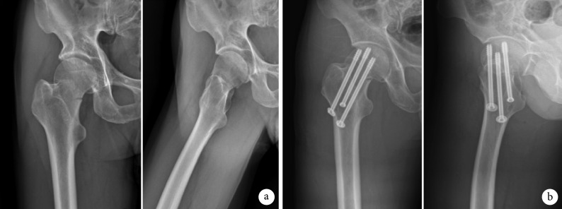
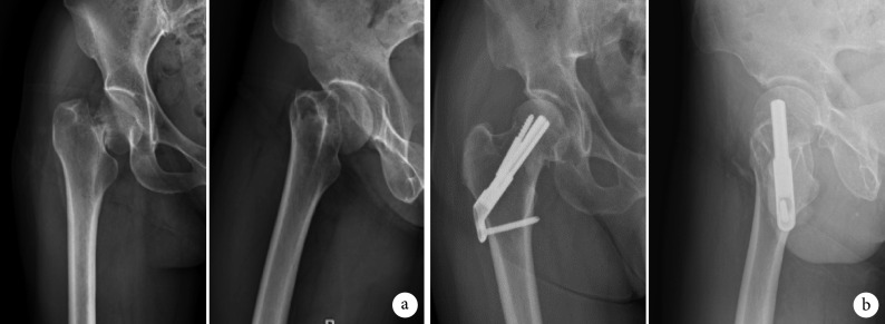
两组患者均顺利完成手术,FNS 组患者手术时间及术后 2 d VAS 评分均低于 CCS 组,差异有统计学意义(P<0.05);两组术中出血量比较差异无统计学意义(t=0.263,P=0.796)。见表 2。两组患者均获随访,CCS 组随访时间 6~18 个月,平均 13.6 个月;FNS 组随访时间 3~12 个月,平均 7.3 个月。两组均未发生内固定物松动并发症,CCS 组发生股骨头坏死 2 例、骨不连 1 例、股骨颈短缩 13 例,FNS 组仅发生 2 例股骨颈短缩,两组并发症发生率(27.6% vs. 8.3%)比较差异有统计学意义(χ2=36.670,P=0.015)。CCS 组 3 例因骨不连、股骨头坏死二期行人工髋关节置换术,余 55 例骨折均达临床愈合;FNS 组中 6 例患者因随访时间不足 6 个月未纳入统计,余 18 例骨折均达临床愈合;两组骨折愈合时间比较差异有统计学意义(t=4.481,P=0.000)。FNS 组术后 6 个月和 9 个月的髋关节 Harris 评分差值显著高于 CCS 组,且两组术后 9 个月 Harris 评分均显著高于术后 6 个月,差异均有统计学意义(P<0.05)。见表 3,图 2、3。
表 2.
Comparison of operation time, intraoperative blood loss, and VAS score at 2 days after operation between the two groups (
 )
)
两组患者手术时间、术中出血量及术后 2 d VAS 评分比较(
 )
)
| 组别
Group |
例数
n |
手术时间(min)
Operation time(min) |
术中出血量(mL)
Intraoperative blood loss(mL) |
术后 2 d VAS 评分
VAS score at 2 days after operation |
| CCS | 58 | 88.38±37.60 | 74.83±42.73 | 5.2±1.2 |
| FNS | 24 | 58.92±6.97 | 79.17±50.17 | 3.3±1.0 |
| 统计值
Statistic |
t=4.055
P=0.000 |
t=0.263
P=0.796 |
t=4.694
P=0.000 |
表 3.
Comparison of fracture healing time and postoperative Harris score of hip joint between the two groups (
 )
)
两组患者骨折愈合时间和术后髋关节 Harris 评分比较(
 )
)
| 组别
Group |
骨折愈合时间(月)
Fracture healing time (months) |
Harris 评分
Harris score |
|||
| 术后 6 个月
Postoperative 6 months |
术后 9 个月
Postoperative 9 months |
差值
Difference |
统计值
Statistic |
||
| CCS | 12.74±5.55 | 80.3±5.2 | 85.0±9.0 | 4.3±2.2 |
t=2.411
P=0.020 |
| FNS | 7.14±1.46 | 90.0±2.3 | 94.7±1.6 | 5.2±2.5 |
t=5.249
P=0.000 |
| 统计值
Statistic |
t=4.481
P=0.000 |
− | − |
t=4.341
P=0.015 |
|
图 2.
Anteroposterior and lateral X-ray films of a 55-year-old female patient with right femoral neck fracture (Garden type Ⅲ, Pauwels type Ⅲ) caused by a fall injury in CCS group
CCS 组患者,女,55 岁,摔伤致右侧股骨颈骨折(Garden Ⅲ型、PauwelsⅢ型)正侧位 X 线片
a. 术前;b. 术后 3 个月示螺钉位置良好,骨折线较清晰
a. Before operation; b. At 3 months after operation, the screw position was good and the fracture lines were clear
图 3.
Anteroposterior and lateral X-ray films of a 49-year-old female patient with right femoral neck fracture (Garden type Ⅲ, Pauwels type Ⅲ) caused by a fall injury in FNS group
FNS 组患者,男,49 岁,摔伤致右侧股骨颈骨折(Garden Ⅲ型、PauwelsⅢ型)正侧位 X 线片
a. 术前;b. 术后 3 个月示钉板系统位置良好,骨折线较模糊
a. Before operation; b. At 3 months after operation, the screw position was good and the fracture lines were vague
3. 讨论
对于中青年股骨颈骨折患者,在排除手术绝对禁忌证后,大多数骨科医生均建议早期行内固定手术治疗[18]。虽然部分研究发现伤后 12 h 以内和以上手术对骨折不愈合及股骨头坏死无明显影响,甚至延迟至 48 h 以上手术对股骨头坏死发生率也无明显影响[8],但越来越多学者建议在伤后 48 h 内进行内固定手术,不仅可减少因围术期过长导致的卧床相关并发症发生,还有利于提高患者舒适度、改善预后[19-21],也符合加速康复理念[22]。
根据不同类型股骨颈骨折的生物力学特点,目前临床上有多种内固定物可用于骨折固定[23]。其中临床使用最广泛的仍是 CCS,该方法手术创伤小、价格相对低廉,可应用于大多数股骨颈骨折。但对于中青年患者而言,股骨颈骨折多为由高能量损伤导致的 Pauwels Ⅲ型,稳定性差,复杂程度高,如果使用传统 CCS 进行内固定则存在较高失效率[24]。王青等[25]在一项临床对比研究中,分别使用 CCS 和滑动髋螺钉(sliding hip screw,SHS)对 Pauwels Ⅲ型中青年股骨颈骨折患者进行内固定治疗,术后经 6~18 个月随访观察显示,CCS 组患者股骨颈骨折不愈合率明显高于 SHS 组,差异有统计学意义(P<0.05)。Kunapuli 等[26]通过生物力学试验对比分析 CCS 和 SHS 内固定治疗股骨颈骨折的强度和稳定性,结果显示在内固定的强度和稳定性方面,CCS 均明显低于 SHS。我们通过总结文献[27-29]认为,CCS 固定股骨颈骨折存在的问题主要是:① 抗剪切力和抗旋转力较差;② 如不能达到 3 枚 CCS 完全平行,易造成应力分布不均,增加术后退钉、骨折移位和再次骨折的可能性;③ 手术操作中需多次透视调整螺钉位置,不仅增加了手术时间和难度,而且多次反复置入导针也会造成骨量丢失,导致内固定稳定性下降。因此,骨科医师们不断探索新的内固定方式,以求达到良好的生物力学稳定性和较高的股骨颈骨折治愈率。
FNS 是一种治疗股骨颈骨折的新型内固定系统,其优势主要体现在:① 微创设计,创伤较小,几乎不会造成软组织激惹。② 具有生物力学优势。Stoffel 等[13]通过对比 FNS、动力髋螺钉及 3 枚 CCS 固定 Pauwels Ⅲ型股骨颈骨折的生物力学稳定性,得出 FNS 稳定性与动力髋螺钉相当,且优于 CCS;Schopper 等[14]通过在尸体标本上对 FNS 与 Hansson Pins 进行比较,也证明 FNS 在股骨颈骨折治疗中比 Hansson Pins 具有更高的稳定性;国内范智荣等[30]应用有限元分析方法研究了 FNS 治疗 Pauwels Ⅲ型股骨颈骨折的生物力学效应,并将其与呈正三角和倒三角的 CCS 进行比较,结果发现 FNS 在治疗 Pauwels Ⅲ型股骨颈骨折中显示出更优的生物力学稳定性。③ 具有抗旋优势,配合防旋钉构成钉中钉的组合方式,可增加抗旋转力和整体的生物力学稳定性,避免了“Z”字效应对股骨头的切割。④ 可滑动加压。刘智[31]在一项综述中指出,在股骨颈骨折治疗中一定要坚持滑动加压原则,任何可能影响骨折端滑动加压的方法都是不合理的,会引起诸多并发症。而 FNS 有 20 mm 的加压空间,而且在骨折愈合过程中由于存在滑动机制,使股骨颈骨折断端的骨质吸收处可以实现再次接触。⑤ 能较好地保存股骨颈内骨量。杨亚军等[32]通过临床回顾性研究比较 34 例股骨颈骨折患者,分别经倒三角平行空心拉力螺钉(19 例)与 FNS(15 例)治疗,结果显示 FNS 创伤小,可减少术中透视次数,并获得了满意近期疗效,但二者骨折愈合时间和术后髋关节功能无明显差异。许新忠等[33]通过回顾性分析 16 例采用 FNS 固定的股骨颈骨折患者,结果显示骨折复位均满意,骨折愈合时间为 3~6 个月,平均 4.5 个月;末次随访时髋关节 Harris 评分达优 10 例,良 5 例,可 1 例。该结果充分说明 FNS 坚强的生物力学固定可以促进股骨颈骨折愈合,使患髋功能恢复良好。
本研究结果显示,FNS 组患者手术时间、骨折愈合时间均短于 CCS 组,术后 6 个月与 9 个月的髋关节 Harris 评分差值优于 CCS 组,术后早期疼痛缓解程度和早期并发症发生率方面也低于 CCS 组。但本研究中 FNS 组患者骨折愈合时间为(7.14±1.46)个月,与既往报道的愈合时间存在差异,我们分析有以下几点原因:① 受患者性别及年龄影响,本研究中 FNS 组大部分为女性患者,且年龄多超过 45 岁。② 与术前受伤程度有关,FNS 组中诊断为 Pauwels Ⅲ型股骨颈骨折患者 18 例,这类患者骨折移位情况复杂,血供破坏大,术中闭合复位次数增加,均可能导致骨折愈合时间延长,甚至降低愈合率。③ 与术后未早期下地及负重有关。④ 本研究纳入患者术后未常规予以抗骨质疏松治疗,这可能也是加快中青年股骨颈骨折患者骨折愈合的因素之一[32]。⑤ 骨折愈合时间是一种主观评价指标,且受随访时间影响,可能造成结果存在一定误差。此外,本研究中 FNS 组有 2 例患者发生股骨颈短缩,我们认为主要原因是骨折未能达到解剖复位。所以无论选择何种内固定方式,良好的解剖复位是关键。
综上述,FNS 固定能缩短股骨颈骨折愈合时间,使患者尽早开始髋关节功能锻炼,改善患者预后。但 FNS 较空心螺钉有更好稳定性的同时,其价格成本也更高,故我们更倾向于将其应用于 Pauwels Ⅲ型骨折患者。本研究存在以下不足,首先,为单中心、小样本回顾性研究;其次,通过 X 线片对股骨颈短缩进行测量,尚不能完全排除患者在摄片时体位不规范对结果产生的影响;另外,仅对患者术后第 6、9 个月行髋关节 Harris 评分,尚需要更长时间随访。FNS 作为一种新兴的股骨颈骨折固定系统,具有较好的临床应用前景,但目前相关报道较少,尚需更多临床数据来验证其价值。
作者贡献:严才平、王星宽负责病例资料收集;严才平、向超负责撰写和修改文章;严才平、邓长弓负责数据收集整理及统计分析;陈骞、李毓灵负责内容构思及文章结构设计;陈路、蒋科负责研究设计、观点形成、文章结构设计,并对文章的知识性内容作批评性审阅。
利益冲突:所有作者声明,在课题研究和文章撰写过程中不存在利益冲突。课题经费支持没有影响文章观点和对研究数据客观结果的统计分析及其报道。
机构伦理问题:研究方案经川北医学院附属医院医学伦理委员会批准[伦理 2021ER(A)050 号]。患者均知情同意。
Funding Statement
四川省医学会课题(2019HR19);四川省科技厅项目(2021YJ0467);南充市2019年市校合作项目(2019SHXZ0120);四川省医学会创新科研项目(Q19069);川北医学院附属医院科研项目(2021ZK001)
Project of the Sichuan Medical Association (2019HR19); Project of the Science and Technology Department of Sichuan Province (2021YJ0467); Program of Cooperation between the Schools and Nanchong City in 2019 (2019SHXZ0120); Innovation Research Project of Sichuan Medical Association (Q19069); Research Project of the Affiliated Hospital of North Sichuan Medical College (2021ZK001)
Contributor Information
科 蒋 (Ke JIANG), Email: 20359251@qq.com.
路 陈 (Lu CHEN), Email: 18057819@qq.com.
References
- 1.林振恩, 郑竑, 陈学生, 等 改良闭合复位技术治疗股骨颈骨折疗效分析. 中国骨伤. 2018;31(2):115–119. doi: 10.3969/j.issn.1003-0034.2018.02.004. [DOI] [PubMed] [Google Scholar]
- 2.张学全, 樊仕才, 黎惠金, 等 带旋髂深血管髂骨瓣和股方肌骨瓣移植治疗青壮年GardenⅢ-Ⅳ型股骨颈骨折的比较. 中国骨伤. 2015;28(9):802–807. [Google Scholar]
- 3.桂景雄, 王小平, 邓志成, 等 闭合复位经皮微创加压空心螺钉内固定治疗股骨颈骨折的临床观察. 创伤外科杂志. 2015;15(4):367–368. [Google Scholar]
- 4.曾剑文, 张华亮, 谢建军, 等 青壮年股骨颈骨折股骨近端空心钉锁定钢板固定效果评价. 武警医学. 2016;27(1):20–22. doi: 10.3969/j.issn.1004-3594.2016.01.007. [DOI] [Google Scholar]
- 5.Slobogean GP, Sprague SA, Scott T, et al Management of young femoral neck fractures: is there a consensus? Injury. 2015;46(3):435–440. doi: 10.1016/j.injury.2014.11.028. [DOI] [PubMed] [Google Scholar]
- 6.Pauyo T, Drager J, Albers A, et al Management of femoral neck fractures in the young patient: A critical analysis review. World J Orthop. 2014;5(3):204–217. doi: 10.5312/wjo.v5.i3.204. [DOI] [PMC free article] [PubMed] [Google Scholar]
- 7.彭方成, 王贤月, 王鹏, 等 股骨近端空心钉锁定板内固定治疗成人股骨颈骨折. 中国骨与关节损伤杂志. 2015;30(12):1305–1306. doi: 10.7531/j.issn.1672-9935.2015.12.025. [DOI] [Google Scholar]
- 8.Yang JJ, Lin LC, Chao KH, et al Risk factors for nonunion in patients with intracapsular femoral neck fractures treated with three cannulated screws placed in either a triangle or an inverted triangle configuration. J Bone Joint Surg (Am) 2013;95(1):61–69. doi: 10.2106/JBJS.K.01081. [DOI] [PubMed] [Google Scholar]
- 9.Kain MS, Marcantonio AJ, Iorio R Revision surgery occurs frequently after percutaneous fixation of stable femoral neck fractures in elderly patients. Clin Orthop Relat Res. 2014;472(12):4010–4014. doi: 10.1007/s11999-014-3957-3. [DOI] [PMC free article] [PubMed] [Google Scholar]
- 10.Han SK, Song HS, Kim R, et al Clinical results of treatment of garden type 1 and 2 femoral neck fractures in patients over 70-year old. Eur J Trauma Emerg Surg. 2016;42(2):191–196. doi: 10.1007/s00068-015-0528-6. [DOI] [PubMed] [Google Scholar]
- 11.Zielinski SM, Keijsers NL, Praet SF, et al Femoral neck shortening after internal fixation of a femoral neck fracture. Orthopedics. 2013;36(7):e849–858. doi: 10.3928/01477447-20130624-13. [DOI] [PubMed] [Google Scholar]
- 12.Zlowodzki M, Ayeni O, Petrisor BA, et al Femoral neck shortening after fracture fixation with multiple cancellous screws: incidence and effect on function. J Trauma. 2008;64(1):163–169. doi: 10.1097/01.ta.0000241143.71274.63. [DOI] [PubMed] [Google Scholar]
- 13.Stoffel K, Zderic I, Gras F, et al Biomechanical evaluation of the femoral neck system in unstable Pauwels Ⅲ femoral neck fractures: A comparison with the dynamic hip screw and cannulated screws. J Orthop Trauma. 2017;31(3):131–137. doi: 10.1097/BOT.0000000000000739. [DOI] [PubMed] [Google Scholar]
- 14.Schopper C, Zderic I, Menze J, et al Higher stability and more predictive fixation with the femoral neck system versus Hansson pins in femoral neck fractures Pauwels Ⅱ. J Orthop Translat. 2020;24:88–95. doi: 10.1016/j.jot.2020.06.002. [DOI] [PMC free article] [PubMed] [Google Scholar]
- 15.Harris WH Traumatic arthritis of the hip after dislocation and acetabular fractures: treatment by mold arthroplasty. An end-result study using a new method of result evaluation. J Bone Joint Surg (Am) 1969;51(4):737–755. doi: 10.2106/00004623-196951040-00012. [DOI] [PubMed] [Google Scholar]
- 16.Dhar SA, Gani NU, Butt MF, et al Delayed union of an operated fracture of the femoral neck. J Orthop Traumatol. 2008;9(2):97–99. doi: 10.1007/s10195-008-0012-8. [DOI] [PMC free article] [PubMed] [Google Scholar]
- 17.Slobogean GP, Stockton DJ, Zeng B, et al Femoral neck fractures in adults treated with internal fixation: A prospective multicenter Chinese cohort. J Am Acad Orthop Surg. 2017;25(4):297–303. doi: 10.5435/JAAOS-D-15-00661. [DOI] [PubMed] [Google Scholar]
- 18.Stacey SC, Renninger CH, Hak D, et al Tips and tricks for ORIF of displaced femoral neck fractures in the young adult patient. Eur J Orthop Surg Traumatol. 2016;26(4):355–363. doi: 10.1007/s00590-016-1745-3. [DOI] [PubMed] [Google Scholar]
- 19.Kim JW, Byun SE, Chang JS The clinical outcomes of early internal fixation for undisplaced femoral neck fractures and early full weight-bearing in elderly patients. Arch Orthop Trauma Surg. 2014;134(7):941–946. doi: 10.1007/s00402-014-2003-y. [DOI] [PubMed] [Google Scholar]
- 20.Jain R, Koo M, Kreder HJ, et al Comparison of early and delayed fixation of subcapital hip fractures in patients sixty years of age or less. J Bone Joint Surg (Am) 2002;84(9):1605–1612. doi: 10.2106/00004623-200209000-00013. [DOI] [PubMed] [Google Scholar]
- 21.Simunovic N, Devereaux PJ, Sprague S, et al Effect of early surgery after hip fracture on mortality and complications: systematic review and meta-analysis. CMAJ. 2010;182(15):1609–1616. doi: 10.1503/cmaj.092220. [DOI] [PMC free article] [PubMed] [Google Scholar]
- 22.赵勇, 秦伟凯 重视股骨颈骨折的评估与内固定治疗的若干问题. 中国骨伤. 2021;34(3):195–199. doi: 10.12200/j.issn.1003-0034.2021.03.001. [DOI] [PubMed] [Google Scholar]
- 23.Panteli M, Rodham P, Giannoudis PV Biomechanical rationale for implant choices in femoral neck fracture fixation in the non-elderly. Injury. 2015;46(3):445–452. doi: 10.1016/j.injury.2014.12.031. [DOI] [PubMed] [Google Scholar]
- 24.Liporace F, Gaines R, Collinge C, et al Results of internal fixation of Pauwels type-3 vertical femoral neck fractures. J Bone Joint Surg (Am) 2008;90(8):1654–1659. doi: 10.2106/JBJS.G.01353. [DOI] [PubMed] [Google Scholar]
- 25.王青, 姜达君, 贾伟涛 Pauwels三型股骨颈骨折不同内固定方式的meta分析. 上海交通大学学报 (医学版) 2018;38(9):1045–1052. [Google Scholar]
- 26.Kunapuli SC, Schramski MJ, Lee AS, et al Biomechanical analysis of augmented plate fixation for the treatment of vertical shear femoral neck fractures. J Orthop Trauma. 2015;29(3):144–150. doi: 10.1097/BOT.0000000000000205. [DOI] [PubMed] [Google Scholar]
- 27.Mei J, Liu S, Jia G, et al Finite element analysis of the effect of cannulated screw placement and drilling frequency on femoral neck fracture fixation. Injury. 2014;45(12):2045–2050. doi: 10.1016/j.injury.2014.07.014. [DOI] [PubMed] [Google Scholar]
- 28.Kloen P, Rubel IF, Lyden JP, et al Subtrochanteric fracture after cannulated screw fixation of femoral neck fractures: a report of four cases. J Orthop Trauma. 2003;17(3):225–229. doi: 10.1097/00005131-200303000-00013. [DOI] [PubMed] [Google Scholar]
- 29.Li J, Wang M, Zhou J, et al. Optimum configuration of cannulated compression screws for the fixation of unstable femoral neck fractures: Finite element analysis evaluation. BioMed research international, 2018, 9: 1271762. doi: 10.1155/2018/1271762.
- 30.范智荣, 苏海涛, 周霖, 等 新型股骨颈内固定系统治疗不稳定性股骨颈骨折的有限元分析. 中国组织工程研究. 2021;25(15):2321–2328. doi: 10.3969/j.issn.2095-4344.3810. [DOI] [Google Scholar]
- 31.刘智 应重视股骨颈骨折的内固定手术治疗. 中国骨伤. 2021;34(3):200–202. [Google Scholar]
- 32.杨亚军, 马涛, 张小钰, 等 股骨颈动力交叉钉系统治疗股骨颈骨折近期疗效. 中国修复重建外科杂志. 2021;35(5):539–543. [Google Scholar]
- 33.许新忠, 常菁, 余水生, 等 股骨颈系统固定治疗股骨颈骨折的近期疗效分析. 中华创伤骨科杂志. 2020;22(7):624–627. doi: 10.3760/cma.j.cn115530-20200618-00410. [DOI] [Google Scholar]


