Abstract
目的
探讨颈椎后路单开门手术中预防性行 C4、5 椎间孔扩大对术后发生 C5 神经根麻痹综合征的影响。
方法
回顾分析 2016 年 3 月—2019 年 3 月收治且符合选择标准的脊髓型颈椎病(颈脊髓受压节段≥3 个)患者临床资料,其中 40 例采用颈椎后路单开门术中预防性行 C4、5 椎间孔扩大治疗(观察组),40 例行单纯颈椎后路单开门手术治疗(对照组)。两组患者性别、年龄、病程、脊髓症状 Nurick 分级及术前 C4、5 椎间孔大小、日本骨科协会(JOA)评分、疼痛视觉模拟评分(VAS)等一般资料比较差异均无统计学意义(P>0.05)。记录并比较两组患者术后发生 C5 神经根麻痹综合征情况,包括发生率、发生麻痹时间、恢复时间、脊髓漂移程度,术前及术后 12 个月采用 VAS、JOA 评分评价疼痛及功能改善情况。
结果
术后两组患者切口均Ⅰ期愈合,无脑脊液漏等术后早期并发症发生。两组患者均获随访,随访时间 12~23 个月,平均 17.97 个月。术后观察组 8 例(右侧 3 例、左侧 5 例)、对照组 2 例(均为右侧)发生 C5 神经根麻痹综合征,两组发生率(20% vs. 5%)差异有统计学意义(χ2=4.114,P=0.043)。除观察组 1 例于术后 5 d 发生 C5 神经根麻痹综合征,其余患者均为术后 1 d 发生;观察组和对照组恢复时间分别为(3.87±2.85)个月和(2.50±0.70)个月,组间差异无统计学意义(t=–0.649,P=0.104)。两组术后 12 个月颈椎 JOA 评分和 VAS 评分均较术前显著改善,差异有统计学意义(P<0.05);组间比较术后 12 个月与术前颈椎 JOA 评分和 VAS 评分差值,以及脊髓漂移程度,差异均无统计学意义(P>0.05)。
结论
预防性行 C4、5 椎间孔扩大并不能有效预防及减少术后 C5 神经根麻痹综合征的发生,相反可能增加其发生概率,临床应用该术式需谨慎。
Keywords: 脊髓型颈椎病, 颈椎后路单开门手术, C4、5 椎间孔扩大 , C5 神经根麻痹综合征
Abstract
Objective
To investigate the effect of prophylactic C4, 5 foraminal dilatation in posterior cervical open-door surgery on postoperative C5 nerve root palsy syndrome.
Methods
The clinical data of patients with cervical spondylotic myelopathy (cervical spinal cord compression segments were more than 3) who met the selection criteria between March 2016 and March 2019 were retrospectively analyzed. Among them, 40 patients underwent prophylactic C4, 5 foraminal dilatation in posterior cervical open-door surgery (observation group) and 40 patients underwent simple posterior cervical open-door surgery (control group). There was no significant difference between the two groups (P>0.05) in gender, age, disease duration, Nurick grade of spinal cord symptoms, and preoperative diameter of C4, 5 intervertebral foramen, Japanese Orthopaedic Association (JOA) score, and visual analogue scale (VAS) score. The occurrence of C5 nerve root paralysis syndrome was recorded and compared between the two groups, including incidence, paralysis time, recovery time, and spinal cord drift. VAS and JOA scores were used to evaluate the improvement of pain and function before operation and at 12 months after operation.
Results
The incisions of the two groups healed by first intention, and there was no early postoperative complications such as cerebrospinal fluid leakage. Patients of both groups were followed up 12-23 months, with an average of 17.97 months. C5 nerve root paralysis syndrome occurred in 8 cases in the observation group (3 cases on the right and 5 cases on the left) and 2 cases in the control group (both on the right). There was significant difference of the incidence (20% vs. 5%) between the two groups (χ2=4.114, P=0.043). Except for 1 case in the observation group who developed C5 nerve root palsy syndrome at 5 days after operation, the rest patients all developed at 1 day after operation; the recovery time of the observation group and the control group were (3.87±2.85) months and (2.50±0.70) months respectively, showing no significant difference between the two groups (t=–0.649,P=0.104). At 12 months after operation, the JOA score and VAS score of cervical spine in the two groups significantly improved when compared with those before operation (P<0.05); there was no significant difference in the difference of the cervical spine JOA score and VAS score between at 12 months after operation and before operation and the degree of spinal cord drift between the two groups (P>0.05).
Conclusion
Prophylactic C4, 5 foraminal dilatation can not effectively prevent and reduce the occurrence of postoperative C5 root palsy, on the contrary, it may increase its incidence, so the clinical application of this procedure requires caution.
Keywords: Cervical spondylotic myelopathy; posterior cervical open-door surgery; prophylactic C4, 5 foraminal dilatation ; C5 nerve root palsy syndrome
随着手术技术及内固定器械的不断发展,手术治疗脊髓型颈椎病取得了越来越肯定的疗效。但术后并发症也逐渐被发现,其中较为严重的有术后四肢瘫、C5 神经根麻痹综合征等。对于颈椎后路手术后发生 C5 神经根麻痹综合征的原因,研究者们提出了不同学说,也进行了多种相关研究,但均未达成共识[1-8]。目前对术后 C5 神经根麻痹综合征的来源和预防策略还不完全清楚,在 C4、5 椎间孔扩大后,是否会增加其“弓弦效应”或是否与术前颈椎原生理弧度密切相关,相关对比分析的文献报道较少见。现回顾分析我科 2016 年 3 月—2019 年 3 月收治的脊髓型颈椎病患者临床资料,比较采用颈椎后路单开门术中预防性行 C4、5 椎间孔扩大与单纯颈椎后路单开门手术的疗效,探讨预防性行 C4、5 椎间孔扩大对术后发生 C5 神经根麻痹综合征的影响。报告如下。
1. 临床资料
1.1. 患者选择标准
纳入标准:① 脊髓型颈椎病,脊髓症状 Nurick 分级≥Ⅲ级;② 颈脊髓受压节段≥3 个;③ 术前 X 线片检查示无颈椎不稳;④ 颈椎后路单开门术中预防性行 C4、5 椎间孔扩大或单纯行颈椎后路单开门手术治疗者;⑤ 有完整影像学资料(手术前后颈椎正侧位及动力位 X 线片、颈椎 CT 及颈椎 MRI)及临床资料者;⑥ 随访时间 1 年以上。排除标准:① 术前已存在 C5 神经根麻痹者;② 骨质疏松者;③ 存在先天性颈椎分节不全者;④ 肿瘤、外伤导致的颈脊髓受损者。2016 年 3 月—2019 年 3 月共 80 例患者符合选择标准纳入研究,其中 40 例采用颈椎后路单开门术中预防性行 C4、5 椎间孔扩大治疗(观察组),40 例行单纯颈椎后路单开门手术治疗(对照组)。
1.2. 一般资料
观察组:男 23 例,女 17 例;年龄 46~79 岁,平均 62.0 岁。症状持续时间 1~10 年,平均 4.55 年。脊髓症状 Nurick 分级:Ⅲ级 34 例,Ⅳ级 4 例,Ⅴ级 2 例。术前 C4、5 椎间孔大小为(2.25±0.22)mm。
对照组:男 22 例,女 18 例;年龄 50~79 岁,平均 62.8 岁。症状持续时间 1~11 年,平均 4.90 年。脊髓症状 Nurick 分级:Ⅲ级 35 例,Ⅳ级 4 例,Ⅴ级 1 例。术前 C4、5 椎间孔大小为(2.31±0.25)mm。
两组性别、年龄、病程、脊髓症状 Nurick 分级及术前 C4、5 椎间孔大小、日本骨科协会(JOA)评分、疼痛视觉模拟评分(VAS)等一般资料比较差异均无统计学意义(P>0.05),具有可比性。见表 1。
表 1.
Comparison of JOA score, VAS score, and spinal cord drift of cervical spine between the two groups before and after operation (n=40,
 )
)
两组患者手术前后颈椎 JOA 评分、VAS 评分以及脊髓漂移程度比较(n=40,
 )
)
| 组别
Group |
JOA 评分
JOA score |
VAS 评分
VAS score |
脊髓漂移程度(mm)
Spinal cord drift (mm) |
|||||||
| 术前
Preoperative |
术后 12 个月
Postoperative at 12 months |
差值
Difference |
统计值
Statistic |
术前
Preoperative |
术后 12 个月
Postoperative at 12 months |
差值
Difference |
统计值
Statistic |
|||
| 观察组
Observation group |
8.7±1.4 | 12.8±2.0 | 4.1±2.4 |
t=−10.372
P=0.008 |
2.9±1.9 | 1.8±1.3 | −0.4±2.3 |
t=0.738
P=0.000 |
4.86±0.98 | |
| 对照组
Control group |
9.1±1.5 | 12.7±1.8 | 3.6±2.1 |
t=−9.503
P=0.008 |
2.4±2.0 | 2.2±1.3 | −1.1±2.2 |
t=2.879
P=0.004 |
4.78±1.04 | |
| 统计值
Statistic |
t=1.359
P=0.510 |
− |
t=−1.051
P=0.322 |
t=0.978
P=0.466 |
− |
t=−1.251
P=0.607 |
t=−0.353
P=0.594 |
|||
1.3. 手术方法
两组均采用 C3~7 共 5 个手术节段的单开门手术方式,手术均由同一组医师完成。内固定器械使用美国美敦力公司的颈椎后路椎板夹固定系统(Centerpiece 系统),两组每例患者均使用 5 块 Centerpiece 椎板夹固定。患者于全麻下取仰卧位,用 Mayfield 头架及 3 颗头钉固定头部于手术床上,采用后正中切口切开皮肤、皮下,显露 C3~7 椎板和关节突关节,注意保护棘间韧带,棘突剪剪除 C6、7 过长棘突。
对照组:选择症状较重的一侧为开门侧,使用磨钻以两侧关节突与椎板交界处为开槽处,开门侧磨至全层骨皮质,门轴侧保留前方靠硬脊膜侧骨皮质;枪状咬骨钳咬除 C2、3、C7~T1 棘突间部分韧带,将开门侧全部翻向门轴侧;C3~7 每个节段使用 1 块 Centerpiece 椎板夹,根据门轴侧椎板厚度选取宽口或窄口椎板夹,3 枚螺钉固定。常规安置引流管,逐层缝合切口。
观察组:神经根探子探查神经根松弛度后,在对照组手术操作基础上,于开门侧使用磨钻去除 C4、5 部分关节突内侧皮质;神经根探子再次探查神经根松弛度,探查过程中密切观察神经电生理监测仪是否有电位改变(波幅下降超过 50% 或潜伏期延长超过 10%),如有改变立即停止手术,麻醉医师配合下行“唤醒试验”及“踝阵挛试验”。
1.4. 术后处理及疗效评价指标
术后 48 h 内引流量低于 30 mL 时拔除引流管;术后使用抗生素 24~48 h,常规镇痛。患者麻醉清醒后立即观察四肢感觉及随意运动情况,并与术前查体进行对比分析,对于发生 C5 神经根麻痹综合征患者常规给予营养神经、高压氧治疗,协助抬肩、上举患肢等功能锻炼。
记录并比较两组患者术后发生 C5 神经根麻痹综合征情况,包括发生率、发生麻痹时间、恢复时间;脊髓漂移程度(术后 12 个月脊髓前间隙与术前的差值);术前及术后 12 个月采用 VAS、JOA 评分评价疼痛及功能改善情况。
1.5. 统计学方法
采用 SPSS22.0 统计软件进行分析。计量资料以均数±标准差表示,组内手术前后比较采用配对 t 检验,组间比较采用独立样本 t 检验;计数资料组间比较采用 χ2 检验;检验水准 α=0.05。
2. 结果
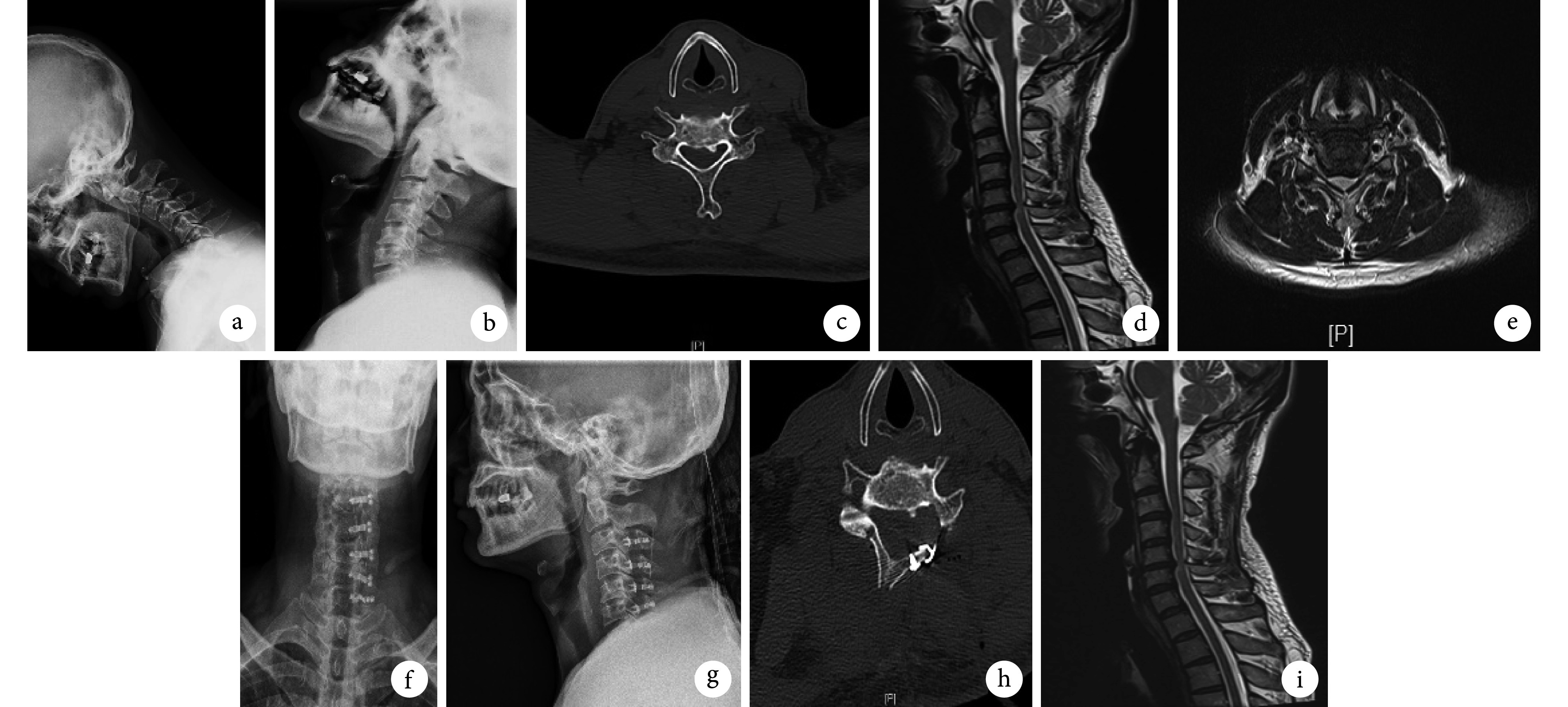
术后两组患者切口均Ⅰ期愈合,无脑脊液漏等术后早期并发症发生。两组患者均获随访,随访时间 12~23 个月,平均 17.97 个月。术后观察组 8 例(右侧 3 例、左侧 5 例)、对照组 2 例(均为右侧)发生 C5 神经根麻痹综合征,两组发生率(20% vs. 5%)差异有统计学意义(χ2=4.114,P=0.043)。除观察组 1 例于术后 5 d 发生 C5 神经根麻痹综合征,其余患者均为术后 1 d 发生;观察组和对照组恢复时间分别为(3.87±2.85)个月和(2.50±0.70)个月,差异无统计学意义(t=–0.649,P=0.104)。两组术后 12 个月颈椎 JOA 评分和 VAS 评分均较术前显著改善,差异有统计学意义(P<0.05);组间比较术后 12 个月与术前颈椎 JOA 评分和 VAS 评分差值,以及脊髓漂移程度,差异均无统计学意义(P>0.05)。见表 1、图 1。
图 1.
A 49-year-old male patient with cervical spondylotic myelopathy in observation group
观察组患者,男,49 岁,脊髓型颈椎病
a、b. 术前过伸、过屈位 X 线片;c. 术前 CT 横断面;d. 术前 MRI 矢状位;e. 术前 MRI 横断面;f、g. 术后 1 d 正侧位 X 线片示椎板夹固定;h. 术后 1 d CT 横断面示骨性椎管扩大;i. 术后 3 d MRI 矢状位示颈椎脊髓向后漂移,脊髓压迫解除
a, b. Preoperative hyperextension and hyperflexion X-ray films; c. Preoperative cross-sectional CT; d. Preoperative sagittal MRI; e. Preoperative cross-sectional MRI; f, g. Anteroposterior and lateral X-ray films at 1 day after operation showed laminar fixation with Centerpiece; h. Cross-sectional CT at 1 day after operation showed osseous spinal canal enlargement; i. Sagittal MRI at 3 days after operation showed posteriorly drift of the cervical spinal cord and release of spinal compression

3. 讨论
对于广泛颈椎管狭窄及多节段脊髓受压,后路椎管扩大成形术是目前较常用且疗效确切的手术方式,其中全椎板切除减压内固定及单开门手术最为常用。随着手术器械的发展,单开门手术越来越受到脊柱医师认可,其保留了患者颈椎大部分活动度,降低了融合技术带来的弊端。但其也有相对适应证[6],术前颈椎过伸过屈位 X 线片示颈椎不稳者不宜采用单开门手术。C5 神经根麻痹综合征是颈椎后路减压术后较为严重的并发症,颈椎前路手术虽然也可能发生,但大宗病例研究发现颈椎前路手术较后路手术发生率低[6, 9]。关于 C5 神经根麻痹综合征,目前较为认可的定义是减压手术后 C5 神经根所支配的三角肌和/或肱二头肌出现无力和麻木等功能障碍,且无术后脊髓功能恶化表现。
发生 C5 神经根麻痹综合征的影响因素众多,目前研究较为深入的有 C4、5 椎间孔大小、颈椎术后漂移程度[10]、术前并发后纵韧带骨化、缺血再灌注损伤[2, 11-13]。本研究中,观察组 1 例发生 C5 神经根麻痹综合征患者术后第 1 天即出现颈部、肩背部、右侧上臂剧烈疼痛,颈部疼痛 VAS 评分为 9 分,给予一般非甾体类止痛药物完全无效后,给予患者杜冷丁止痛后稍好转。此例情况较极端,分析原因主要与 C5 神经根解剖相关,C5 神经根通常位于单开门脊髓减压漂移顶点,在脊髓向后漂移过程中受牵拉距离较长,而其本身较短,故与未减压患者相比麻痹发生更快[14-15],更易形成阻挡效应。
本研究中,手术开门侧均为术前症状相对较重的一侧,磨钻在两侧关节突于椎板交界处开门。研究发现如太靠近外侧可能使关节突关节破坏过多造成颈椎不稳[16],从而导致内固定失败;如太偏内侧,脊髓减压效果可能不佳[17],术后功能改善不明显。观察组 8 例发生了 C5 神经根麻痹综合征,均为预防性减压者,分析原因是术中椎间孔扩大到哪种程度足够并不明确,术后因局部水肿或伪影无法单纯通过 MRI 判断椎间孔与神经根毗邻情况[18],更多是依靠主刀医师神经根探子的探查。另外,术中为了增加手术安全性,虽然使用神经电生理监测仪,但目前对于电生理监测技术并无统一标准[2]。一般术中将电极放置于三角肌和肱二头肌,采用经颅电刺激运动诱发电位及自发肌电图进行监测[19-20],波幅下降超过 50% 或潜伏期延长超过 10% 时立即通知主刀医师停止手术,而仪器的敏感度可能也是干预结果的影响因素之一。
本研究中,10 例发生 C5 神经根麻痹综合征者多见于开门侧,分析原因可能为开门侧发生脊髓后移距离较大。但在预防性行 C4、5 椎间孔扩大患者中发生率更高,分析原因可能与术中体位有关,术中采用 Mayfield 头架及 3 颗头钉固定头部,正常 C4、5 位于颈椎生理性前凸顶点,此时固定头部后未能及时关注患者术前长期维持的生理曲度,而过度追求颈椎正常生理曲度,虽然单开门为非融合技术,但术后颈椎生理曲度仍有改变。尤其在 C4、5 椎间孔扩大后,因颈椎生理曲度恢复正常,C5 神经根更容易随脊髓向后漂移而形成“弓弦效应”,神经根近脊髓端无法适应快速向后牵拉,椎间孔外侧附近的神经根形成卡压,造成神经根快速水肿,本研究发生 C5 神经根麻痹综合征者大多数发生于术后第 1 天。经过长期临床实践,我们建议在术中固定头部后利用 C 臂 X 线机拍摄标准颈椎侧位 X 线片,然后与术前标准颈椎侧位 X 线片对比,通过调节 Mayfield 头架调整颈椎弧度至与术前相当,避免过度颈椎前凸。本研究观察组发生 C5 神经根麻痹综合征的患者中,存在颈椎管极度狭窄或后纵韧带骨化等情况[21],但患者术前并无根性症状。是否考虑不预防性行 C4、5 椎间孔扩大术,尚需进一步大样本、多中心研究明确。
综上述,预防性行 C4、5 椎间孔扩大并不能有效预防及减少术后 C5 神经根麻痹综合征的发生,相反可能增加其发生概率,因此临床应用该术式需谨慎。但本研究为回顾性研究,缺乏随机对照组,同时病例数相对较少,随访时间相对较短。随着研究的深入以及手术技术和器械的精细化,未来希望能进行多中心随机对照研究明确其远期效果。
作者贡献:袁心伟负责研究设计、数据整理与分析、论文撰写;万仑、胡豇、张伟负责研究设计与论文修改及审核。
利益冲突:所有作者声明,在课题研究和文章撰写过程中不存在利益冲突。
机构伦理问题:研究方案经四川省医学科学院·四川省人民医院医学伦理委员会批准[伦审(研)2020 年第 424 号]。患者术前均签署知情同意书。
References
- 1.Wang H, Tang ZR, Li W, et al. Prediction of the risk of C5 palsy after posterior laminectomy and fusion with cervical myelopathy using a support vector machine: an analysis of 184 consecutive patients. J Orthop Surg Res, 2021, 16(1): 332. doi: 10.1186/s13018-021-02476-5.
- 2.Yoshihara H, Margalit A, Yoneoka D Incidence of C5 Palsy: Meta-analysis and potential etiology . World Neurosurg. 2019;122:e828–e837. doi: 10.1016/j.wneu.2018.10.159. [DOI] [PubMed] [Google Scholar]
- 3.Hirabayashi S, Kitagawa T, Yamamoto I, et al Postoperative C5 palsy: Conjectured causes and effective countermeasures . Spine Surg Relat Res. 2018;3(1):12–16. doi: 10.22603/ssrr.2018-0016. [DOI] [PMC free article] [PubMed] [Google Scholar]
- 4.Lee DH, Cho JH, Hwang CJ, et al Multilevel posterior foraminotomy with laminoplasty versus laminoplasty alone for cervical spondylotic myelopathy with radiculopathy: a comparative study. Spine J. 2018;18(3):414–421. doi: 10.1016/j.spinee.2017.08.222. [DOI] [PubMed] [Google Scholar]
- 5.Witiw CD, Smieliauskas F, O’Toole JE, et al Comparison of anterior cervical discectomy and fusion to posterior cervical foraminotomy for cervical radiculopathy: Utilization, costs, and adverse events 2003 to 2014. Neurosurgery. 2019;84(2):413–420. doi: 10.1093/neuros/nyy051. [DOI] [PubMed] [Google Scholar]
- 6.Guday E, Bekele A, Muche A Anatomical study of prefixed versus postfixed brachial plexuses in adult human cadaver. ANZ J Surg. 2017;87(5):399–403. doi: 10.1111/ans.13534. [DOI] [PubMed] [Google Scholar]
- 7.Wang T, Wang H, Liu S, et al. Incidence of C5 nerve root palsy after cervical surgery: A meta-analysis for last decade. Medicine (Baltimore), 2017, 96(45): e8560. doi: 10.1097/MD.0000000000008560.
- 8.Hitchon PW, Moritani T, Woodroffe RW, et al C5 palsy following posterior decompression and instrumentation in cervical stenosis: Single center experience and review . Clin Neurol Neurosurg. 2018;174:29–35. doi: 10.1016/j.clineuro.2018.08.028. [DOI] [PubMed] [Google Scholar]
- 9.Tetreault L, Ibrahim A, Côté P, et al A systematic review of clinical and surgical predictors of complications following surgery for degenerative cervical myelopathy. J Neurosurg Spine. 2016;24(1):77–99. doi: 10.3171/2015.3.SPINE14971. [DOI] [PubMed] [Google Scholar]
- 10.Lee JY, Sharan A, Baron EM, et al Quantitative prediction of spinal cord drift after cervical laminectomy and arthrodesis. Spine (Phila Pa 1976) 2006;31(16):1795–1798. doi: 10.1097/01.brs.0000225992.26154.d0. [DOI] [PubMed] [Google Scholar]
- 11.Cherian J, Mayer RR, Haroun KB, et al Contribution of lordotic correction on C5 palsy following cervical laminectomy and fusion . Neurosurgery. 2016;79(6):816–822. doi: 10.1227/NEU.0000000000001199. [DOI] [PubMed] [Google Scholar]
- 12.Yoshihara H, Pivec R, Naam A Positioning-related neuromonitoring change during anterior cervical discectomy and fusion. World Neurosurg. 2018;117:238–241. doi: 10.1016/j.wneu.2018.06.116. [DOI] [PubMed] [Google Scholar]
- 13.Pan FM, Wang SJ, Ma B, et al. C5 nerve root palsy after posterior cervical spine surgery. J Orthop Surg (Hong Kong), 2017, 25(1): 2309499016684502. doi: 10.1177/2309499016684502.
- 14.Dombrowski ME, Morales-Restrepo A, Fourman MS, et al Prophylactic perioperative dexamethasone decreases the incidence of postoperative C5 palsies after a posterior cervical laminectomy and fusion . Spine J. 2019;19(2):253–260. doi: 10.1016/j.spinee.2018.05.031. [DOI] [PubMed] [Google Scholar]
- 15.Macki M, Alam R, Kerezoudis P, et al Manual muscle test at C5 palsy onset predicts the likelihood of and time to C5 palsy resolution . J Clin Neurosci. 2016;24:112–116. doi: 10.1016/j.jocn.2015.09.003. [DOI] [PubMed] [Google Scholar]
- 16.Rodriguez-Feo JA, Leas D, Odum SM, et al Reoperation rates following open-door cervical laminoplasty. Int J Spine Surg. 2018;12(6):751–756. doi: 10.14444/5094. [DOI] [PMC free article] [PubMed] [Google Scholar]
- 17.Boniello A, Petrucelli P, Kerbel Y, et al Short-term outcomes following cervical laminoplasty and decompression and fusion with instrumentation. Spine (Phila Pa 1976) 2019;44(17):E1018–E1023. doi: 10.1097/BRS.0000000000003057. [DOI] [PubMed] [Google Scholar]
- 18.Katsumi K, Yamazaki A, Watanabe K, et al Can prophylactic bilateral C4/C5 foraminotomy prevent postoperative C5 palsy after open-door laminoplasty?: a prospective study. . Spine (Phila Pa 1976) 2012;37(9):748–754. doi: 10.1097/BRS.0b013e3182326957. [DOI] [PubMed] [Google Scholar]
- 19.Hirai T, Yoshii T, Sakai K, et al Long-term results of a prospective study of anterior decompression with fusion and posterior decompression with laminoplasty for treatment of cervical spondylotic myelopathy. J Orthop Sci. 2018;23(1):32–38. doi: 10.1016/j.jos.2017.07.012. [DOI] [PubMed] [Google Scholar]
- 20.Yang XJ, Sang HX, Liu CX, et al A new method for calculating the desired laminoplasty opening size based on the target sagittal canal diameter before single-door cervical laminoplasty. Eur Spine J. 2019;28(10):2302–2310. doi: 10.1007/s00586-019-05992-5. [DOI] [PubMed] [Google Scholar]
- 21.Nakagawa H, Okazaki T, Saito K Surgical strategies in management of cervical spondylotic myelopathy. World Neurosurg. 2015;84(2):220–221. doi: 10.1016/j.wneu.2015.04.005. [DOI] [PubMed] [Google Scholar]


