Abstract
Leiomyomas are benign lesions of the uterine smooth muscles that contain various amounts of fibrous connective tissue. Hystrosalpingography is not a method of diagnosing uterine fibroids, and other methods such as ultrasound and MRI are preferred, but during hystrosalpingography, especially in infertile females, uterine fibroids may be seen frequently. Leiomyomas have a wide range of appearances depending on their number, size and location. Leiomyomas may enlarge, elongate, displace, distort or rotate the uterine cavity and can be detected by such changes showing in hysterosalpingograms. These changes may be symmetric or asymmetric. Leiomyomas may result in uterine atony which can be locolized or generalized. Leiomyomas also may appear as one or multiple filling defects in different sizes which can be smooth or irregular. Some of the noted findings may create similar and frequent appearances looking like some patterns in nature and can be considered “excellent signs” for better detecting and enabling differential diagnosis. This study aims to improve the process of training on the diagnostic appearances of leiomyomas in hysterosalpingography by aligning the images with patterns found in nature that can be easily remembered by radiologists.
Introduction
Leiomyomas are benign lesions of the uterine smooth muscles that contain various amounts of fibrous connective tissue.1–5 These lesions are found in approximately 20–40% of females in reproductive ages and are therefore considered the most common pelvic masses.1–4 Several terms are used to refer to leiomyomas, including myomas, fibromas and fibroids.1–6
Leiomyomas range in size and location and may be found as solitary or multiple.4 The common symptoms of uterine fibroids include abnormal uterine bleeding, pelvic pressure and pain, constipation and urinary symptoms.1–4,7–10
According to their location, leiomyomas are classified into three groups1–4,6,7:
Subserosal leiomyoma: subserosal leiomyoma externally extends to the serosa and may protrude into the pelvic cavity or be seen as a pedunculated fibroma.
Intramural leiomyoma: intramural leiomyoma is the most prevalent type of leiomyoma located in the myometrium.
Submucosal leiomyoma: submucosal leiomyoma internally protrudes into the endometrial cavity. Given that it causes severe symptoms and infertility; this class of leiomyoma is considered the most significant type of uterine fibroids.
Munro has offered another classification scheme, as shown in Figure 1.11
Figure 1.
Munro fibroid classification 2011.
Transabdominal and transvaginal ultrasound scan are initially used for the evaluation of the female pelvis, particularly in the case of uterine leiomyomas.1–4 MRI is a highly accurate modality for differentiating leiomyomas in the case of an enlarged uterus that has a reported accuracy of 99%.1 Although HSG is not a diagnostic method of fibroids, but due to the great importance and numerous use of this method in infertile females investigation, the signs of fibroid can be seen in. Uterine leiomyoma are common findings in the hysterosalpingograms of females in reproductive ages, particularly those with infertility. Leiomyomas have a wide range of appearances in HSG depending on the number, size and location of the tumor relative to the uterine cavity.12–15
Just as we can see images of the face of humans or animals or shapes of things in the objects around us, a similar phenomenon can occur in hysterosalpingography images. Radiologists have an artistic eye that helps them resemble the shape of fibrosis in an HSG to shapes commonly found in nature during their inspections and diagnosis of grayscale radiology images.
Fibroids have various appearances on HSG, however other uterine lesions such as polyps and gestational sac may cause the same appearance as well. This study aims to improve the process of teaching to trace diagnostic signs of leiomyomas in HSG by aligning the images with patterns found in nature that can be easily remembered by radiologists. Knowing that final diagnosis requires further investigation.
Hystrosalpingography findings
Uterine leiomyoma cannot be identified on plain radiography unless calcified degeneration occurs. Irregular coarse calcifications are more common with subserosal lesions and are mostly found in tumors with pedicles. Sometimes, a large soft-tissue mass may be seen in the pelvis as representing large fibroids.
Uterine leiomyoma has a wide range of appearances depending on the number, size and location of the tumor relative to the uterine cavity. Leiomyomas may enlarge, elongate, displace, distort or rotate the uterine cavity and can be detected by such changes in the HSG; however, detecting their exact location is not possible with this method and it requires other methods, including ultrasound. These changes may be symmetric or asymmetric. Leiomyomas may result in uterine atony, which can be localized or generalized. The reduction of normal uterine tonicity produces a flabby and sac-like appearance. Some leiomyomas may appear as one or multiple filling defects in different sizes that can be smooth or irregular. The endometrium overlying leiomyomas is often thin or necrotic, causing mucosal irregularity and venous myometrial intravasation. Sometimes, a set of these features may be seen in an HSG. These changes can create similar and frequent appearances resembling some of the patterns found in nature, and some of them have formerly been named according to these resemblances, such as the crescent sign. This article presents similar samples of very common images of fibroids in HSG and names them based on their similarities in the effort to introduce new diagnostic signs. The diagnosis of leiomyoma was confirmed in most patients by one of the methods of 2D or 4D ultrasound, hysterosonography and MRI. These patients who referred for infertility evaluation often had one or more of the following symptoms and some had no symptoms. The most common symptoms are: menorrhagia, metrorrhagia, spotting, and urinary symptoms with feeling of pressure and heaviness in the pelvis in the case of huge leiomyomas. Depending on the type of leiomyomas, they were treated by laparascopy, laparotomy or hysteroscopy methods, and sometimes their infertility treatment was started without surgical intervention.
I. The crescent sign: this appearance of the uterus is a result of the asymmetrical enlargement of the uterine cavity caused by a large leiomyoma (Figures 2 and 3). This sign has already been discussed in radiology books.15
Figure 2.
The crescent or moon sign; asymmetrical elongation and enlargement of the right uterine wall due to the extensive pressure of a large leiomyoma. (a) HSG, (b) Schematic, (c) 3D Hysterosonography, coronal view.
Figure 3.
Similar views of the moon sign in different patients.
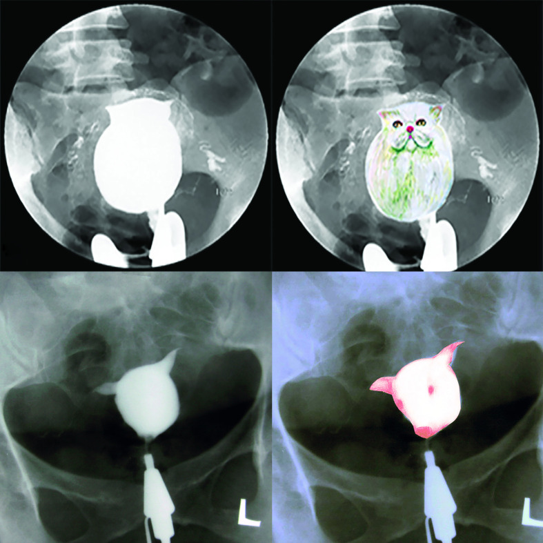
II. The ball and lamp appearance: when the volume of the uterine cavity increases considerably due to generalized uterine atony, the shape appearing is frequently a ball or lamp. Since these appearances are very common, we named them the “Ball and Lamp appearance” (Figures 4 and 5).
Figure 4.
Fibromatosis uterine; the absence of normal uterine tonicity produces a rugbi ball appearance (a) HSG, (b) Schematic, (c) Hysterosonography, sagittal view.
Figure 5.
Fibromatosis uterine; the absence of normal uterine tonicity produces a lamp, or ball appearance.
Some uncommon appearances caused by generalized atony are also presented in this article (Figure 6).
Figure 6.
The loss of tonicity in the body and isthmus looks like a cat or the head of a pig.
III. The fish appearance: asymmetrical uterine atony in one cornua often creates a shape resembling fish. This view is very common and can be considered a diagnostic sign and we like to call it the “Fish appearance” (Figures 7–9).
Figure 7.
Unilateral atonicity of cornua due to leiomyoma. (a) HSG, (b) Schematic, (c) 3D TVS, coronal view.
Figure 8.
Various views of cornual atonicity.
Figure 9.
Interesting views of different types of fishes frequently observed.
IV. The flower appearance: some leiomyomas may have a compressive effect on the uterine wall and change its shape. Since the compressive effect of leiomyoma on the fundal portion of the uterine is a frequently-observed appearance, we named it the “Flower appearance” (Figure 10).
Figure 10.
A leiomyoma with a compression effect on the fundus with a fading defect creates a flower appearance (calla lily). (a) HSG, (b) Schematic, (c) 3D TVS, coronal view.
The shape observed due to the compressive effect of leiomyoma on the other portions of the uterine cavity resembles a different type of flower which is not common but is interesting to note (Figure 11).
Figure 11.
Compressive effect of multiple myomas with asymmetric atonicity zones and multiple air bubbles created a flower-like image (rose). (a) HSG, (b) Schematic, (c) 3D TVS, coronal view.
V. The sail appearance: sometimes, leiomyomas may create no filling defects and just cause the displacement of the cavity with a compressive effect on the uterine wall. This appearance is considered one of the most common views of the leiomyoma and can be called the “Sail appearance” (Figure 12).
Figure 12.
The normal triangular shape of uterus shifted to the left side-of the pelvis with the elongation of the right wall is observed, resembling a sail. (a) HSG, (b) Schematic, (c) 3D TVS, coronal view.
Intramural leiomyoma with a submucosal component with asymmetrical atonicity has caused the distortion of the uterine cavity in different directions that produces asymmetric and deformity of cavity. These changes can create various fascinating views. Some of them are shown in Figures 13–15.
Figure 13.
The filling defect caused by the myoma in the body, overshadowed by contrast medium and two fine defects due to air bubbles, resembling a botton. (a) HSG, (b) Schematic, (c) 3D TVS, coronal view.
Figure 14.
The elongation and displacement of the right uterine wall caused by a large myoma can be observed, resembling a giraffe.
Figure 15.
Uterine atonicity with the pressure effect of the intramural myoma with a submocusal component on the fondus has increased the distance between the two cornuas. With the spillage of contrast media, these features resembled an angel resting on crescent moon. (a) HSG, (b) Schematic, (c) 3D TVS, coronal view.
Conclusion
This article presented highly common images in HSG that resemble an image found in nature and has made a connection between them. Studying and focusing on HSG images helps make a link between the virtual and visible worlds and reveal the truth of the statement “God inspires radiologists with signs to enable better perceptions, detections, differential diagnoses and rejections of diseases”. To the best of the researchers’ knowledge, some of these common appearances have never been noted in any studies as diagnostic signs.
The results of this study can help improve the sensitivity and accuracy of gynecological disease detection.
Footnotes
Acknowledgements: We are indepted to Dr Gholamreza Shahrzad for his images archive. We would like to thank the talents of Ms Nasrin Nooshfar and Ms. Zahra Jafari Aghdaee for their excellent drawings on radiograms. We appreciate the able assistance of our radiology technologists.
Contributors: All the authors contributed to conception and design of the work (article), drafting and revision of important intellectual content, gathering images provided in the manuscript, reviewing and approving the final version. And they agree to be accountable for ensuring that questions related to the accuracy or integrity of any part of the work are appropriately investigated and resolved.
Contributor Information
Firoozeh Ahmadi, Email: dr.ahmadi1390@gmail.com.
Fereshteh Hosseini, Email: hosseini.f1346@gmail.com.
Maryam Javam, Email: maryam_javam@yahoo.com.
Fattaneh Pahlavan, Email: midwifer.esfahan@gmail.com.
REFERENCES
- 1.Wilde S, Scott-Barrett S, Sue W, Sarah S-B. Radiological appearances of uterine fibroids. Indian J Radiol Imaging 2009; 19: 222. doi: 10.4103/0971-3026.54887 [DOI] [PMC free article] [PubMed] [Google Scholar]
- 2.Baird DD, Dunson DB, Hill MC, Cousins D, Schectman JM. High cumulative incidence of uterine leiomyoma in black and white women: ultrasound evidence. Am J Obstet Gynecol 2003; 188: 100–7. doi: 10.1067/mob.2003.99 [DOI] [PubMed] [Google Scholar]
- 3.Novak E. Berek & Novak’s gynecology. Philadelphia: Lippincott Williams & Wilkins; 2007. [Google Scholar]
- 4.Parker WH, Etiology PWH. Etiology, symptomatology, and diagnosis of uterine myomas. Fertil Steril 2007; 87: 725–36. doi: 10.1016/j.fertnstert.2007.01.093 [DOI] [PubMed] [Google Scholar]
- 5.Wallach EE, Vlahos NF. Uterine myomas: an overview of development, clinical features, and management. Obstet Gynecol 2004; 104: 393–406. doi: 10.1097/01.AOG.0000136079.62513.39 [DOI] [PubMed] [Google Scholar]
- 6.Fielding JR, Brown DL, Thurmond AS. Gynecologic imaging E-Book: expert radiology series (expert consult premium Edition-Enhanced online features and print. Amsterdam: Elsevier Health Sciences; 2011. [Google Scholar]
- 7.Murase E, Siegelman ES, Outwater EK, Perez-Jaffe LA, Tureck RW. Uterine leiomyomas: histopathologic features, MR imaging findings, differential diagnosis, and treatment. Radiographics 1999; 19: 1179–97. doi: 10.1148/radiographics.19.5.g99se131179 [DOI] [PubMed] [Google Scholar]
- 8.Hutchins FL. Uterine fibroids. diagnosis and indications for treatment. Obstet Gynecol Clin North Am 1995; 22: 659–65. [PubMed] [Google Scholar]
- 9.Coronado GD, Marshall LM, Schwartz SM. Complications in pregnancy, labor, and delivery with uterine leiomyomas: a population-based study. Obstet Gynecol 2000; 95: 764–9. doi: 10.1016/s0029-7844(99)00605-5 [DOI] [PubMed] [Google Scholar]
- 10.Lippman SA, Warner M, Samuels S, Olive D, Vercellini P, Eskenazi B. Uterine fibroids and gynecologic pain symptoms in a population-based study. Fertil Steril 2003; 80: 1488–94. doi: 10.1016/S0015-0282(03)02207-6 [DOI] [PubMed] [Google Scholar]
- 11.Munro MG, Critchley HOD, Fraser IS, .FIGO Menstrual Disorders Working Group . The FIGO classification of causes of abnormal uterine bleeding in the reproductive years. Fertil Steril 2011; 95: 2204–8. doi: 10.1016/j.fertnstert.2011.03.079 [DOI] [PubMed] [Google Scholar]
- 12.Botwe BO, Bamfo-Quaicoe K, Hunu E, Anim-Sampong S. Hysterosalpingographic findings among Ghanaian women undergoing infertility work-up: a study at the Korle-Bu teaching hospital. Fertil Res Pract 2015; 1: 9. doi: 10.1186/s40738-015-0001-6 [DOI] [PMC free article] [PubMed] [Google Scholar]
- 13.Ahmadi F, Haghighi H, Akhbari F. Hysterosalpingography. Middle East Fertil Soc J 2012; 17: 210–4. doi: 10.1016/j.mefs.2012.07.001 [DOI] [Google Scholar]
- 14.Chalazonitis A, Tzovara I, Laspas F, Porfyridis P, Ptohis N, Tsimitselis G. Hysterosalpingography: technique and applications. Curr Probl Diagn Radiol 2009; 38: 199–205. doi: 10.1067/j.cpradiol.2008.02.003 [DOI] [PubMed] [Google Scholar]
- 15.Ahmadi F, Zafarani F, Niknejadi M, Vosough A, Ahmadi F, Zafarani F. Uterine leiomyoma: Hysterosalpingographic appearances. Int J Fertil Steril 2007; 1: 137–44. [Google Scholar]


