Abstract
Physiological/normal tooth mobility may be defined as the slight displacement of the clinical crown of a tooth, which is allowed by the resilience of an intact and healthy periodontium, under the application of a moderate force. The factors influencing the success and longevity of dental splinting are the type of material used for the splint, the type of composite resin, the number and location of the dental units included for splinting (maxillary or mandibular arch). In periodontology, the term ‘splint’ is defined as the joining of two or more teeth into a rigid unit through restorations or fixed or removable devices. The purpose of using periodontal splints for tooth immobilization is to provide a period of rest in the areas where the healing process has begun and to allow normal functioning there where the tissues alone would not be able to withstand occlusal forces. The aim of the present study was to evaluate comparatively, by means of energy dispersive electron spectrometry (EDX), the chemical composition in the case of in vitro samples that structurally reproduce direct periodontal immobilization systems using fiberglass strips and fluid composite resins. The differences in the chemical composition of the investigated materials can influence their physiochemical properties, but also their biological and toxicological reliability. Elements such as C, O, Al, Si and Ba were identified as common elements among the resins tested in the present study. The only difference in terms of the component elements was found in the case of the fluid composite Clearfil Majesty ES Flow, which, apart from the elements mentioned, also presented Co in its structure.
Keywords: periodontal splints, dental mobility, flowable composites, EDX spectrometer, periodontal disease, G-aenial Flo X, GrandioSO Heavy Flow, Clearfil Majesty ES Flow
Introduction
Periodontal disease is a term that includes a wide range of inflammatory diseases that affect the periodontium, an entity that consists of a set of structures that support the tooth, gingiva, cementum, periodontal ligament and alveolar bone. Thus, any damage to adjacent tissues can lead to increased tooth mobility over time (1).
Physiological or normal dental mobility can be defined as a slight displacement of the clinical dental crown, due to the application of a moderate force, the displacement being allowed by the resilience of a healthy and intact periodontium.
Findings of previous studies conducted primarily on animals showed that the main cause of dental mobility is the laxity of periodontal ligaments and/or the destruction of supporting periodontal tissue, resulting from the presence of active or treated periodontitis, whether or not combined with occlusal trauma (2). In addition, occlusal forces play an important role, as they can exacerbate a pre-existing periodontal injury when they exceed the strength threshold of a compromised supporting tissue (3).
In practice, it is accepted that dental splinting, especially of the lower central incisors, is performed in order to maintain the patient's natural dentition for a longer period, as well as to reduce the patient's discomfort and improve the masticatory and aesthetic functions (4). The factors influencing the success and longevity of dental splinting are the type of material used for the splint, the type of composite resin, and the number and location of the dental units included for splinting (maxillary or mandibular arch) (5).
In periodontology, the term ‘splint’ is defined as the joining of two or more teeth into a rigid unit through restorations or fixed or removable devices. The purpose of using periodontal splints is to provide a period of rest in the areas where the healing process has begun (to reduce stress on the affected teeth) and to allow normal functioning where the tissues alone would not be able to withstand occlusal forces (6). Currently, many types of splints for immobilization are available, their diversity depending on their purpose and duration of use, the position of the teeth on the mandible, but also of the manufacturing material. Various materials have been used for splints, such as composite resin in combination with adhesive systems, orthodontic systems or composites reinforced with pre-impregnated fibers in combination with composite resins. An important aspect for selecting the type of splint is the mechanical interaction between the material from which the splint is made and the underlayment of the tooth (6). Fluid composites are low-viscosity composites, and this makes them more fluid than universal composites, with high tooth humectability and low elasticity (6).
The aim of the present study was to evaluate comparatively, by means of energy dispersive electron spectrometry (EDX), the chemical composition in the case of in vitro samples that structurally reproduce direct periodontal immobilization systems using fiberglass strips and fluid composite resins.
Materials and methods
Materials
In order to achieve the proposed aim, 54 samples of teeth were selected with the following dimensions: 15 mm in length, 4 mm in width and 2.5 mm in thickness, using a heat-resistant silicone conformer.
For the construction of the immobilization systems, the same fiberglass tape, Interlig® (Angelus®, Brazil) and three different fluid composites were used: G-aenial Flo X® (GC Corporation®, Japan), GrandioSO Heavy Flow® (Voco®, Germany) and Clearfil Majesty ES Flow® (Kuraray Noritake Dental®, Japan). The number of samples produced and later subject to elemental investigation was equal, namely 18 for each composite resin. The photopolymerization of the samples was carried out using a Celalux 2® type lamp (Voco).
Subsequently, all 54 periodontal splint samples were grouped, and their microstructure was analyzed using a Quanta 200 3D scanning electron microscope (FEI, The Netherlands) in the laboratory of the Faculty of Mechanics from the ‘Gheorghe Asachi’ Technical University of Iasi, Romania.
EDX spectrometer
The EDX spectrometer is an instrument that determines quantitatively the elements in a sample by irradiating it with X-rays and then analyzing the re-emitted X-rays. It represents a standard method to identify and quantify element compositions of very small samples of material (even a few cubic micrometers). The EDX elemental analysis was performed for each type of composite separately, G-aenial Flo X (GC Corp.), GrandioSO Heavy Flow (Voco) and Clearfil Majesty ES Flow (Kuraray), in three different areas of each sample.
The data were automatically corrected for atomic number, absorption and fluorescent excitation effects using the ZAF correction method for each investigated element. Following the investigations, we analyzed, for each element, both the mass percentage (wt%) and the atomic percentage (at%).
Statistical analysis
The statistical analysis was performed using SPSS 25.0 (IBM Corp.) The Kolmogorov-Smirnov test was used to test the distribution of the numerical variables. The average value, the standard error and the standard deviation of the numerical variables were calculated. The Kruskal-Wallis and Mann-Whitney non-parametric tests were used to compare the variables with non-normal distributions. P<0.05 was considered to indicate statistically significant results.
Results
Carbon (C) content
The values found for carbon were significantly different between the three types of materials, in the case of both measurements, both globally and comparatively between materials two by two. For example, for wt% determinations, the highest C content was recorded for G-aenial Flo X (21.7644±1.32468) and the lowest for GrandioSO Heavy Flow (5.9611±0.87104); the C content for Clearfil Majesty ES being intermediate (12.9989±3.44164). However, the differences between the three materials were statistically significant in all combinations. A similar behavior was observed in the case of at% determinations (Fig. 1).
Figure 1.
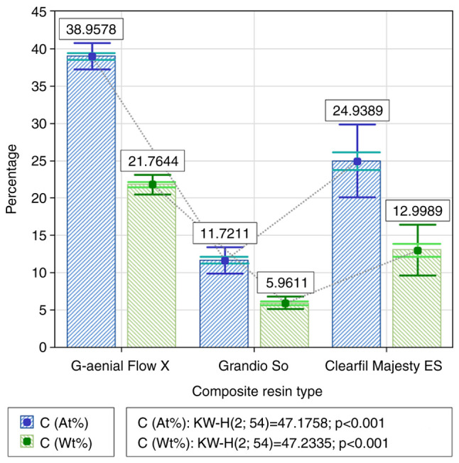
Statistical analysis of the mass (Wt%) and atomic percentage (At%) of the carbon (C) content for the three types of materials.
Oxygen (O) content
The O content was also significantly different between the three types of materials, in both measurements, both globally and comparatively between materials two by two. In the case of the wt% measurement, the highest O content was observed for GrandioSO Heavy Flow (33.878±1.74974), followed by Clearfil Majesty ES (30.1978±2.91155), and lastly by G-aenial Flo X (28.5767±2.05314). The observed differences were considered statistically significant (Table I). The same results were observed in the case of at% measurements (Fig. 2).
Table I.
Statistical analysis of the mass (wt%) and atomic percentage (at%) for all analyzed elements in the three types of materials.
| Mann-Whitney test (2 samples) P-value | ||||
|---|---|---|---|---|
| Mass and atomic percentage of the elements | Kruskal-Wallis test (3 samples) P-value | G-aenial Flo X vs. Grandio SO Heavy Flow | G-aenial Flo X vs. Clearfil Majesty ES | Grandio SO Heavy Flow vs. Clearfil Majesty ES |
| C (wt%) | <0.001b | <0.001b | <0.001b | <0.001b |
| C (at%) | <0.001b | <0.001b | <0.001b | <0.001b |
| O (wt%) | <0.001b | <0.001b | 0.037a | <0.001b |
| O (at%) | <0.001b | <0.001b | <0.001b | <0.001b |
| Al (wt%) | <0.001b | <0.104 | <0.001b | 0.027a |
| Al (at%) | <0.001b | <0.001b | <0.001b | 0.293 |
| Si (wt%) | <0.001b | <0.001b | <0.001b | <0.001b |
| Si (at%) | <0.001b | <0.001b | <0.001b | <0.001b |
| Ba (wt%) | <0.001b | <0.001b | 0.864 | <0.001b |
| Ba (at%) | 0.211 | 0.719 | 0.293 | 0.068 |
aStatistically significant, P<0.05;
bhighly statistically significant, P<0.001. C, carbon; O, oxygen; Al, aluminum; Si, silicium; Ba, barium.
Figure 2.
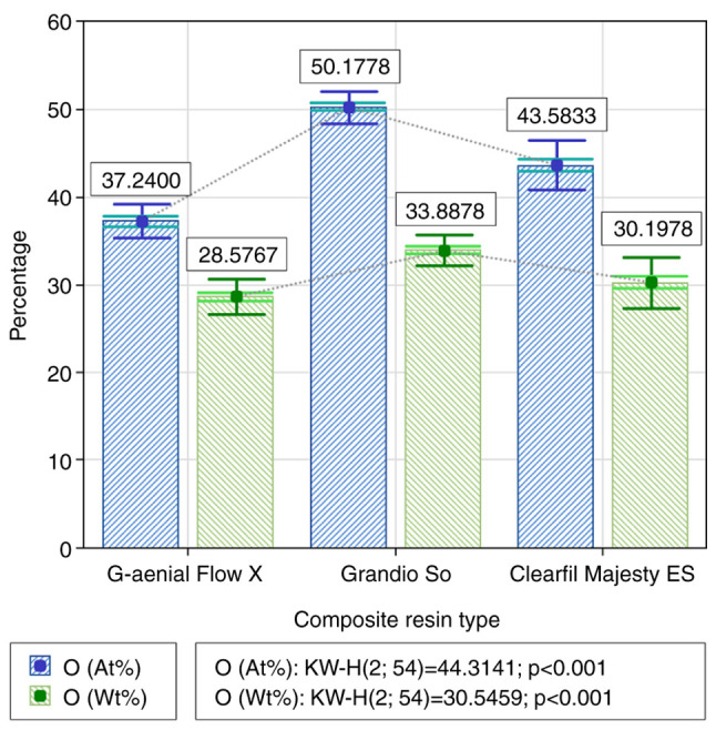
Statistical analysis of the mass (Wt%) and atomic percentage (At%) of the oxygen (O) content for the three types of materials.
Cobalt (Co) content
The average Co content was observed only for the Clearfil Majesty ES material, being 1.4167±0.33941 in the case of the wt% measurement and 0.5356±0.15523 in the case of the at% measurement, respectively (Fig. 3).
Figure 3.
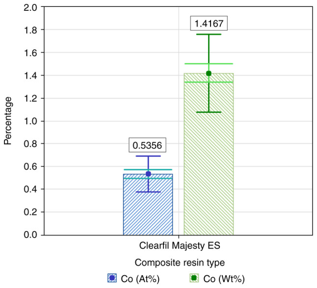
Statistical analysis of the mass (Wt%) and atomic percentage (At%) of the cobalt (Co) content for the three types of materials.
Aluminum (Al) content
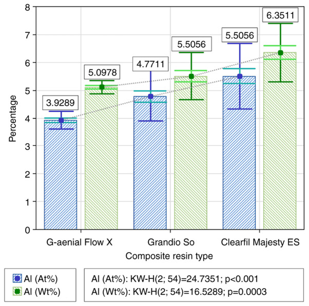
The Al content was also significantly different between the three types of materials, in both measurements, but only globally. In the case of the wt% measurement, the highest Al content was observed for Clearfil Majesty ES (6.3511±1.04642), followed by Grandio So (5.5056±0.83701) and lastly, by G-aenial Flo X (5.0978±0.26932). The Al content of Clearfil Majesty ES was statistically significantly higher than that of the other two materials tested; however, the differences between the latter two (G-aenial Flo X and Grandio So) were small and had no statistical significance (Fig. 4). A similar behavior was observed in the case of at% measurements, except that in this case the difference between Clearfil Majesty ES and Grandio So was smaller, not statistically significant, while other differences between varying combinations of materials were important and statistically significant (Table I).
Figure 4.
Statistical analysis of the mass (Wt%) and atomic percentage (At%) of the aluminum (Al) content for the three types of materials.
Silicium (Si) content
The Si content was also significantly different between the three types of materials, in the case of both measurements, both globally and comparatively between materials, two by two. In the case of the wt% measurement, the highest Si content was observed again for GrandioSo Heavy flow (35.6133±1.23026), followed by Clearfil Majesty ES (26.4044±1.93859) and lastly, by G-aenial Flo X (22.1044±0.64334), the observed differences being large enough to be statistically significant. The same phenomenon was observed in the case of at% measurements (Fig. 5).
Figure 5.
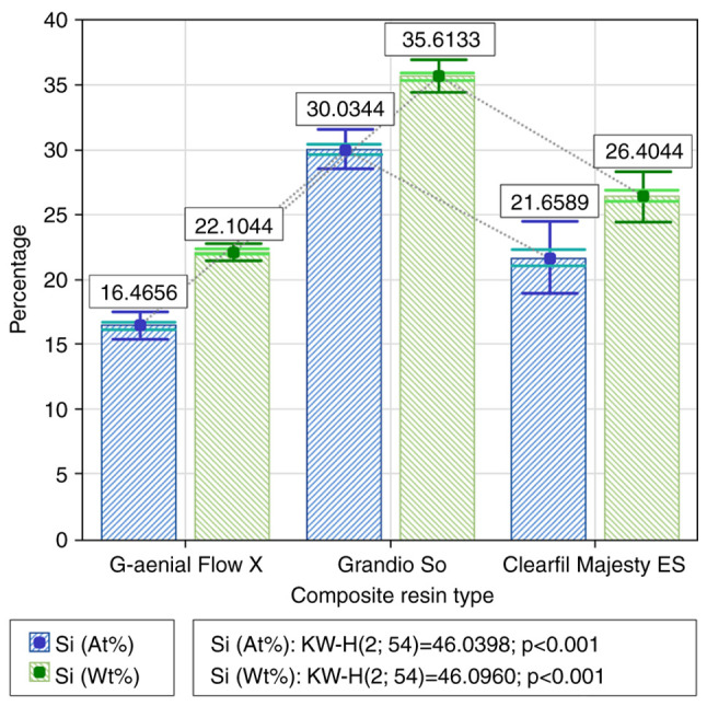
Statistical analysis of the mass (Wt%) and atomic percentage (At%) of the silicone (Si) content for the three types of materials.
Barium (Ba) content
The Ba content was significantly different between the three types of materials, but only globally and only in the case of the wt% measurement. In the case of the wt% measurement, the highest Ba content was observed for Clearfil Majesty ES (22.6356±3.09334), followed by G-aenial Flo X (22.4600±2.11290) at a very small difference and without statistical significance and, at a difference that was higher and with low statistical significance, by Grandio So (19.0311±0.92871) (Table I). The order of the three materials was the same in the case of at% measurements in terms of Ba content, only in this case the recorded values were close to each other, without statistically significant differences between the analyzed materials (Fig. 6).
Figure 6.
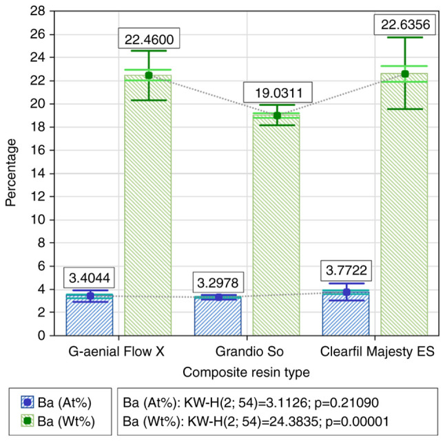
Statistical analysis of the mass (Wt%) and atomic percentage (At%) of the barium (Ba) content for the three types of materials.
Discussion
The composition of composite resins is complex and from the perspective of energy dispersive electron spectrometry (EDX) analysis, general information was obtained regarding the structural elements of resins (7). Elements such as C, O, Al, Si and Ba were identified as common elements among the resins tested in the present study. The only difference in terms of the component elements was found in the case of the fluid composite Clearfil Majesty ES flow, which, apart from the elements mentioned, also presented Co in its structure.
Clearfil Majesty ES flow is a light-curable, radio-opaque composite resin containing nanoparticles and a high refraction matrix (8). It has a light diffusion property very similar to the structure of natural teeth, which makes it versatile for almost any situation in which a composite must be used. The high refraction matrix also offers another key benefit: a very small change in transparency after photopolymerization/light curing (9).
GrandioSO Heavy Flow is a nanohybrid composite with high viscosity which, due to its high filling rate, has exceptional physical properties including stability, compressive strength, adaptability, and flexural strength. It is capable of withstanding abrasion, shrinkage, wear and polishing, thus giving the dentist many solutions to the challenges posed by practice. With an initial shrinkage of less than 3% and flexural strength above 400 MPa, GrandioSO Heavy Flow creates varied opportunities for use compared to other flow composites. Compared to the other composites used in the present study, G-aenial Flo X is a composite with high strength and radio-opacity, specially designed for optimal handling in microcavities and cracks.
There are two basic properties by which fibers can increase the effectiveness of a composite resin. Fibers act as a stress-bearing component. This enhances the otherwise brittle effect of the composite matrix. Secondly, fibers have a fracture-arresting property or a crack deflection mechanism, which in turn increases the strength of the material. It is important for the clinician to manufacture a system able to withstand all failure mechanisms in the most favorable manner possible. All these mechanisms depend on the direction in which the stress is applied to the device (10).
Fiberglass-reinforced composite resins are a type of material that combine a polymeric matrix and reinforcing fibers. When pressure is applied to the material, the intervening fibers are the composite fibers, which lend the material strength and hardness. The reinforced fibers can be unidirectional continuous (rovings), bidirectional continuous (weaves), randomly oriented continuous (matte) or they can be discontinuous and randomly oriented (11).
Currently, many materials are used for a dental immobilization system, including metal wires or fiber-reinforced composites. In the medical field, composite materials are used more often because they are chemically stable and do not induce negative effects, as the human body tolerates them easily. The most widely used materials today for making fiber-reinforced composite resin systems are glass and polyethylene fibers. The fibers have been developed in order to be able to strengthen the dental composite resins, thus forming strong, but thin structures (12). Based on the results of their study, Juloski et al concluded that the reinforcement of fibers with fluid composite does not affect the shear strength of the enamel, and that the flexural strength of the fibers is significantly influenced by their composition and model (13).
An important factor in the success of periodontal splinting is the biomechanical behavior of these systems on dental units. The type and material of the immobilization system also play an important role in the success of the treatment. There are differences between composites reinforced with polyethylene fibers and those made of metal wires. Thus, the stresses on the bone in the case of incisors were higher in the polyethylene fiber tapes compared to those containing metal wires, this being attributed to the more elastic behavior of the polyethylene fibers. When the force was applied to the canines, higher stresses were identified in the case of metal immobilization systems (14).
The capacity of the material used must be evaluated primarily according to the durability and efficiency it offers. These two characteristics are also influenced by the consistency of the composite material used. Other findings on the influence of composite resins on the success rate of periodontal immobilization have concluded that high viscosity flow composite resins showed the best treatment success rate (15). Compared to these, solid composites showed an acceptable success rate, the failures being closely related to the consequences of difficult handling of this type of material on the lingual face of the teeth. At the opposite end, avoidance of classical flow composites in periodontal immobilization is recommended, since their mechanical strength is not sufficient for the stress to which they are subjected (16).
The differences in the chemical composition of the investigated materials can influence their physiochemical properties, but also their biological and toxicological reliability. Local and systemic toxic effects of dental materials can appear if these materials are placed in the oral cavity for a long period of time (17). The general opinion of the scientific community is that no material can be 100% safe biologically, and that most adverse effects of materials are mediated by substances released from the material during corrosion (18).
In vitro studies performed on cultured mouse fibroblasts revealed changes in the growth and morphology of cells exposed to cobalt. Co exposure was found to be associated with the depression of the cell growth rate, concluding that increased concentrations of cobalt could affect the normal reconstructive activity of fibroblasts (19). However, significant health effects (neurological, cardiovascular and endocrine deficits) are unlikely to occur at blood Co concentrations under 300 µg/l in healthy individuals, and chronic exposure to acceptable doses is not expected to pose considerable health hazards (20).
Although a higher percentage of aluminum present in the material is associated with low material density, good malleability and good ductility, it is important to take into account the fact that aluminum is a toxic metal, and its release in high quantities can lead to unwanted adverse effects (pro-oxidant, inflammagen, immunogen), which is involved in human neurodegenerative diseases (21). However, Meryon and Jakeman investigated the release of aluminum from several materials and its in vitro toxicity towards culture cells, but the aluminum concentrations released in the medium did not exceed 15 ppm, while levels of 40 ppm were usually associated with toxic effects on fibroblasts and macrophages (22).
Based on various property measurements, silica-filled cured composites showed highly desirable comprehensive performances including dielectric, breakdown, mechanical, thermal and mass stability properties. Thus, low permittivity, low dielectric loss, high electric breakdown strength, high ageing breakdown strength, high shock strength, high thermal conductivity and low weight loss percentage were obtained in micro-silica loaded composites, leading to their promising application in electrical insulation cases. The large band gap and relatively high deformation ability of silica particles could contribute to favorable breakdown and mechanical properties of composites. Silica-filled cured composites exhibited a breakdown strength of ca. 48 MV m-1 and shock strength of ca. 9950 J m-2. This study may open the door towards the large-scale manufacture of high-performance epoxy composite potting-adhesives (23).
To enhance the penetration of dental adhesives, the enhancement of hydrophilicity and homogeneity of adhesive formula is the most commonly used strategy.
Silica fillers are hydrophilic, so there is a restriction in their affinity to hydrophobic resins. Through surface treatment with silane-coupling agents, the mixing of hydrophilic silica filler and the interaction between filler and organic components could be improved. Kim et al hypothesized that an increased hydrophilicity of the fillers can increase the even dispersion within the adhesive layer under moist conditions, thus increasing the bond strength (24).
Carefully modified handling property is helpful in producing a uniform adhesive layer at a feasible thickness, thus leading to a satisfactory bonding performance. In addition to texture, viscosity is an important parameter in evaluating the handling properties of dental adhesives (25).
Bonding performance is one of the most important aspects for the evaluation of dental adhesives. High-bond strength of the adhesion joint is always an aim in the development of dental adhesives.
In the current study, the highest amount of Si was identified in the samples produced using GrandioSO Heavy flow, which suggests a higher biomechanical strength of this product by comparison with the other samples included in the study. The presence of cobalt was identified only in the samples of Clearfil Majesty ES Flow and from the point of view of our results is irrelevant to the oral toxicity. The issue of metal ions release over time in the oral medium must be taken into account and evaluated frequently. The acid environment in the mouth leads to erosion, and cobalt release from dental materials has been associated with allergic reactions (26,27).
The statistically significantly higher amount of aluminum was determined in the samples made using the Clearfil Majesty ES composite resin, which can thus be associated with low material density, good malleability and good ductility.
The high amount of carbon identified in the samples that involved the use of G-aenial Flo X may indicate a slightly higher abrasive potential of the material in the clinical context.
Although the materials included in the present study can be successfully used for periodontal splinting, their behavior towards the adjacent tissues should also be evaluated. For this reason, the future research direction could assess the cytotoxicity of these materials and potentially establish a link between the presence of certain chemical elements, material strength and biocompatibility.
Thus, further studies are necessary and must be focused on the biological compatibility of the composite resins used for periodontal splinting in order to minimize the toxicological hazards at the level of oral tissues.
Acknowledgements
Professional editing, linguistic and technical assistance performed by Irina Radu, Individual Service Provider.
Funding Statement
Funding: No funding was received.
Availability of data and materials
The data used and/or analyzed during the current study are available from the corresponding authors on reasonable request.
Authors' contributions
AG, AJ, IM, SMS, LF, MT and IL designed the study and interpreted the data. IL supervised and coordinated the research project. AG, AJ and IL wrote the main manuscript. BI made the experimental determinations. CGD, IȚ, AM and DCKN contributed to the data analysis and data interpretation and edited the final form of the manuscript. All authors read and approved the final manuscript for publication. AG, BI and IL confirm the authenticity of all the data.
Ethics approval and consent to participate
Not applicable.
Patient consent for publication
Not applicable.
Competing interests
The authors declare that they have no competing interests, and none of the authors have any affiliations with GC Corporation, Voco and Kuraray Noritake Dental.
References
- 1.Cardoso EM, Reis C, Manzanares-Céspedes MC. Chronic periodontitis, inflammatory cytokines, and interrelationship with other chronic diseases. Postgrad Med. 2018;130:98–104. doi: 10.1080/00325481.2018.1396876. [DOI] [PubMed] [Google Scholar]
- 2.Giannakoura A, Pepelassi E, Kotsovilis S, Nikolopoulos G, Vrotsos I. Tooth mobility parameters in chronic periodontitis patients prior to periodontal therapy: A cross-sectional study. Dent Oral Craniofac Res. 2019;5:1–8. [Google Scholar]
- 3.Soares PB, Fernandes Neto AJ, Magalhães D, Versluis A, Soares CJ. Effect of bone loss simulation and periodontal splinting on bone strain: Periodontal splints and bone strain. Arch Oral Biol. 2011;56:1373–1381. doi: 10.1016/j.archoralbio.2011.04.002. [DOI] [PubMed] [Google Scholar]
- 4.Kathariya R, Devanoorkar A, Golani R, Shetty N, Vallakatla V, Bhat MY. To splint or not to splint: The current status of periodontal splinting. J Int Acad Periodontol. 2016;18:45–56. [PubMed] [Google Scholar]
- 5.Littlewood SJ, Millett DC, Doubleday B, Bearn DR, Worthington HV. Orthodontic retention: A systematic review. J Orthod. 2006;33:205–212. doi: 10.1179/146531205225021624. [DOI] [PubMed] [Google Scholar]
- 6.Liu X, Zhang Y, Zhou Z, Ma S. Retrospective study of combined splinting restorations in the aesthetic zone of periodontal patients. Br Dent J. 2016;220:241–247. doi: 10.1038/sj.bdj.2016.178. [DOI] [PMC free article] [PubMed] [Google Scholar]
- 7.Vilchis RJ, Hotta Y, Yamamoto K. Examination of six orthodontic adhesives with electron microscopy, hardness tester and energy dispersive X-ray microanalyzer. Angle Orthod. 2008;78:655–661. doi: 10.2319/0003-3219(2008)078[0655:EOSOAW]2.0.CO;2. [DOI] [PubMed] [Google Scholar]
- 8.Scougall-Vilchis RJ, Hotta M, Hotta M, Idono T, Yamamoto K. Examination of composite resins with electron microscopy, microhardness tester and energy dispersive X-ray microanalyzer. Dent Mater J. 2009;28:102–112. doi: 10.4012/dmj.28.102. [DOI] [PubMed] [Google Scholar]
- 9.Karadas M. The effect of different beverages on the color and translucency of flowable composites. Scanning. 2016;38:701–709. doi: 10.1002/sca.21318. [DOI] [PubMed] [Google Scholar]
- 10.Özcan M, Kumbuloglu O. Periodontal and trauma splints using fiber reinforced resin composites. In: Clinical Guide to Principles of Fiber-Reinforced Composites in Dentistry. 1st edition. Woodhead Publishing, pp111-130, 2017. [Google Scholar]
- 11.Vallittu PK. An overview of development and status of fiber-reinforced composites as dental and medical biomaterials. Acta Biomater Odontol Scand. 2018;4:44–55. doi: 10.1080/23337931.2018.1457445. [DOI] [PMC free article] [PubMed] [Google Scholar]
- 12.Bechir ES, Pacurar M, Hantoiu TA, Bechir A, Smatrea O, Burcea A, Gioga C, Monea M. Aspects in effectiveness of glass- and polyethylene-fibre reinforced composite resin in periodontal splinting. Mater Plast. 2016;53:104–109. [Google Scholar]
- 13.Juloski J, Beloica M, Goracci C, Chieffi N, Giovannetti A, Vichif A, Vulicevic ZR, Ferrari M. Shear bond strength to enamel and flexural strength of different fiber-reinforced composites. J Adhes Dent. 2013;15:123–130. doi: 10.3290/j.jad.a28362. [DOI] [PubMed] [Google Scholar]
- 14.Vieriu RM, Tanculescu O, Mocanu F, Solomon SM, Savin C, Bosinceanu DG, Doloca A, Iordache C, Ifteni G, Saveanu I. In vitro study regarding the biomechanical behaviour of bone, fibre reinforced polymer and wire composite perio-dontal splints. II. model analysis. Mater Plast. 2020;57:253–262. [Google Scholar]
- 15.Luchian I, Nanu S, Martu I, Teodorescu C, Pasarin L, Solomon S, Martu MA, Tatarciuc M, Martu S. The influence of highly viscous flowable composite resins on the survival rate of periodontal splints. Rom J Oral Rehabilitation. 2018;10:63–69. [Google Scholar]
- 16.Luchian I, Nanu S, Martu I, Martu MA, Nichitean G, Nitescu DC, Gurau C, Victorita S, Pasarin L, Tatarciuc M, Solomon SM. The influence of the composite resin material on the clinical working time in fiberglass reinforced periodontal splints. Mater Plast. 2020;57:316–320. [Google Scholar]
- 17.Shahi S, Özcan M, Maleki Dizaj S, Sharifi S, Al-Haj Husain N, Eftekhari A, Ahmadian E. A review on potential toxicity of dental material and screening their biocompatibility. Toxicol Mech Methods. 2019;29:368–377. doi: 10.1080/15376516.2019.1566424. [DOI] [PubMed] [Google Scholar]
- 18.Wataha JC. Predicting clinical biological responses to dental materials. Dent Mater. 2012;28:23–40. doi: 10.1016/j.dental.2011.08.595. [DOI] [PubMed] [Google Scholar]
- 19.Polyzois GL. In vitro evaluation of dental materials. Clin Mater. 1994;16:21–60. doi: 10.1016/0267-6605(94)90088-4. [DOI] [PubMed] [Google Scholar]
- 20.Leyssens L, Vinck B, Van Der Straeten C, Wuyts F, Maes L. Cobalt toxicity in humans-A review of the potential sources and systemic health effects. Toxicology. 2017;387:43–56. doi: 10.1016/j.tox.2017.05.015. [DOI] [PubMed] [Google Scholar]
- 21.Exley C. Human exposure to aluminium. Environ Sci Process Impacts. 2013;15:1807–1816. doi: 10.1039/c3em00374d. [DOI] [PubMed] [Google Scholar]
- 22.Meryon S, Jakeman J. Aluminium and dental materials-a study in vitro of its potential release and toxicity. Int Endod J. 1987;20:16–19. doi: 10.1111/j.1365-2591.1987.tb00582.x. [DOI] [PubMed] [Google Scholar]
- 23.Hu JB. High-performance ceramic/epoxy composite adhesives enabled by rational ceramic bandgaps. Sci Rep. 2020;10(484) doi: 10.1038/s41598-019-57074-7. [DOI] [PMC free article] [PubMed] [Google Scholar]
- 24.Kim IJ, Kim D, Ahn B, Lee HJ, Kim HJ, Kim W. Vulcanizate structures of SBR compounds with silica and carbon black binary filler systems at different curing temperatures. Polymers (Basel) 2020;12(2343) doi: 10.3390/polym12102343. [DOI] [PMC free article] [PubMed] [Google Scholar]
- 25.Wang J, Yu Q, Yang Z. Effect of hydrophobic surface treated fumed silica fillers on a one-bottle etch and rinse model dental adhesive. J Mater Sci Mater Med. 2017;29(10) doi: 10.1007/s10856-017-6015-3. [DOI] [PubMed] [Google Scholar]
- 26.Kostić M, Pejcic A, Igić M, Gligorijević N. Adverse reactions to denture resin materials. Eur Rev Med Pharmacol Sci. 2017;21:5298–5305. doi: 10.26355/eurrev_201712_13909. [DOI] [PubMed] [Google Scholar]
- 27.Bationo R, Ablassé R, Diarra A, Beugré-Kouassi ML, Jordana F, Beugré JB. In vitro Assessment of cytotoxicity of orthodontic and dental composite resins using human gingival fibroblast. Sch J Dent Sci. 2019;6:352–355. [Google Scholar]
Associated Data
This section collects any data citations, data availability statements, or supplementary materials included in this article.
Data Availability Statement
The data used and/or analyzed during the current study are available from the corresponding authors on reasonable request.


