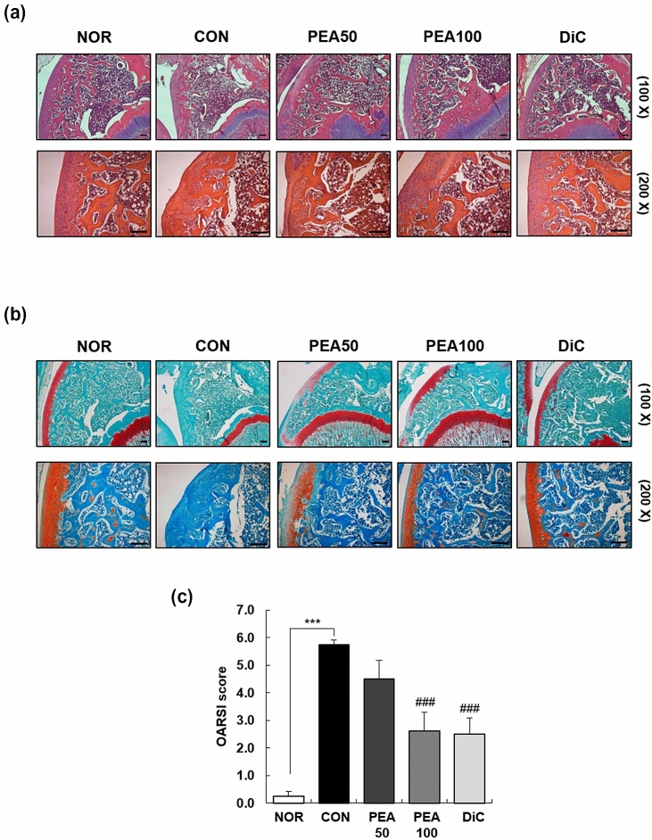
Fig. 2.
Effect of PEA on histological changes in the articular cartilage in MIA-induced OA rats. MIA-induced OA rats were administered either PEA (50 or 100 mg/kg BW/day) or diclofenac (6 mg/kg BW/day) for 4 weeks. Articular cartilage was stained with H&E (a) and safranin O (b), and subjected to Osteoarthritis Research Society International (OARSI) scoring (c). Representative staining images are shown. Scale bar, 50 μm. Each bar represents mean ± SEM (n = 5). *P < 0.01, **P < 0.05, and ***P < 0.001 significantly different from the NOR group. #P < 0.01, ##P < 0.05, and ###P < 0.001 significantly different from the CON group. PEA palmitoylethanolamide, OA osteoarthritis, MIA monosodium iodoacetate, BW body weight, H&E hematoxylin and eosin, NOR normal control group (injected with saline + treated with phosphate-buffered saline (PBS)), CON control group (injected with MIA + treated with PBS), PEA50 or PEA100 50 or 100 mg/kg body weight (BW)/day PEA-treated group (injected with MIA + treated with 50 or 100 mg/kg BW/day), DiC positive control group (injected with MIA + treated with 6 mg of diclofenc/kg BW/day)

