


Abstract
Background
Root canal therapy is a sequence of treatments involving root canal cleaning, shaping, decontamination, and obturation. It is conventionally performed through a hole drilled into the crown of the affected tooth, namely orthograde root canal therapy. When it fails, retrograde filling, which seals the root canal from the root apex, is a good alternative. Many materials are used for retrograde filling. Since none meets all the criteria an ideal material should possess, selecting the most efficacious material is of utmost importance. This is an update of a Cochrane Review first published in 2016.
Objectives
To determine the effects of different materials used for retrograde filling in children and adults for whom retrograde filling is necessary in order to save the tooth.
Search methods
An Information Specialist searched five bibliographic databases up to 21 April 2021 and used additional search methods to identify published, unpublished, and ongoing studies. We also searched four databases in the Chinese language.
Selection criteria
We selected randomised controlled trials (RCTs) that compared different retrograde filling materials, with the reported success rate that was assessed by clinical or radiological methods for which the follow‐up period was at least 12 months.
Data collection and analysis
Records were screened in duplicate by independent screeners. Two review authors extracted data independently and in duplicate. Original trial authors were contacted for any missing information. Two review authors independently assessed the risk of bias of the included studies. We followed Cochrane's statistical guidelines and assessed the certainty of the evidence using GRADE.
Main results
We included eight studies, all at high risk of bias, involving 1399 participants with 1471 teeth, published between 1995 and 2019, and six comparisons of retrograde filling materials.
‐ Mineral trioxide aggregate (MTA) versus intermediate restorative material (IRM): there may be little to no effect of MTA compared to IRM on success rate at one year, but the evidence is very uncertain (risk ratio (RR) 1.09, 95% confidence interval (CI) 0.97 to 1.22; I2 = 0%; 2 studies; 222 teeth; very low‐certainty evidence).
‐ MTA versus super ethoxybenzoic acid (Super‐EBA): there may be little to no effect of MTA compared to Super‐EBA on success rate at one year, but the evidence is very uncertain (RR 1.03, 95% CI 0.96 to 1.10; 1 study; 192 teeth; very low‐certainty evidence).
‐ Super‐EBA versus IRM: the evidence is very uncertain about the effect of Super‐EBA compared with IRM on success rate at 1 year, with results indicating Super‐EBA may reduce or have no effect on success rate (RR 0.90, 95% CI 0.80 to 1.01; 1 study; 194 teeth; very low‐certainty evidence).
‐ Dentine‐bonded resin composite versus glass ionomer cement: compared to glass ionomer cement, dentine‐bonded resin composite may increase the success rate of the treatment at 1 year, but the evidence is very uncertain (RR 2.39, 95% CI 1.60 to 3.59; 1 study; 122 teeth; very low‐certainty evidence). Same result was obtained when considering the root as unit of analysis at one year (RR 1.59, 95% CI 1.20 to 2.09; 1 study; 127 roots; very low‐certainty evidence).
‐ Glass ionomer cement versus amalgam: the evidence is very uncertain about the effect of glass ionomer cement compared with amalgam on success rate at one year, with results indicating glass ionomer cement may reduce or have no effect on success rate (RR 0.98, 95% CI 0.86 to 1.12; 1 study; 105 teeth; very low‐certainty evidence).
‐ MTA versus root repair material (RRM): there may be little to no effect of MTA compared to RRM on success rate at one year, but the evidence is very uncertain (RR 1.00, 95% CI 0.94 to 1.07; I2 = 0%; 2 studies; 278 teeth; very low‐certainty evidence).
Adverse events were not assessed by any of the included studies.
Authors' conclusions
Based on the present limited evidence, there is insufficient evidence to draw any conclusion as to the benefits of any one material over another for retrograde filling in root canal therapy. We conclude that more high‐quality RCTs are required.
Keywords: Adult, Child, Humans, Crowns, Glass Ionomer Cements, Root Canal Therapy
Plain language summary
What are the benefits and risks of different materials used for retrograde filling in people who need this treatment in order to save a tooth?
Key messages
‐ Due to a lack of robust evidence, the benefits and risks of different materials used for retrograde filling in root canal therapy are unclear.
‐ The evidence is not robust enough to determine which material is best to use in retrograde filling.
‐ We need future studies to strengthen the evidence.
What is retrograde filling in root canal therapy?
The living part of the tooth, also known as the tooth pulp, can become permanently swollen because of damage or bacterial infection due to tooth decay. To deal with this problem, the dentist has to drill a hole on the top of the crown of the tooth to access the inner space of the tooth, the root canal system. The dentist will then remove the infected tissue and bacteria by a combination of mechanical cleaning and irrigation.
After this is done, the dentist fills the space with an inactive packing material and seals the opening. This treatment is known as root canal therapy. Although results are generally good, a small number of failures can happen. This can be because the root canal system is complex and it is not always easy to completely eliminate all bacteria. These can spread and the infection around the root can last indefinitely.
When root canal therapy fails, a retreatment called retrograde filling is a good alternative to save the tooth. During retrograde filling the dentist cuts a flap in the gum and creates a hole in the bone to get access to the bottom tip of the root of the tooth. After cutting off the tip, followed by thorough preparation, the apex is sealed (the apical seal), and the hole made by the dentist filled with a dental material. This sealing process is considered the most important factor in achieving success in a retrograde root filling.
What materials can be used for retrograde filling?
Many materials have been developed to seal the root tip, for example, mineral trioxide aggregate (MTA), intermediate restorative material (IRM), super ethoxybenzoic acid (Super‐EBA), dentine‐bonded resin composite, glass ionomer cement, amalgam, and root repair material (RRM). However, there is no agreement on which material is best.
What did we want to find out?
We wanted to find out which material works better for retrograde filling in root canal therapy, and whether they are associated with any unwanted (adverse) effects.
What did we do?
We searched for studies that compared different materials used for retrograde filling in root canal therapy. We compared and summarised the results of the studies and rated our confidence in the evidence, based on factors such as study methods and sizes.
What did we find?
We found eight studies with a minimum duration of 12 months that involved 1399 people (1471 teeth) over 17 years of age undergoing retrograde filling using different types of filling material.
The evidence:
‐ is not robust enough to determine which material is best to use in retrograde filling.
No studies investigated the unwanted effects of any of the materials.
What are the limitations of the evidence?
The main limitations of the evidence are that studies:
‐ were very small;
‐ were conducted in ways that may have introduced errors into their results; and
‐ there were not enough studies to be certain about the results.
Due to these limitations, we have little confidence in the evidence.
How up to date is this evidence?
The evidence is up to date to April 2021.
Summary of findings
Summary of findings 1. MTA versus IRM for retrograde filling in root canal therapy.
| MTA versus IRM for retrograde filling in root canal therapy | ||||||
| Patient or population: patients needing retrograde filling in root canal therapy Settings: UK and the Netherlands Intervention: MTA versus IRM | ||||||
| Outcomes | Illustrative comparative risks* (95% CI) | Relative effect (95% CI) | Number of teeth (studies) | Certainty of the evidence (GRADE) | Comments | |
| Assumed risk | Corresponding risk | |||||
| IRM | MTA | |||||
| Success rate ‐ 1‐year outcome (tooth as unit of analysis) Assessed by combination of clinical and radiological methods Follow‐up: mean 1 year | 806 per 1000 | 879 per 1000 (782 to 983) | RR 1.09 (0.97 to 1.22) | 222 (2 studies) | ⊕⊝⊝⊝ verylowa | There may be little to no effect of MTA compared to IRM on success rate at 1 year but the evidence is very uncertain |
| Adverse events | Outcome was not assessed by the included studies | |||||
|
*The basis for the assumed risk (e.g. the median control group risk across studies) is provided in footnotes. The corresponding risk (and its 95% CI) is based on the assumed risk in the comparison group and the relative effect of the intervention (and its 95% CI) CI: confidence interval; IRM: intermediate restorative material; MTA: mineral trioxide aggregate; RR: risk ratio | ||||||
|
GRADE Working Group grades of evidence High certainty: we are very confident that the true effect lies close to that of the estimate of the effect Moderate certainty: we are moderately confident in the effect estimate: the true effect is likely to be close to the estimate of the effect, but there is a possibility that it is substantially different Low certainty: our confidence in the effect estimate is limited: the true effect may be substantially different from the estimate of the effect Very low certainty: we have very little confidence in the effect estimate: the true effect is likely to be substantially different from the estimate of effect | ||||||
aDowngraded by 3 levels due to high risk of bias (downgraded by 1 level; both studies had the personnel unblinded due to the nature of the study design, and Chong 2003 had incomplete outcome data reported) and imprecision (downgraded by 2 levels; small number of participants and wide confidence intervals including both benefit and no benefit).
Summary of findings 2. MTA versus Super‐EBA for retrograde filling in root canal therapy.
| MTA versus Super‐EBA for retrograde filling in root canal therapy | ||||||
| Patient or population: patients needing retrograde filling in root canal therapy Settings: South Korea Intervention: MTA versus Super‐EBA | ||||||
| Outcomes | Illustrative comparative risks* (95% CI) | Relative effect (95% CI) | Number of teeth (studies) | Certainty of the evidence (GRADE) | Comments | |
| Assumed risk | Corresponding risk | |||||
| Super‐EBA | MTA | |||||
| Success rate ‐ 1‐year outcome (tooth as unit of analysis) Assessed by combination of clinical and radiological methods Follow‐up: mean 1 year | 931 per 1000 | 959 per 1000 (894 to 1000) | RR 1.03 (0.96 to 1.10) | 192 (1 study) | ⊕⊝⊝⊝ very lowa | There may be little to no effect of MTA compared to Super‐EBA on success rate at 1 year but the evidence is very uncertain |
| Adverse events | Outcome was not assessed by the included studies | |||||
|
*The basis for the assumed risk (e.g. the median control group risk across studies) is provided in footnotes. The corresponding risk (and its 95% CI) is based on the assumed risk in the comparison group and the relative effect of the intervention (and its 95% CI) CI: confidence interval; MTA: mineral trioxide aggregate; RR: risk ratio; Super‐EBA: super ethoxybenzoic acid | ||||||
|
GRADE Working Group grades of evidence High certainty: we are very confident that the true effect lies close to that of the estimate of the effect Moderate certainty: we are moderately confident in the effect estimate: the true effect is likely to be close to the estimate of the effect, but there is a possibility that it is substantially different Low certainty: our confidence in the effect estimate is limited: the true effect may be substantially different from the estimate of the effect Very low certainty: we have very little confidence in the effect estimate: the true effect is likely to be substantially different from the estimate of effect | ||||||
aDowngraded by 3 levels due to high risk of bias (downgraded by 1 level; the included study (Kim 2016) had high risk of bias regarding incomplete data reporting and personnel blinding) and imprecision (downgraded by 2 levels; small population might cause serious imprecision).
Summary of findings 3. Super‐EBA versus IRM for retrograde filling in root canal therapy.
| Super‐EBA versus IRM for retrograde filling in root canal therapy | ||||||
| Patient or population: patients needing retrograde filling in root canal therapy Settings: Sweden Intervention: Super‐EBA versus IRM | ||||||
| Outcomes | Illustrative comparative risks* (95% CI) | Relative effect (95% CI) | Number of teeth (studies) | Certainty of the evidence (GRADE) | Comments | |
| Assumed risk | Corresponding risk | |||||
| IRM | Super‐EBA | |||||
| Success rate ‐ 1‐year outcome (tooth as unit of analysis) Assessed by combination of clinical and radiological methods Follow‐up: mean 1 year | 906 per 1000 | 815 per 1000 (725 to 915) | RR 0.90 (0.80 to 1.01) | 194 (1 study) | ⊕⊝⊝⊝ very lowa | The evidence is very uncertain about the effect of Super‐EBA compared with IRM on success rate at 1 year, with results indicating Super‐EBA may reduce or have no effect on success rate |
| Adverse events | Outcome was not assessed by the included studies | |||||
|
*The basis for the assumed risk (e.g. the median control group risk across studies) is provided in footnotes. The corresponding risk (and its 95% CI) is based on the assumed risk in the comparison group and the relative effect of the intervention (and its 95% CI) CI: confidence interval; IRM: intermediate restorative material; RR: risk ratio; Super‐EBA: super ethoxybenzoic acid | ||||||
|
GRADE Working Group grades of evidence High certainty: we are very confident that the true effect lies close to that of the estimate of the effect Moderate certainty: we are moderately confident in the effect estimate: the true effect is likely to be close to the estimate of the effect, but there is a possibility that it is substantially different Low certainty: our confidence in the effect estimate is limited: the true effect may be substantially different from the estimate of the effect Very low certainty: we have very little confidence in the effect estimate: the true effect is likely to be substantially different from the estimate of effect | ||||||
aDowngraded by 3 levels due to high risk of bias (downgraded by 1 level; high risk of bias existed in the only included study (Wälivaara 2011) on allocation concealment and personnel blinding) and imprecision (downgraded by 2 levels; small number of participants and wide confidence intervals including both benefit and no benefit).
Summary of findings 4. Dentine‐bonded resin composite versus glass ionomer cement for retrograde filling in root canal therapy.
| Dentine‐bonded resin composite versus glass ionomer cement for retrograde filling in root canal therapy | ||||||
| Patient or population: patients needing retrograde filling in root canal therapy Settings: Denmark Intervention: dentine‐bonded resin composite versus glass ionomer cement | ||||||
| Outcomes | Illustrative comparative risks* (95% CI) | Relative effect (95% CI) | Number of teeth/roots (studies) | Certainty of the evidence (GRADE) | Comments | |
| Assumed risk | Corresponding risk | |||||
| Glass ionomer cement | Dentine‐bonded resin composite | |||||
| Success rate ‐ 1‐year outcome PP analysis (tooth as unit of analysis) Assessed by combination of clinical and radiological methods Follow‐up: mean 1 year | 306 per 1000 | 731 per 1000 (490 to 1000) | RR 2.39 (1.60 to 3.59) | 122 teeth (1 study) | ⊕⊝⊝⊝ very lowa | Compared to glass ionomer cement, dentine‐bonded resin composite may increase the success rate of the treatment at 1 year but the evidence is very uncertain |
| Success rate ‐ 1‐year outcome PP analysis (root as unit of analysis) Assessed by radiological methods Follow‐up: mean 1 year | 519 per 1000 | 825 per 1000 (623 to 1000) | RR 1.59 (1.20 to 2.09) | 127 roots (1 study) | ⊕⊝⊝⊝ very lowa | Compared to glass ionomer cement, dentine‐bonded resin composite may increase the success rate of the treatment at 1 year but the evidence is very uncertain |
| Adverse events | Outcome was not assessed by the included studies | |||||
|
*The basis for the assumed risk (e.g. the median control group risk across studies) is provided in footnotes. The corresponding risk (and its 95% CI) is based on the assumed risk in the comparison group and the relative effect of the intervention (and its 95% CI). CI: confidence interval; PP: per‐protocol; RR: risk ratio | ||||||
|
GRADE Working Group grades of evidence High certainty: we are very confident that the true effect lies close to that of the estimate of the effect Moderate certainty: we are moderately confident in the effect estimate: the true effect is likely to be close to the estimate of the effect, but there is a possibility that it is substantially different Low certainty: our confidence in the effect estimate is limited: the true effect may be substantially different from the estimate of the effect Very low certainty: we have very little confidence in the effect estimate: the true effect is likely to be substantially different from the estimate of effect | ||||||
aDowngraded by 3 levels due to high risk of bias (downgraded by 1 level; the included study (Jensen 2002) did not have participants, personnel, or outcome assessors blinded) and imprecision (downgraded by 2 levels; small population might cause serious imprecision).
Summary of findings 5. Glass ionomer cement versus amalgam for retrograde filling in root canal therapy.
| Glass ionomer cement versus amalgam for retrograde filling in root canal therapy | ||||||
| Patient or population: patients needing retrograde filling in root canal therapy Settings: Sweden Intervention: glass ionomer cement versus amalgam | ||||||
| Outcomes | Illustrative comparative risks* (95% CI) | Relative effect (95% CI) | Number of teeth (studies) | Certainty of the evidence (GRADE) | Comments | |
| Assumed risk | Corresponding risk | |||||
| Amalgam | Glass ionomer cement | |||||
| Success rate ‐ 1‐year outcome (tooth as unit of analysis) Assessed by combination of clinical and radiological methods Follow‐up: mean 1 year | 904 per 1000 | 886 per 1000 (777 to 1000) | RR 0.98 (0.86 to 1.12) | 105 (1 study) | ⊕⊝⊝⊝ very lowa | The evidence is very uncertain about the effect of glass ionomer cement compared with amalgam on success rate at 1 year, with results indicating glass ionomer cement may reduce or have no effect on success rate |
| Adverse events | Outcome was not assessed by the included studies | |||||
|
*The basis for the assumed risk (e.g. the median control group risk across studies) is provided in footnotes. The corresponding risk (and its 95% CI) is based on the assumed risk in the comparison group and the relative effect of the intervention (and its 95% CI) CI: confidence interval; RR: risk ratio | ||||||
|
GRADE Working Group grades of evidence High certainty: we are very confident that the true effect lies close to that of the estimate of the effect Moderate certainty: we are moderately confident in the effect estimate: the true effect is likely to be close to the estimate of the effect, but there is a possibility that it is substantially different Low certainty: our confidence in the effect estimate is limited: the true effect may be substantially different from the estimate of the effect Very low certainty: we have very little confidence in the effect estimate: the true effect is likely to be substantially different from the estimate of effect | ||||||
aDowngraded by 3 levels due to high risk of bias (downgraded by 1 level; the included study (Jesslen 1995) had the personnel unblinded due to the nature of the study design) and imprecision (downgraded by 2 levels; small number of participants and wide confidence intervals including both benefit and no benefit).
Summary of findings 6. MTA versus RRM for retrograde filling in root canal therapy.
| MTA versus RRM for retrograde filling in root canal therapy | ||||||
|
Patient or population: patients needing retrograde filling in root canal therapy Settings: China Intervention: MTA versus RRM | ||||||
| Outcomes | Illustrative comparative risks* (95% CI) | Relative effect (95% CI) | Number of teeth (studies) | Certainty of the evidence (GRADE) | Comments | |
| Assumed risk | Corresponding risk | |||||
| RRM | MTA | |||||
|
Success rate ‐ 1‐year outcome (tooth as unit of analysis) Assessed by combination of clinical and radiological methods Follow‐up: 1 year |
933 per 1000 | 896 per 1000 (840 to 970) | RR 1.00 (0.94 to 1.07) | 278 (2 studies) | ⊕⊝⊝⊝ very lowa | There may be little to no effect of MTA compared to RRM on success rate at 1 year but the evidence is very uncertain |
| Adverse events | Outcome was not assessed by the included studies | |||||
|
*The basis for the assumed risk (e.g. the median control group risk across studies) is provided in footnotes. The corresponding risk (and its 95% confidence interval) is based on the assumed risk in the comparison group and the relative effect of the intervention (and its 95% CI) CI: confidence interval; MTA: mineral trioxide aggregate; RR: risk ratio; RRM: root repair material | ||||||
|
GRADE Working Group grades of evidence High certainty: we are very confident that the true effect lies close to that of the estimate of the effect Moderate certainty: we are moderately confident in the effect estimate: the true effect is likely to be close to the estimate of the effect, but there is a possibility that it is substantially different Low certainty: our confidence in the effect estimate is limited: the true effect may be substantially different from the estimate of the effect Very low certainty: we have very little confidence in the effect estimate: the true effect is likely to be substantially different from the estimate of effect | ||||||
aDowngraded by 3 levels due to high risk of bias (downgraded by 1 level; the included studies (Safi 2019; Zhou 2017) had high risk of bias regarding personnel blinding) and imprecision (downgraded by 2 levels; small number of participants and wide confidence intervals including both benefit and no benefit).
Background
Description of the condition
Root canal therapy is a sequence of treatments for the infected pulp of a tooth which results in the elimination of that infection and the subsequent protection of the decontaminated tooth from future microbial invasion (Cohen 2006). It involves the removal of the infected pulp, the subsequent shaping, cleaning, decontamination of the hollow tooth core and obturation. Traditionally, treatment is carried out through a hole drilled on the top of the crown of the tooth, and it is known as orthograde root canal therapy (Figure 1 (A and B)). With the development of new materials and techniques, orthograde root canal therapy has been demonstrated to provide satisfactory results for patients in most cases. However, because of the well‐known complexity of the root canal system and the acknowledged difficulty of completely eliminating all bacteria, their by‐products and toxins, from the canal system, failures occur at a reported rate of 4% to 15% (Sjogren 1990; Swartz 1983; Wong 2004). A higher failure rate was ascertained by Eriksen's review of multiple clinical studies of success and failure (Eriksen 1991). There are many causes for such failures, such as untreated canals, ledges formation, perforations, and overextensions of root‐filling materials. To plan treatment effectively, the clinician may place the aetiological factors into four groups (Sundqvist 1998):
1.
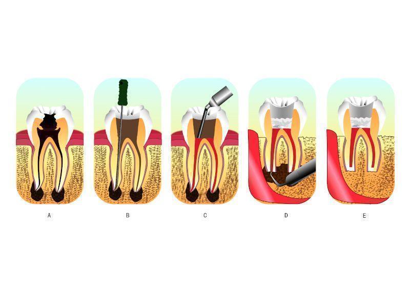
A: an infected tooth. The inner space of the tooth is the root canal system, where the pulp is located. The pulp of this tooth is irreversibly inflamed from bacterial infection due to decay.
B and C: the process of root canal therapy. B: a hole has been drilled from the top of the crown of the tooth. The dentist could then remove the infected tissues and toxic irritants by a combination of mechanical cleaning and irrigation in the root canal system through the hole. C: after cleaning and irrigation, the dentist fills the space with an inert packing material and seals the opening.
D and E: the process of retrograde filling. D: when retrograde filling is indicated, the dentist needs to cut a flap in the gum and creates a hole in the bone to get access to the bottom tip of the root. E: after cutting off the tip, then thorough preparation, the apex is sealed (the apical seal) and the hole made by the dentist filled with a dental material.
persistent or reintroduced intraradicular micro‐organisms;
extraradicular infection;
foreign body reaction;
true cysts.
For these treatment failures, conventional orthograde endodontic retreatment is always the first choice. Although it is a highly predictable option in most cases, periradicular surgery may be indicated for teeth with persistent periradicular pathosis that have not responded to non‐surgical approaches (Lee 2004). Sometimes, apical surgery is preferred to orthograde treatment for expediency or if a tight‐fitting post, especially a fibre post, is present. At this circumstance, conventional retreatment needs to remove more dentine to acquire a pathway into the original root canal, which may cause root perforation or root fracture.
Periradicular surgery should be considered as an extension to non‐surgical treatment, because the underlying aetiology of the disease process and the treatment objectives are the same: the prevention or elimination of apical periodontitis (Hargreaves 2015). Periradicular surgery, also called retrograde filling, always requires root‐end preparation and obturation (Figure 1 (D and E)). The former aims to expose the apical root via flap elevation and bone removal prior to cavity preparation on the apical root where the apical foramina is located. Following that, materials are placed in the cavity for apical sealing. Harty reported that the apical seal was the single most important factor in achieving success in such surgery (Harty 1970). This apical seal is established by retrograde filling materials obturated between the root canal system and the surrounding tissues (Gutmann 1991). Thus, subsequent studies have evaluated many retrograde materials so as to determine which is most efficacious.
Description of the intervention
Amalgam has been used as a retrograde filling material for many years. Its earliest use as a root‐end filling following resection has been reported in 1884 (Vasudev 2003). It has the advantages of being easily available, inexpensive, and easy to handle. Therefore, for many years, amalgam was accepted as the material of choice for root‐end filling, and the clinical application of amalgam was well documented in several clinical studies with a reported success rate of 50% to 80% (Dalal 1983; Finne 1977; Grung 1990; Hirsch 1979; Persson 1974; Rud 1972). However, in recent years, the efficacy of amalgam has been questioned due to initial marginal leakage, corrosion, moisture sensitivity, mercury contamination of periapical tissue and the potential hazards associated with mercury‐containing materials (Eley 1993; Gartner 1992). The disadvantages associated with amalgam and the potential long‐term damage to the environment has led to the research and development of alternative materials.
In the past decades, amalgam has slowly given way to zinc oxide eugenol (ZOE) containing materials, such as intermediate restorative material (IRM), which has 20%, by weight, polymethacrylate added to the base zinc oxide powder, and super ethoxybenzoic acid (Super‐EBA), which contains ethoxybenzoic acid. In vitro leakage studies, animal studies, and retrospective in vivo studies indicate that these ZOE‐containing materials are superior to amalgam in terms of sealability and biocompatibility (Dorn 1990; Kim 2006; King 1990). Shortcomings of the currently available ZOE‐containing cements are their mild to moderate toxicity, when freshly mixed, and their radiopacity, which is similar to that of gutta‐percha (Johnson 1999).
In recent years a promising new root‐end filling material, mineral trioxide aggregate (MTA), developed at Loma Linda University, California, USA (Torabinejad 1993) has received widespread attention (Lee 1993). Its major components are similar to Portland cement, a mixture of dicalcium silicate, tricalcium silicate, tricalcium aluminate, gypsum, and tetracalcium aluminoferrite (Camilleri 2005). Although it is an expensive material and requires additional skill and equipment to use satisfactorily, the clinician can handle it satisfactorily after suitable training (Wang 2010). MTA has major advantages, including excellent biocompatibility (Camilleri 2006), ideal adherence to cavity walls, low solubility (Poggio 2007), and the ability of inducing cementogenesis at the root surface, with deposition of new cementum onto the exposed dentine and MTA surfaces (Baek 2005). MTA is an excellent bioactive material. When it is placed in direct contact with human tissues, it will form calcium hydroxide that releases calcium ions for cell attachment and proliferation (Takita 2006), modulates cytokine production (Koh 1998), and encourages proliferation and migration of progenitors followed by their differentiation into odontoblast‐like cells (Kuratate 2008). However, the mean setting time of MTA is 165 ± 5 minutes, which is longer than amalgam, Super‐EBA, and IRM (Torabinejad 1995), which is potentially problematic in endodontic surgery.
In addition to polymers, glass ionomer cements, polycarboxylate cements, zinc phosphate cements, calcium phosphate cements and composite resins have all been employed and several cases reported (Hauman 2003). A new material, Biodentine, which is reported to have reparative dentine synthesis properties (Laurent 2012), is awaiting clinical evaluation as a possible retrograde filling material. All these materials have different characteristics and are potential alternatives to traditional materials, although potential harm should be carefully considered before widespread use is considered.
How the intervention might work
Studies have proved that the main contributory factor in endodontic failure is persistent microbial infection in the root canal system and periapical region (Siqueira Jr 2003). Chemical and mechanical preparation may not reach every corner of the complex root canal system. Bacteria in isthmuses, ramifications, irregularities and dentinal tubules may persist and some necrotic tissue debris may also remain. Bacteria may gain access to the periapical region if complete sealing is not achieved, leading to pathological lesions (Lin 1991; Siqueira 2001).
To control microbial infection is always a high priority in periradicular surgery. The surgery removes the pathogenic agents and establishes an environment facilitating the regeneration of damaged tissue first. Then the procedure usually involves root‐end exposure and resection, in addition to preparing a Class I cavity and retrofilling with packing materials (Torabinejad 1995). Hence, these materials can form a proper seal of the internal root canal contents from the external periradicular tissues and therefore repair root defects (Chong 2004).
Why it is important to do this review
Periapical surgery is the last resort to save a tooth in endodontics. If it is not successful, the tooth might be lost. The use of proven retrograde filling materials is critical for apical sealing, which is the single most important factor in achieving success in periradicular surgery. To maintain a perfect apical seal, an ideal endodontic retrograde filling material is required to adhere to the tooth structure, be insoluble in tissue fluids, be dimensionally stable, non‐resorbable, radiopaque, and exhibit biocompatibility, if not bioactivity (Johnson 1999; Kratchman 2004). In order to find the best, if not ideal, retrograde filling material, many clinical trials have been conducted in an attempt to evaluate the efficacy and safety of different materials. Secondly, the sample size of most clinical trials is small and some of the results conflict with one another. Therefore, the purpose of our systematic review, through the use of strict criteria to integrate small sample size trials, was to clarify the clinical effect and safety of different materials for retrograde filling in root canal therapy. This is an update of the Cochrane Review first published in 2016 (Ma 2016).
Objectives
To determine the effects of different materials used for retrograde filling in children and adults for whom retrograde filling is necessary in order to save the tooth.
Methods
Criteria for considering studies for this review
Types of studies
This review included randomised controlled trials (RCTs), including cluster, split‐mouth and cross‐over RCTs.
Types of participants
We included participants for whom orthograde root canal filling or retreatment was not possible and where periapical surgery was used to save the tooth. There were no age or gender limitations.
Types of interventions
Intervention group: retrograde obturation with any material.
Control group: retrograde obturation with any materials other than those used in the intervention group.
Types of outcome measures
Primary outcomes
Success rate: this was assessed by either clinical or radiological methods, or a combination of the two. Minimum follow‐up was 12 months.
Clinical methods included the assessment of clinical symptoms including pain, pain only on percussion or palpation, tenderness, increased tooth mobility, sinus tract formation or any other subjective discomfort. Any one of these was counted as treatment failure.
Radiological methods were used to detect periapical bone regeneration on medical images. Lack of apical bone regeneration compared to the baseline was counted as treatment failure.
The combination of clinical and radiological methods was used to assess the success of retrograde fillings in this review, unless specifically stated otherwise.
All cases not assessed as treatment failure were considered as successes.
Secondary outcomes
Adverse events.
Search methods for identification of studies
Electronic searches
Cochrane Oral Health's Information Specialist conducted systematic searches in the following databases for randomised controlled trials and controlled clinical trials without language or publication status restrictions:
Cochrane Oral Health's Trials Register (to 21 April 2021) (Appendix 1);
the Cochrane Central Register of Controlled Trials (CENTRAL; 2021, Issue 3) in the Cochrane Library (searched 21 April 2021) (Appendix 2);
MEDLINE Ovid (1946 to 21 April 2021) (Appendix 3);
Embase Ovid (1980 to 21 April 2021) (Appendix 4);
LILACS BIREME Virtual Health Library (Latin American and Caribbean Health Science Information database; from 1982 to 21 April 2021) (Appendix 5);
OpenSIGLE (1980 to 2005) (Appendix 6).
Searches of OpenSIGLE were discontinued due to poor yield.
We also searched the following databases:
Chinese BioMedical Literature Database (in Chinese, 1978 to 21 April 2021) (Appendix 7);
VIP (in Chinese, 1989 to 21 April 2021) (Appendix 8);
China National Knowledge Infrastructure (in Chinese, 1994 to 21 April 2021) (Appendix 9).
Subject strategies were modelled on the search strategy designed for MEDLINE Ovid. Where appropriate, they were combined with subject strategy adaptations of the highly sensitive search strategies designed by Cochrane for identifying randomised controlled trials and controlled clinical trials (as described in the Cochrane Handbook for Systematic Reviews of Interventions, Version 6.1, (Lefebvre 2021)).
Searching other resources
Cochrane Oral Health's Information Specialist searched the following trial registries for ongoing studies:
US National Institutes of Health Ongoing Trials Register ClinicalTrials.gov (clinicaltrials.gov; searched 21 April 2021) (Appendix 10);
World Health Organization International Clinical Trials Registry Platform (apps.who.int/trialsearch; searched 21 April 2021) (Appendix 10).
We also searched Sciencepaper Online (in Chinese, to 21 April 2021) (Appendix 11).
The following journals were handsearched:
Chinese Journal of Stomatology (January 2001 to April 2021)
Stomatology (January 2001 to April 2021)
West China Journal of Stomatology (January 2001 to April 2021)
Journal of Practical Stomatology (January 2001 to April 2021)
Journal of Clinical Stomatology (January 2001 to April 2021)
Journal of Comprehensive Stomatology (January 2001 to April 2021)
Journal of Modern Stomatology (January 2001 to April 2021)
Chinese Journal of Conservative Dentistry (January 2001 to April 2021)
Journal of Maxillofacial Surgery (January 2001 to April 2021)
Shanghai Journal of Stomatology (January 2001 to April 2021)
Chinese Journal of Dental Material and Devices (January 2001 to April 2021)
Beijing Journal of Stomatology (January 2001 to April 2021)
Chinese Journal of Dental Prevention and Treatment (January 2001 to April 2021)
Chinese Journal of Orthodontics (January 2001 to April 2021)
Chinese Journal of Implantology (January 2001 to April 2021)
Journal of International Stomatology (January 2001 to April 2021)
Chinese Journal of Prosthodontics (January 2001 to April 2021)
China Journal of Oral and Maxillofacial Surgery (2003 to April 2021)
Chinese Journal of Geriatric Dentistry (2002 to April 2021).
The reference lists from the included studies were also searched. We contacted the authors of eligible studies to see if there was any additional published or unpublished studies. Related manufacturers of different materials were contacted to identify if there were any unpublished trials on the material.
We did not perform a separate search for adverse effects of interventions used, we considered adverse effects described in included studies only.
We checked that none of the included studies in this review were retracted due to error or fraud.
Data collection and analysis
Selection of studies
Two review authors (Xiangyu Ma (XM) and Zhiyong Guo (ZG)) reviewed independently and in duplicate the titles and abstracts (if available) of the articles identified by the search to locate articles that met the inclusion criteria. If eligibility could not be assessed from the title and abstract, the full‐text article was obtained and further reviewed. Any disagreement was resolved by discussion among the review authors. Non‐English and non‐Chinese papers were examined with the help of Cochrane Oral Health (COH).
Data extraction and management
Two review authors (Honglin Li (HL) and ZG) independently extracted the data from relevant articles with the help of a data extraction form designed specifically for this review. These forms had been piloted on several papers and modified as required before use.
For each study, the following data were recorded.
Basic information: date of the study, year of publication, country of origin.
Study type: details of sequence generation, allocation concealment, and blinding.
Participants: inclusion and exclusion criteria, characteristics of the participants, sample size calculation.
Intervention: characteristics of the interventions (instruments used, cavity form prepared, and material used).
Outcome: outcome measures and detailed follow‐up information.
Any disagreement was resolved by discussion. The study authors were contacted for clarification if necessary.
Assessment of risk of bias in included studies
The risk of bias assessment was delivered by two review authors (HL and ZG), also independently and in duplicate, according to the guidelines in the Cochrane Handbook for Systematic Reviews of Interventions (Higgins 2011). Any disagreement was resolved by discussion. Seven domains were considered for the risk of bias assessment.
Random sequence generation (selection bias).
Allocation concealment (selection bias).
Blinding of participants and personnel (performance bias).
Blinding of outcome assessment (detection bias).
Incomplete outcome data (attrition bias).
Selective reporting (reporting bias).
Other bias which is not covered by the first six (including confounding bias, baseline imbalance, co‐intervention, and contamination).
For each study included in the review, the risk of bias in every domain was judged as either low, high, or unclear, according to the included studies or from correspondence with the author.
The summary assessment was also prepared according to the Cochrane Handbook for Systematic Reviews of Interventions (Higgins 2011) and presented graphically.
| Risk of bias | Interpretation | Within a study | Across studies |
| Low risk of bias | Plausible bias unlikely to seriously alter the results | Low risk of bias for all key domains | Most information is from studies at low risk of bias |
| Unclear risk of bias | Plausible bias that raises some doubt about the results | Unclear risk of bias for one or more key domains | Most information is from studies at low or unclear risk of bias |
| High risk of bias | Plausible bias that seriously weakens confidence in the results | High risk of bias for one or more key domains | The proportion of information from studies at high risk of bias is sufficient to affect the interpretation of results |
Measures of treatment effect
The success rate and adverse events were classified as dichotomous data. For dichotomous outcomes, we expressed the estimate of effect of an intervention as risk ratios (RRs) together with 95% confidence intervals (CIs).
Unit of analysis issues
Generally, we considered each participant as the unit of analysis in this review. However, where everyone was only analysed with one tooth or each tooth was analysed, each tooth could also be considered the unit of analysis. For success rate evaluated solely by radiological methods, the root could be considered as the unit of analysis if clearly stated.
Dealing with missing data
For papers with missing data, we contacted the first and corresponding authors in an attempt to retrieve such data. Failing that, we followed guidance included in the Cochrane Handbook for Systematic Reviews of Interventions (Higgins 2011). For missing patients, we analysed only the available data ignoring the missing data for the per‐protocol (PP) analyses. We would perform sensitivity analyses to assess how sensitive results were to reasonable changes in the assumptions that were made. Here, we performed intention‐to‐treat (ITT) analyses ('worst‐case scenario' analysis: all lost to follow‐up participants in the intervention group were considered as failed cases; all lost to follow‐up participants in the control group were considered successful cases).
Assessment of heterogeneity
If meta‐analyses were performed, we assessed the possible presence of heterogeneity visually by inspecting the point estimates and confidence intervals on the forest plots; if the confidence intervals had poor overlap then heterogeneity was considered to be present. We used the Chi2 test to calculate statistical heterogeneity. Statistical heterogeneity was classified into four categories according to the I2 statisticwhich was used to describe the percentage of the statistical variability in effect estimates. P < 0.1 indicated statistical heterogeneity:
0% to 40% implied slight heterogeneity,
30% to 60% moderate heterogeneity,
50% to 90% substantial heterogeneity,
75% to 100% considerable heterogeneity.
Assessment of reporting biases
Publication and other reporting biases were planned to be assessed with the help of a funnel plot. If the funnel plot appeared asymmetric, bias was further planned to be investigated via Begg's test (Begg 1994).
Data synthesis
A meta‐analysis was only performed if there were studies of similar comparisons reporting the same outcomes and there was not considerable heterogeneity (I2 > 75%). A fixed‐effect model was considered if the number of studies was smaller than four. Otherwise, a random‐effects model was used.
Subgroup analysis and investigation of heterogeneity
If clinical or methodological heterogeneity existed, subgroup analysis was performed. Such analysis would have been based on the different materials used (e.g. mineral trioxide aggregate (MTA), intermediate restorative material (IRM), super ethoxybenzoic acid (Super‐EBA), dentine‐bonded resin composite, glass ionomer, and amalgam cement, and with the development of materials science, any other materials used in the future for refilling), and short‐term and long‐term observations. Short‐term observations should be one year. It is necessary to follow up for at least one year because incomplete healing could be regarded as success at the one‐year follow‐up. Long‐term observations should be more than two years when 80% of the teeth rated as successful after one year remained successful.
If unexplained heterogeneity existed, metaregression would have been adopted to investigate it.
Sensitivity analysis
Sensitivity analyses were planned to be performed on the basis of risk of bias by excluding studies from the analysis which exhibited high and unclear risk of bias.
Summary of findings and assessment of the certainty of the evidence
For each comparison, we created a summary of findings table, which reflected the certainty assessment of the body of evidence for each outcome under each comparison. The certainty of the evidence included assessment of the risk of bias at study level, directness of the evidence, heterogeneity, precision of effect estimates, and risk of publication bias.
We adopted the GRADE system for evaluating the certainty of the evidence with the help of GRADEpro GDT software (GRADEpro GDT). The certainty of a body of evidence was classified into four categories: high, moderate, low, and very low (Atkins 2004).
Results
Description of studies
See Characteristics of included studies; Characteristics of excluded studies; and Characteristics of ongoing studies tables.
Results of the search
For this update of the review, a total of 402 articles were identified after removing duplicates. Only seven studies were considered to be possibly eligible and were further assessed. Finally, two new studies (Safi 2019; Zhou 2017), one additional report of a previously included study (Kim 2016), and one ongoing study (CTRI/2020/02/023443) were included.
The process of study selection is presented in Figure 2.
2.
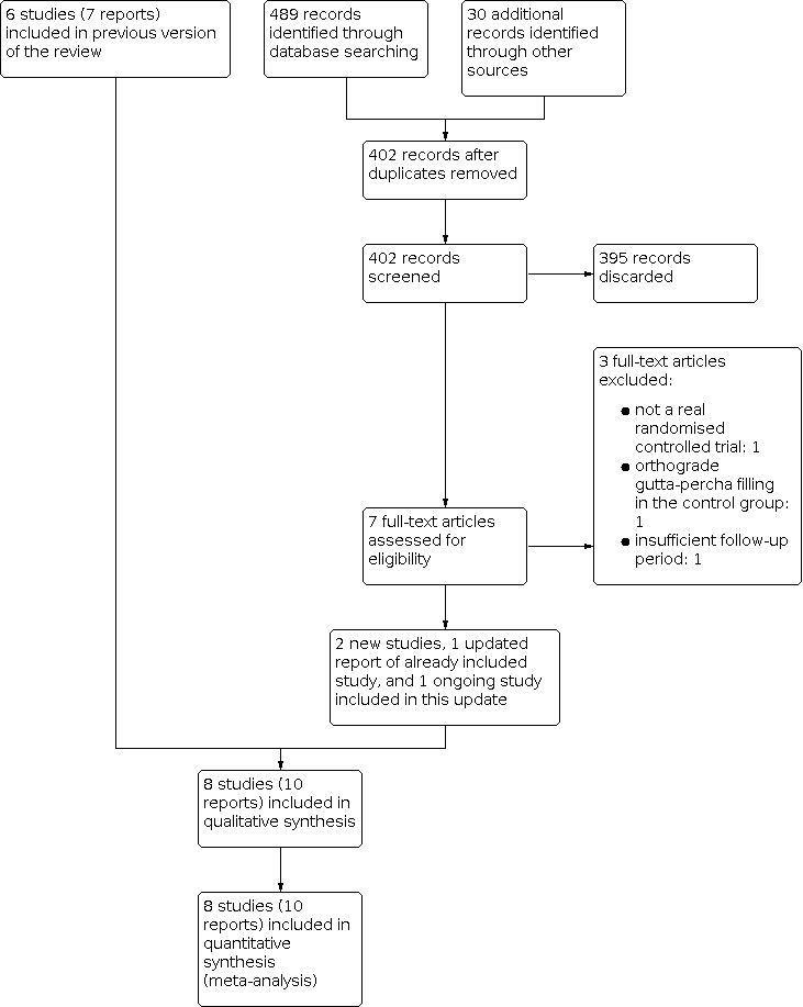
Study flow diagram.
Included studies
This review included eight randomised controlled trials (RCTs) (Chong 2003; Jensen 2002; Jesslen 1995; Kim 2016; Lindeboom 2005; Safi 2019; Wälivaara 2011; Zhou 2017), which were published between 1995 and 2019. The details of the included studies are listed in the Characteristics of included studies tables.
Characteristics of the trial designs and settings
All included studies used a parallel design. Five studies were conducted in Europe, one in America, and two in Asia.
Europe: United Kingdom (Chong 2003), Denmark (Jensen 2002), Sweden (Jesslen 1995; Wälivaara 2011), and the Netherlands (Lindeboom 2005).
America: USA (Safi 2019).
Three studies performed sample size calculations (Chong 2003; Kim 2016; Safi 2019). Chong 2003 calculated sample size assuming a 15% difference in success rate between the groups with the power of 80%, the significance level of 5%, and 20% attrition at the two‐year follow‐up. Also, Kim 2016 calculated the sample size according to the method that the power of the study of 80% was sufficient to detect on average a 10% difference in success rate between the groups at the 0.05 significance level (Walters 2004). The minimum sample size of Safi 2019 was determined based on a 20% mean difference in success rate between the groups and power 5 = of 0.80 and significance level of 0.05. Zhou 2017 seems to state that the sample size calculation was performed when in fact, the authors extrapolated the maximum number of patients they could enrol based on the dynamics of endodontic surgery at the Department of Conservative Dentistry, West China Hospital of Stomatology, while envisaging a response rate of 80%. So we decided to remove Zhou 2017 from the list of studies that performed sample size calculations. The remaining four studies did not mention sample size calculation at all (Jensen 2002; Jesslen 1995; Lindeboom 2005; Wälivaara 2011).
Six studies did not state their funding sources (Jensen 2002; Jesslen 1995; Lindeboom 2005; Safi 2019; Wälivaara 2011; Zhou 2017) and the remaining two studies stated that they received non‐industry funding (Chong 2003; Kim 2016).
Characteristics of the participants
This review involved 1399 participants and 1471 teeth.
| Studies | Targeted disease | Periodontal condition | Previous treatment | Age | Sex ratio (male/female) | Follow‐up period | Lost to follow‐up ratio |
| Chong 2003 | Teeth had clear periapical lesions and required retrograde filling | No serious periodontitis or apicomarginal communication | Root canal treatment before | Unclear | Unclear | 1 year and 2 years | < 10% |
| Jensen 2002 | Unclear | Mean age of 49 years | 48/86 | 1 year | < 10% | ||
| Jesslen 1995 | Unclear | Unclear | Unclear | Unclear | 1 year and 5 years | 10% to 20% | |
| Lindeboom 2005 | No serious periodontitis or apicomarginal communication | Root canal treatment before | 17 to 64 years | 33/57 | 1 year | 0% | |
| Kim 2016 | Unclear | Unclear | Unclear | 1 year and 4 years | 20% | ||
| Wälivaara 2011 | Unclear | Unclear | 65/99 | 1 year | < 10% | ||
| Zhou 2017 | Root canal treatment before | Unclear | Unclear | 1 year | > 30% | ||
| Safi 2019 | Previous endodontic treatment | Unclear | Unclear | at least 1 year | > 50% |
Characteristics of the interventions
The interventions used in the included studies were:
mineral trioxide aggregate (MTA);
intermediate restorative material (IRM);
super ethoxybenzoid acid (Super‐EBA);
dentine‐bonded resin composite;
glass ionomer cement;
amalgam;
root repair material (RRM).
These interventions were all used singly. We evaluated the following comparisons:
MTA versus IRM (Chong 2003; Lindeboom 2005);
MTA versus Super‐EBA (Kim 2016);
Super‐EBA versus IRM (Wälivaara 2011);
dentine‐bonded resin composite versus glass ionomer cement (Jensen 2002);
glass ionomer cement versus amalgam (Jesslen 1995);
The preparation of the root tip differed between studies. Three studies (Jensen 2002; Jesslen 1995; Zhou 2017) prepared the root tip with cone or diamond burs. Other studies (Chong 2003; Kim 2016; Lindeboom 2005; Wälivaara 2011) used ultrasonic apical preparation. Three studies (Kim 2016; Wälivaara 2011; Zhou 2017) used magnification equipment. The lengths of apical root resection were 2 to 3 mm (Jensen 2002; Kim 2016), 3 mm (Lindeboom 2005; Zhou 2017), and 3 to 4 mm (Wälivaara 2011). The details of the surgical procedure in Safi 2019 were not provided.
Characteristics of the outcomes
The primary outcome in our review was success rate which was assessed via clinical or radiological methods or a combination of the two. It was defined as no pain, pain on percussion or palpation, tenderness, increased tooth mobility, sinus tract formation or any other subjective discomfort; and no presence of apical absorption or no bony change compared to the baseline indicated treatment failure.
All included studies followed the definition of success rate using the specified criteria provided by Molven 1987 and Rud 1972 (Chong 2003; Jensen 2002; Kim 2016; Lindeboom 2005; Safi 2019; Wälivaara 2011; Zhou 2017) or Zetterqvist 1991 (Jesslen 1995). Via these criteria, treatment results were classified into four types:
complete healing;
incomplete or scar healing (improvement);
uncertain healing (no improvement);
unsatisfactory healing.
As described in these specified criteria, the rate of complete healing, incomplete or scar healing (improvement) were all considered as meeting the definition of success rate used in our review; the other two (uncertain healing (no improvement), unsatisfactory healing) were considered to be failures.
All included studies reported the success rate assessed via the combination of clinical and radiological methods. And only Jensen 2002 reported the success rate solely assessed by radiological methods.
Adverse events, secondary outcome of this review, were not assessed by any of the included studies.
Excluded studies
We have listed all the studies that were excluded and the reasons for their exclusion in the Characteristics of excluded studies tables.
We excluded three studies (Pantchev 2009; Rud 1991; Schwartz‐Arad 2003) because they were retrospective. We excluded von Arx 2012 because it was a cohort study; Wälivaara 2009 was excluded because it was a quasi‐RCT. Tang 2019 was excluded because it was not a real RCT. For Christiansen 2009, the control group did not have retrograde obturation with any material. For Kruse 2016, the control group received orthograde gutta‐percha filling. For Hou 2008, no radiological outcome was recorded. And for Platt 2004, the methods used to prepare the cavity differed in both randomised groups. Two studies (NCT04198298; Silva 2016) were excluded because the following‐up period was too short (only six months). We excluded four studies (Burstein 2001; Nordenram 1970; Pantschev 1994; Rud 1996) because the authors did not mention randomisation. We have tried to contact the study authors, but no response was obtained.
Risk of bias in included studies
All eight studies were assessed as being at high overall risk of bias (Figure 3; Figure 4). A detailed explanation is presented in the Characteristics of included studies table.
3.
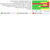
Risk of bias graph: review authors' judgements about each risk of bias item presented as percentages across all included studies.
4.
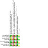
Risk of bias summary: review authors' judgements about each risk of bias item for each included study.
Allocation
Sequence generation
Seven out of the eight studies reported adequate methods of randomisation and were graded as at low risk of bias in this domain: Chong 2003; Lindeboom 2005; and Zhou 2017 used sealed envelopes randomly picked from a pack; Kim 2016 used a "minimization method" described by Pocock (Pocock 1983); Wälivaara 2011 allocated according to a randomisation table; Jensen 2002 used a computer‐generated random table; and Safi 2019 applied the University of Pennsylvania Web server for randomisation. Only Jesslen 1995 did not report a clear method and was graded as at unclear risk of bias.
Allocation concealment
Three studies (Chong 2003; Lindeboom 2005; Zhou 2017) used sealed envelopes, so that the allocation was adequately concealed, and were judged to be at low risk of bias for this domain. Safi 2019 used the University of Pennsylvania Web server which also allowed for adequate allocation concealment. Three studies had unclear allocation concealment and were therefore graded as at unclear risk of bias (Jensen 2002; Jesslen 1995; Kim 2016). One study was judged at high risk of bias (Wälivaara 2011), the study author confirming via correspondence that the allocation process was not concealed.
Blinding
Blinding of participants and personnel (performance bias)
It was not possible to blind the surgeons, so all the included studies were ranked as at high risk in terms of performance bias. In Jensen 2002; Kim 2016; Lindeboom 2005; and Wälivaara 2011 surgeons were not blinded according to authors' correspondence. Safi 2019 mentioned that the operator was aware what he or she was using after the material was dispensed to him or her, and therefore was also ranked as at high risk of performance bias. The other studies (Chong 2003; Jesslen 1995; Zhou 2017) did not mention the blinding of participants and personnel so they were ranked as at high risk of performance bias.
Blinding of outcome assessment (detection bias)
Five studies had low risk of detection bias (Chong 2003; Kim 2016; Lindeboom 2005; Safi 2019; Zhou 2017) as they clearly reported the blinding of the outcome assessors. Two studies had unclear risk (Jesslen 1995; Wälivaara 2011) and one had high risk (Jensen 2002) as the author replied that the assessors were not blinded to the treatment.
Although some studies reported the blinding of outcome assessment, different materials may be identifiable via radiological assessment and this might influence the risk of bias.
Incomplete outcome data
In Chong 2003; Jesslen 1995; Kim 2016; Safi 2019; and Zhou 2017 too many participants were lost during follow‐up and the study authors did not do an intention‐to‐treat (ITT) analysis.
Selective reporting
Selective data reporting was not detected in the studies included in the review as they all fully reported the outcomes they stated in the methods section.
Other potential sources of bias
In Chong 2003; Jesslen 1995; Safi 2019; and Zhou 2017 the issue of whether baseline demographic characteristics were comparable was not clearly reported and therefore we classified them as at unclear risk of other bias. For the other four studies, there was no other potential source of bias detected.
Effects of interventions
See: Table 1; Table 2; Table 3; Table 4; Table 5; Table 6
Generally, each participant was considered the unit of analysis in this review, or if that was not possible, each tooth. But for success rate evaluated solely by radiological methods, the root could be considered the unit of analysis if clearly stated.
Comparison 1: MTA versus IRM
Both Chong 2003 and Lindeboom 2005 compared mineral trioxide aggregate (MTA) versus intermediate restorative material (IRM). There was no significant clinical heterogeneity, and we pooled their data. The two studies were assessed as at high risk of bias.
Success rate
Both studies reported this outcome. Chong 2003 described this in one‐ and two‐year follow‐ups, and Lindeboom 2005 reported only the one‐year follow‐up outcome.
One‐year outcome
Data from the two studies at high risk of bias with 222 teeth were pooled, with 114 teeth in the MTA group and 108 teeth in the IRM group. Both studies in this comparison used a sample with only one tooth per patient. Also, in that context, there is no unit‐of‐analysis error due to clustering of teeth within a patient, and the results could be presented as per tooth or per patient. To describe the results more accurately, we chose the tooth as the unit of analysis. Results showed there may be little to no effect of MTA compared to IRM on success rate at one year (risk ratio (RR) 1.09, 95% confidence intervals (CI) 0.97 to 1.22; I2 = 0%; 2 studies; 222 teeth; very low‐certainty evidence; Analysis 1.1), but the evidence is very uncertain due to imprecision and high risk of bias.
1.1. Analysis.
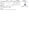
Comparison 1: MTA versus IRM, Outcome 1: Success rate ‐ 1‐year outcome (tooth as unit of analysis)
Two‐year outcome
Only one study (Chong 2003) assessed as at high risk of bias was included in this analysis. As explained above, we chose the tooth as the unit of analysis. In this study, 45 patients were missing at the 2‐year review, among whom 22 patients were healed at 12 months. Thus the 22 patients were regarded as the complete healing category by the authors, 8 in the IRM group and 14 in the MTA group. Evidence showed there may be little to no effect of MTA compared to IRM on success rate at two years (RR 1.05, 95% CI 0.92 to 1.20; 1 study; 108 teeth; Analysis 1.2).
1.2. Analysis.
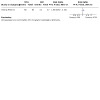
Comparison 1: MTA versus IRM, Outcome 2: Success rate ‐ 2‐year outcome (tooth as unit of analysis)
Adverse events
Adverse events were not assessed by the included studies.
Comparison 2: MTA versus Super‐EBA
Only Kim 2016 compared MTA with super ethoxybenzoic acid (Super‐EBA). The study was assessed as at high risk of bias. We regarded each tooth as the unit of analysis.
Success rate
One‐year outcome
After one year, 192 teeth were followed up (90 in the MTA group and 102 in the Super‐EBA group). In the MTA group, 86 teeth were successfully treated, while the Super‐EBA group had 95 teeth. The detailed data were recorded in the early study by Song et al in 2012. Results showed there may be little to no effect of MTA compared to Super‐EBA on success rate at one year (RR 1.03, 95% CI 0.96 to 1.10; 1 study; 192 teeth; very low‐certainty evidence; Analysis 2.1). The evidence is very uncertain due to high risk of bias and imprecision.
2.1. Analysis.
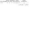
Comparison 2: MTA versus Super‐EBA, Outcome 1: Success rate ‐ 1‐year outcome (tooth as unit of analysis)
Four‐year outcome
In the fourth follow‐up year, 182 teeth were included (83 in the MTA group and 99 in the Super‐EBA group). There were 76 teeth in the MTA group with complete or partial healing. In the Super‐EBA group, 89 teeth were successfully treated. Results showed there may be little to no effect of MTA compared to Super‐EBA on success rate at four years (RR 1.02, 95% CI 0.93 to 1.12; 1 study; 182 teeth; Analysis 2.2).
2.2. Analysis.
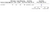
Comparison 2: MTA versus Super‐EBA, Outcome 2: Success rate ‐ 4‐year outcome (tooth as unit of analysis)
Adverse events
Adverse events were not assessed by the included studies.
Comparison 3: Super‐EBA versus IRM
Only Wälivaara 2011 compared Super‐EBA with IRM. The study was assessed as at high risk of bias. We regarded each tooth as the unit of analysis.
Success rate
One‐year outcome
At the one‐year follow‐up, 194 teeth were considered (98 in the Super‐EBA group and 96 in the IRM group). The evidence is very uncertain about the effect of Super‐EBA compared with IRM on success rate at 1 year, with results indicating Super‐EBA may reduce or have no effect on success rate (RR 0.90, 95% CI 0.80 to 1.01; 1 study; 194 teeth; very low‐certainty evidence; Analysis 3.1). The certainty of the evidence was very low because of imprecision and high risk of bias.
3.1. Analysis.
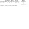
Comparison 3: Super‐EBA versus IRM, Outcome 1: Success rate ‐ 1‐year outcome (tooth as unit of analysis)
Adverse events
Adverse events were not assessed by the included studies.
Comparison 4: dentine‐bonded resin composite versus glass ionomer cement
Only Jensen 2002 compared dentine‐bonded resin composite with glass ionomer cement. The study was assessed as at high risk of bias.
Success rate
One‐year outcome
In this comparison, we regarded each tooth as unit of analysis because only one tooth selected at random was included in the material in this study. Both per‐protocol (PP) and intention‐to‐treat (ITT) analyses ('worst‐case scenario' analysis: all lost to follow‐up participants in the intervention group were considered as failed cases) were adopted.
Within the dentine‐bonded resin composite group (60 teeth), 44 teeth were successfully healed. In the glass ionomer cement group (62 teeth), only 19 teeth were considered as clinical and radiological success. Compared to glass ionomer cement, dentine‐bonded resin composite may increase the success rate of the treatment at one year (RR 2.39, 95% CI 1.60 to 3.59; 1 study; 122 teeth; very low‐certainty evidence; Analysis 4.1), but the evidence is very uncertain due to imprecision and high risk of bias.
4.1. Analysis.
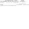
Comparison 4: Dentine‐bonded resin composite versus glass ionomer cement, Outcome 1: Success rate ‐ 1‐year outcome PP analysis (tooth as unit of analysis)
For the ITT analysis we used the data we extracted from the article ourselves, and there was no presentation in the original text. The ITT analysis results also supported that the resin may result in an increase in the opportunity of successful treatment of a tooth at one year compared to glass ionomer cement (RR 1.83, 95% CI 1.27 to 2.64; 1 study; 134 teeth; Analysis 4.2) with 67 teeth in each group.
4.2. Analysis.
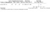
Comparison 4: Dentine‐bonded resin composite versus glass ionomer cement, Outcome 2: Success rate ‐ 1‐year outcome ITT analysis (tooth as unit of analysis)
We also collected data using the root of the teeth as the unit of analysis. However, instead of using combined methods for evaluation, only the radiological method was adopted in this case. As there was a clear description of the participants lost to follow‐up, both PP and ITT analyses ('worst‐case scenario' analysis) were performed. Results from the PP analysis showed that, compared to glass ionomer cement, dentine‐bonded resin composite may increase the success rate of the treatment at one year (RR 1.59, 95% CI 1.20 to 2.09; 1 study; 127 roots; very low‐certainty evidence; Analysis 4.3). However, the evidence is very uncertain due to imprecision and high risk of bias.
4.3. Analysis.
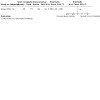
Comparison 4: Dentine‐bonded resin composite versus glass ionomer cement, Outcome 3: Success rate ‐ 1‐year outcome PP analysis (root as unit of analysis)
A total of 178 roots included were treated in Jensen 2002, of which 127 roots could be evaluated radiographically for PP analysis. 31 roots showed either signs of a loose retrograde filling or were reoperated before one year, which were regarded as failures in our analysis. The remaining 20 roots, 12 roots in the dentine‐bonded resin composite group and 8 roots in the glass ionomer cement group, were the real missing roots for ITT analysis. The ITT analysis results also supported that compared to glass ionomer cement, dentine‐bonded resin composite may increase the opportunity to successfully treat a root at one year (RR 1.62, 95% CI 1.22 to 2.16; 1 study; 178 roots; Analysis 4.4).
4.4. Analysis.
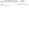
Comparison 4: Dentine‐bonded resin composite versus glass ionomer cement, Outcome 4: Success rate ‐ 1‐year outcome ITT analysis (root as unit of analysis)
Adverse events
Adverse events were not assessed by the included studies.
Comparison 5: glass ionomer cement versus amalgam
Only Jesslen 1995 compared glass ionomer cement with amalgam. The study was assessed as at high risk of bias. We regarded each tooth as the unit of analysis.
Success rate
One‐year outcome
After the first year, 105 teeth were followed up (53 in the glass ionomer cement group and 52 in the amalgam group). Each group had 47 teeth that had complete or partial healing and were therefore considered a treatment success. The evidence is very uncertain about the effect of glass ionomer cement compared with amalgam on success rate at one year, with results indicating glass ionomer cement may reduce or have no effect on success rate (RR 0.98, 95% CI 0.86 to 1.12; 1 study; 105 teeth; very low‐certainty evidence; Analysis 5.1). The certainty of the evidence was very low because of imprecision and high risk of bias.
5.1. Analysis.
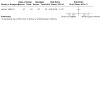
Comparison 5: Glass ionomer cement versus amalgam, Outcome 1: Success rate ‐ 1‐year outcome (tooth as unit of analysis)
Five‐year outcome
In the fifth follow‐up year, 82 teeth were included (41 in the glass ionomer group and 41 in the amalgam group). There were 35 teeth in each group with complete or partial healing. The evidence suggests that there may be little to no effect of glass ionomer cement compared to amalgam on success rate at five years (RR 1.00, 95% CI 0.84 to 1.20; 1 study; 82 teeth; Analysis 5.2).
5.2. Analysis.
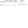
Comparison 5: Glass ionomer cement versus amalgam, Outcome 2: Success rate ‐ 5‐year outcome (tooth as unit of analysis)
Adverse events
Adverse events were not assessed by the included studies.
Comparison 6: MTA versus RRM
Both Zhou 2017 and Safi 2019 compared MTA with root repair material (RRM). Both studies were at high risk of bias. We regarded each tooth as the unit of analysis.
Success rate
One‐year outcome
At the one‐year follow‐up, 278 teeth were included in total (144 in the MTA group and 134 in the RRM group). In the MTA group, 130 teeth were considered complete or partial healing, while the RRM group had 125 teeth. Evidence suggests that there may be little to no effect of MTA compared to RRM on success rate at one year (RR 1.00, 95% CI 0.94 to 1.07; I2 = 0%; 2 studies; 278 teeth; very low‐certainty evidence; Analysis 6.1), but the evidence is very uncertain due to imprecision and high risk of bias.
6.1. Analysis.
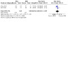
Comparison 6: MTA versus RRM, Outcome 1: Success rate ‐ 1‐year outcome (tooth as unit of analysis)
Adverse events
Adverse events were not assessed by the included studies.
Discussion
Summary of main results
The aim of this review was to compare the outcomes of different retrograde filling materials. Eight studies (Chong 2003; Jensen 2002; Jesslen 1995; Kim 2016; Lindeboom 2005; Safi 2019; Wälivaara 2011; Zhou 2017) with six comparisons of retrograde filling materials were finally considered to be eligible for inclusion in this systematic review. We assessed the certainty of the body of evidence using GRADE (Atkins 2004) and created summary of findings tables for the main comparisons where we were able to perform quantitative analysis as follows:
mineral trioxide aggregate (MTA) versus intermediate restorative material (IRM) (Table 1)
MTA versus super ethoxybenzoic acid (Super‐EBA) (Table 2)
Super‐EBA versus IRM (Table 3)
dentine‐bonded resin composite versus glass ionomer cement (Table 4)
glass ionomer cement versus amalgam (Table 5)
MTA versus root repair material (RRM) (Table 6).
There was little pooled data and very little evidence for each comparison. Results suggested that there may be little to no effect of MTA compared to IRM on success rate at one year, but the evidence from two studies is very uncertain due to imprecision and high risk of bias. The long setting time (165 ± 5 minutes) is potentially a problem in this type of surgery. Many investigations have been performed to overcome it, such as using a chelating agent or sodium phosphate dibasic (Na2HPO4) setting accelerator (Ber 2007; Huang 2008). Some new and potentially promising materials have been available for root‐end filling, such as bioceramics (Damas 2011) and biodentine (Soundappan 2014), but more clinical studies are needed to testify their properties. Evidence also suggests that there may be little to no effect of MTA compared to RRM on success rate at one year, but the evidence from two studies is very uncertain due to imprecision and high risk of bias. In Zhou 2017, iRoot BP Plus root repair material (BP‐RRM) was defined as control group. Bioceramic root repair material (BCRRM) was used to compared with MTA in Safi 2019. Because BP‐RRM and BCRRM are similar, we pooled data from these two studies.
The remaining results were based on single studies. There was insufficient evidence, from these single studies (at high risk of bias and mostly with small sample sizes), to determine differences on success rate at one year between the following comparisons: MTA versus Super‐EBA, Super‐EBA versus IRM, dentine‐bonded resin composite versus glass ionomer cement, and glass ionomer cement versus amalgam. The certainty of the evidence was very low for all these comparisons because of imprecision and high risk of bias.
Adverse events were not assessed by any of the included studies.
Overall completeness and applicability of evidence
One of the studies included in the review was conducted in the UK (Chong 2003), one in Denmark (Jensen 2002), two in Sweden (Jesslen 1995; Wälivaara 2011), one in South Korea (Kim 2016), one in China (Zhou 2017), one in the USA (Safi 2019), and one in the Netherlands (Lindeboom 2005). 1399 participants with 1471 teeth were randomised. The participants were of different gender and age (the youngest reported was 17 (Lindeboom 2005) and the oldest was 64 (Lindeboom 2005)). The studies included different types of teeth. The applicability of the results could be adopted to different demographics of patients and different types of teeth.
The detail of surgical procedure was not provided in Safi 2019. The rest of studies followed a rigid surgical procedure. The procedures were of little difference except for some detail in the root resection and form of cavity. The lengths of apical root resection were 2 to 3 mm (Jensen 2002; Kim 2016), 3 mm (Lindeboom 2005; Zhou 2017), and 3 to 4 mm (Wälivaara 2011). There was no complete agreement on how much of the root should be resected to satisfy biological principles. Gilheany 1994 suggested that at least 2 mm be removed to minimise bacterial leakage from the canals. An anatomical study of the root apex showed that at least 3 mm of the root‐end must be removed to reduce 98% of apical ramifications and 93% of lateral canals (Kim 2001). Besides the length of resection, the choice of root‐end bevel angle was still in question. Traditionally, the bevel angle which provided best access and visibility was used. But the bevel may open up many channels of communication between the infected canal system and surrounding tissue, allowing the intradental infection to create persistent inflammation. Recent studies indicate that a right angle to the long axis of the root is preferable. In the studies included in this review, only one adopted such a right angle (Chong 2003). Zhou 2017 conducted resection without a bevel. In the other five studies, a slightly oblique resection of the root was performed. Jensen 2002 resected at a mean angle of 35 degrees, Lindeboom 2005 10 to 25 degrees, and Kim 2016 had a bevel angle of 0 to 10 degrees.
Recently the use of a microscope has become popular (Christiansen 2009). In the studies within this review, Kim 2016 and Zhou 2017 used a microscope and in two studies (Lindeboom 2005; Wälivaara 2011) the dental surgeon used magnification loupes to acquire a better visual field. Following root resection, cavities were prepared in two ways: four studies used ultrasonic equipment while three used a traditional bur. Jensen 2002 prepared slightly concave cavities, and Jesslen 1995 prepared box‐type cavities. The depth of the cavity was 2 to 3 mm in Lindeboom 2005, 3 mm in Kim 2016 and Wälivaara 2011. The condition of the root‐end varied. The modern concept of an ideal root‐end preparation is defined as a Class I cavity at least 3 mm into the root dentine, with walls parallel to and coincident with the anatomic outline of the root canal space, which would achieve the aim of removing the intracanal filling material and associated irritants so as to create a cavity that could be properly filled. From this description, we could consider that the results of the systematic review may not be seriously influenced by heterogeneity of performance of the surgery.
Quality of the evidence
The body of evidence that we identified does not allow for any robust conclusions about which retrograde filling material is best. Eight studies, which analysed a total of 1399 participants (1471 teeth), were included. All the studies had a high risk of bias. All of the eight pieces of evidence produced from the studies were of very low certainty according to GRADE (Petrisor 2006). When such risk of bias issues were considered alongside the fact that the studies in each comparison/outcome were either single small studies (leading to serious imprecision) or had 95% confidence intervals that prevented the intervention being favoured over the control, the evidence was rated very low certainty. These GRADE ratings can be interpreted as indicating that there is a lack of confidence in the effect estimates and further research is highly likely to change the estimates, and our confidence in them.
Potential biases in the review process
To discover as many relevant studies as possible, articles were identified irrespective of publication language, status, and date through electronic searches and handsearching. The reference lists from each identified article, reviews and related textbooks were also searched. However, we still failed to acquire the data from a potentially relevant study, entitled '18‐month clinical trial of endodontic surgical retrofilling materials' which was presented at the American Association of Endodontists' 58th Annual Session. We have tried all means to contact the author and the American Association of Endodontists, but no reply was received. Another four potentially eligible studies (Burstein 2001; Nordenram 1970; Pantschev 1994; Rud 1996) were excluded because their study type could not be determined from the reports and the authors failed to reply to our contact.
Agreements and disagreements with other studies or reviews
To compare marginal adaptation of MTA with other filling materials in root‐end cavities, Selen Kucukkaya Eren and his colleagues searched five databases and included 20 studies, which were all of in vitro design (Kucukkaya Eren 2019). Since the design of included studies varied greatly, the authors only described the results qualitatively. Among the included studies, seven of them showed a better performance of MTA compared to other materials, while two associated it with worse marginal adaptation. The rest of the studies found no significant difference between MTA and other filling materials. They concluded that MTA performed better than other materials in marginal adaptation. However, we could not determine the benefits of MTA when compared to any other material. Results from in vitro studies should also be interpreted with caution.
Another published systematic review (Tang 2010) compared the clinical outcomes of MTA used as root‐end filling with other materials in endodontic surgery. They searched studies conducted on human teeth in vivo, regardless of whether they were prospective studies. Five studies, two that compared MTA with IRM, one that compared MTA with gutta‐percha, and two that compared MTA with amalgam, were selected. The analysis suggested that MTA as a root‐end filling is better than amalgam but similar to IRM, which were similar to the results indicated in our review. However, the authors of Tang 2010 had included some studies which seemed non‐existent (we have tried to retrieve the studies they included and discovered at least two studies to which they referred did not actually exist in the corresponding issues of those journals, and no studies with similar titles could be found).
Richard Niederman and his colleagues have carried out two systematic reviews focused on retrograde filling materials. The first one (Niederman 2003) was conducted to identify randomised controlled trials (RCTs) and controlled clinical trials (CCTs), cohort studies and case‐control studies, which were conducted on humans, in vivo. The languages were limited to English, German, and French. Only two RCTs (Jesslen 1995; Zetterqvist 1991), which were also analysed in our review, were included in this study. They concluded that glass ionomer cement is almost as effective as amalgam. Furthermore, the other six CCTs and six case‐control studies selected in Niederman's review indicated that EBA cement, composite with Gluma and gold leaf, as well as orthograde gutta‐percha, may be more effective than retrograde amalgam filling. The second review (Theodosopoulou 2005) tested the characteristics of retrograde filling materials in vitro. 34 studies met all their inclusion and validity criteria. The results indicated that, beyond 10 days in vitro, the most effective retrofilling materials, when measured by dye/ink penetration were: composites, followed by glass ionomer cement, amalgam, orthograde gutta‐percha, and EBA. The results of these in vitro studies are not congruent with in vivo study results, suggesting a need to re‐evaluate the clinical validity and importance of in vitro studies.
Authors' conclusions
Implications for practice.
Based on the present limited evidence, we do not have sufficient evidence to determine the benefits of any one material over another for retrograde filling in root canal therapy.
Implications for research.
The present results call for further research. We hope future studies could address and answer the following issues.
Participants: studies with a large number of participants from different races.
Intervention and comparison: trials focusing on the materials considered in this systematic review are still needed, and trials using new materials are also required. Many investigations have tried to use a chelating agent or accelerator to overcome the main drawback of mineral trioxide aggregate (MTA), its long setting time, which is also worth more research.
Follow‐up: increasing the follow‐up period to observe the long‐term effects and safety.
To improve the quality of future evidence, we recommend blinding the outcome assessors. Most studies only have the participants and the statistical assessor blinded. But Christiansen 2009 figured out an easy method to blind the radiograph observers. They exported radiographs from the Digora system in TIFF format to Adobe Photoshop format, masking the apical root filling with grey patches. Future studies could make use of this methodology.
What's new
| Date | Event | Description |
|---|---|---|
| 1 July 2021 | New citation required but conclusions have not changed | Review update includes 2 new trials bringing the total to 8 included studies. Conclusions remain the same. New co‐authors |
| 1 July 2021 | New search has been performed | Searches updated 21 April 2021 |
History
Protocol first published: Issue 4, 2005 Review first published: Issue 12, 2016
| Date | Event | Description |
|---|---|---|
| 5 September 2008 | Amended | Converted to new review format. |
Acknowledgements
Our thanks go to the Cochrane Oral Health editorial team and external peer reviewers for their help in conducting this systematic review. We would like to thank in particular Anne Littlewood, Information Specialist at Cochrane Oral Health, for developing the electronic search strategy and running the electronic searches. We would also like to thank Luisa Fernandez Mauleffinch (Managing Editor and Copy Editor, Cochrane Oral Health), Professor Ana Jeroncic (University of Split School of Medicine, Croatia), Professor Alison Qualtrough, and Philip Riley (Editor, Cochrane Oral Health), for their kind help and guidance in preparing and revising the review. We thank Dr Liuhe Jia and Wenwen Liu who participated in the preparing and writing of the previous version.
The review authors also send their thanks to Dr Raphael Freitas de Souza and Dr Anette Bluemle for their help in screening the titles and abstracts which were not in English or Chinese. Great thanks should also be sent to Dr Jerome AH Lindeboom, Dr Simon Storgård Jensen, Dr Minju Song, and Dr Dan‐Åke Wälivaara, who were the initial investigators of four randomised trials included in this systematic review, for their assistance in providing valuable information about the studies. And thanks to Dr Chenyang Xiang, Wenhang Dong, Qiushi Wang, Zhaoyang Ban, and Feng Li from West China College of Stomatology, Sichuan University, for their assistance with handsearching.
Appendices
Appendix 1. Cochrane Oral Health's Trials Register search strategy
Cochrane Oral Health's Trials Register is available via the Cochrane Register of Studies. For information on how the register is compiled, see oralhealth.cochrane.org/trials
From March 2015, searches of Cochrane Oral Health's Trials Register for this review were undertaken using the Cochrane Register of Studies and the search strategy below:
1 ((apicoectom* or apicectom* or "root canal" or periapical* or periradicular* or endodont* or apical*):ti,ab) AND (INREGISTER) 2 ((retrograd* or retrofill* or retro‐fill* or retroseal* or retro‐seal* or "apical* seal*" or "apical* prepar*" or retroprepar*):ti,ab) AND (INREGISTER) 3 (#1 and #2) AND (INREGISTER)
Previous searches of the Register were undertaken using the Procite software and the search strategy below:
((apicoectom* or apicectom* or "root canal" or periapical* or periradicular* or endodont* or apical*) AND (retrograd* or retrofill* or retro‐fill* or retroseal* or retro‐seal* or "apical* seal*" or "apical* prepar*" or retroprepar*))
Appendix 2. Cochrane Central Register of Controlled Trials (CENTRAL) search strategy
#1 MeSH descriptor Apicoectomy this term only #2 MeSH descriptor Root Canal Therapy explode all trees #3 (apicect* in All Text or apicoect* in All Text) #4 ("root canal therapy" in All Text or ("root canal*" in All Text near/6 treatment in All Text) or ("root canal*" in All Text near/6 filling* in All Text) or ("root canal*" in All Text near/6 restor* in All Text) ) #5 MeSH descriptor Tooth apex this term only #6 MeSH descriptor Periapical diseases explode all trees #7 endodontic* in All Text #8 ((apex in All Text or apical* in All Text) and (surgery in All Text or surgical in All Text)) #9 (periapical* in All Text or periradicular in All Text) #10 MeSH descriptor Retrograde obturation explode all trees #11 ((retrograde in All Text near/6 fill* in All Text) or retrofill* in All Text or retro‐fill* in All Text or retroseal* in All Text or retro‐seal* in All Text or (retro* in All Text near/6 seal* in All Text) or retro‐seal* in All Text or (root next end in All Text near/6 fill* in All Text) or (root‐end in All Text near/6 fill* in All Text) or (root next end in All Text near/6 seal* in All Text) or (root‐end in All Text near/6 seal* in All Text) or apical* next seal* in All Text or (apical* in All Text near/6 prepar* in All Text) or retroprepar* in All Text or retro‐prepar* in All Text or retrograd* in All Text or (reverse in All Text near/6 obturat* in All Text) or (reverse in All Text near/6 fill* in All Text)) #12 (#10 or #11) #13 (#1 or #2 or #3 or #4 or #5 or #6 or #7 or #8 or #9) #14 (#12 and #13)
Appendix 3. MEDLINE Ovid search strategy
Apicoectomy/
exp Root Canal Therapy/
(apicect$ or apicoect$).mp.
("root canal$ therap$" or ("root canal$" adj6 treatment$) or ("root canal$" adj6 filling$) or ("root canal$" adj6 restor$)).mp.
Tooth apex/
exp Periapical diseases/
(periapical$ or periradicular).mp.
endodont$.mp.
((apex or apical$) and (surgery or surgical$)).mp.
or/1‐9
((retrograde adj6 fill$) or retrofill$ or retro‐fill$ or retroseal$ or retro‐seal$ or (retro adj6 seal$) or ("root end" adj6 fill$) or (root‐end adj6 fill$) or ("root end" adj6 seal$) or (root‐end adj6 seal$) or "apical$ seal$" or "apical$ prepar$" or retroprepar$ or retrograd$ or (reverse adj6 obturat$) or (reverse adj6 fill$)).mp.
Retrograde obturation/
11 or 12
10 and 13
The above subject search was linked to the Cochrane Highly Sensitive Search Strategy (CHSSS) for identifying randomised trials (RCTs) in MEDLINE: sensitivity maximising version (2009 revision) as referenced in (Lefebvre 2021).
randomized controlled trial.pt.
controlled clinical trial.pt.
randomized.ab.
placebo.ab.
drug therapy.fs.
randomly.ab.
trial.ab.
groups.ab.
or/1‐8
exp animals/ not humans.sh.
9 not 10
Appendix 4. Embase Ovid search strategy
Endodontics/
exp Root Canal Filling Material/
(apicect$ or apicoect$).mp.
((root adj canal adj therapy) or (root adj canal$ adj6 treatment) or (root adj canal$ adj6 filling$) or (root canal$ adj6 restor$)).mp.
Tooth Root Canal/ or Tooth root/
Tooth Periapical Disease/
(periapical$ or periradicular or peri‐radicular).mp.
endodontic$.mp.
(apex or apical$).mp.
((retrograde adj6 fill$) or (retrograde adj obturat$) or retrofill$ or retro‐fill$ or retroseal$ or retro‐seal$ or (retro$ adj6 seal$) or retro‐seal$ or (root adj end adj6 fill$) or (root‐end adj6 fill$) or (root adj end adj6 seal$) or (root‐end adj6 seal$) or apical$ next seal$ or apical$ near prepar$ or retroprepar$ or retro‐prepar$ or retrograd$ or (reverse adj6 obturat$) or (reverse adj6 fill$)).mp.
or/1‐9
10 and 11
The above subject search was linked with the highly sensitive search strategy designed by Cochrane for identifying randomised controlled trials and controlled clinical trials in Embase (as described in Lefebvre 2021, box 3e).
Randomized controlled trial/
Controlled clinical study/
random$.ti,ab.
randomization/
intermethod comparison/
placebo.ti,ab.
(compare or compared or comparison).ti.
((evaluated or evaluate or evaluating or assessed or assess) and (compare or compared or comparing or comparison)).ab.
(open adj label).ti,ab.
((double or single or doubly or singly) adj (blind or blinded or blindly)).ti,ab.
double blind procedure/
parallel group$1.ti,ab.
(crossover or cross over).ti,ab.
((assign$ or match or matched or allocation) adj5 (alternate or group$1 or intervention$1 or patient$1 or subject$1 or participant$1)).ti,ab.
(assigned or allocated).ti,ab.
(controlled adj7 (study or design or trial)).ti,ab.
(volunteer or volunteers).ti,ab.
human experiment/
trial.ti.
or/1‐19
random$ adj sampl$ adj7 ("cross section$" or questionnaire$1 or survey$ or database$1)).ti,ab. not (comparative study/ or controlled study/ or randomi?ed controlled.ti,ab. or randomly assigned.ti,ab.)
Cross‐sectional study/ not (randomized controlled trial/ or controlled clinical study/ or controlled study/ or randomi?ed controlled.ti,ab. or control group$1.ti,ab.)
(((case adj control$) and random$) not randomi?ed controlled).ti,ab.
(Systematic review not (trial or study)).ti.
(nonrandom$ not random$).ti,ab.
"Random field$".ti,ab.
(random cluster adj3 sampl$).ti,ab.
(review.ab. and review.pt.) not trial.ti.
"we searched".ab. and (review.ti. or review.pt.)
"update review".ab.
(databases adj4 searched).ab.
(rat or rats or mouse or mice or swine or porcine or murine or sheep or lambs or pigs or piglets or rabbit or rabbits or cat or cats or dog or dogs or cattle or bovine or monkey or monkeys or trout or marmoset$1).ti. and animal experiment/
Animal experiment/ not (human experiment/ or human/)
or/21‐33
20 not 34
Appendix 5. LILACS BIREME Virtual Health Library search strategy
Mh apicoectomy or Mh root canal therapy or apicoect$ or apicect$ or "root canal$ therap$" or "root canal$ treat$" or "root canal fill$" or "root canal restror$" or Mh Tooth apex or Mh Periapical diseases or periapical$ or periradicular or endodont$)) [Words] and (Mh Retrograde obturation or (retrograde and fill$) or retrofill$ or retro‐fill$ or "retro seal$" or "retro‐seal$" or retroseal$ or "root end fill$" or "root‐end fill$" or "root end seal$" or "root‐end seal$" or "apical seal$" or "apical prepar$" or retroprepar$ or retrograd$ or (reverse and obturat$ or (reverse and fill$))
The above subject search was linked to the Brazilian Cochrane Center filter for LILACs via BIREME:
Pt randomized controlled trial OR Pt controlled clinical trial OR Mh randomized controlled trials OR Mh random allocation OR Mh double‐blind method OR Mh single‐blind method) AND NOT (Ct animal AND NOT (Ct human and Ct animal)) OR (Pt clinical trial OR Ex E05.318.760.535$ OR (Tw clin$ AND (Tw trial$ OR Tw ensa$ OR Tw estud$ OR Tw experim$ OR Tw investiga$)) OR ((Tw singl$ OR Tw simple$ OR Tw doubl$ OR Tw doble$ OR Tw duplo$ OR Tw trebl$ OR Tw trip$) AND (Tw blind$ OR Tw cego$ OR Tw ciego$ OR Tw mask$ OR Tw mascar$)) OR Mh placebos OR Tw placebo$ OR (Tw random$ OR Tw randon$ OR Tw casual$ OR Tw acaso$ OR Tw azar OR Tw aleator$) OR Mh research design) AND NOT (Ct animal AND NOT (Ct human and Ct animal)) OR (Ct comparative study OR Ex E05.337$ OR Mh follow‐up studies OR Mh prospective studies OR Tw control$ OR Tw prospectiv$ OR Tw volunt$ OR Tw volunteer$) AND NOT (Ct animal AND NOT (Ct human and Ct animal)))and not (Ct ANIMAL AND NOT (Ct HUMAN and Ct ANIMAL)))
Appendix 6. OpenSIGLE search strategy
(("root canal") AND (fill* or restor*? or therap* or treatment*))
((endodont*) AND (fill* or restor*? or therap* or treatment*))
((retrograd*) AND (fill* or restor* or seal* or prepar*))
apicectom* or apicoectom*
Appendix 7. Chinese BioMedical Literature Database search strategy
"根尖手术"[常用字段:智能] OR "倒根充"[常用字段:智能]
Appendix 8. VIP search strategy
M=根尖手术 OR M=倒根充
Appendix 9. China National Knowledge Infrastructure search strategy
(主题:根尖手术)OR(主题:倒根充)
Appendix 10. US National Institutes of Health Ongoing Trials Register (ClinicalTrials.gov) and World Health Organization International Clinical Trials Registry Platform search strategies
retrograde AND filling
retrograde AND seal
retrograde AND sealant
Appendix 11. Sciencepaper Online search strategy
主题=根尖手术 或 主题=倒根充
Data and analyses
Comparison 1. MTA versus IRM.
| Outcome or subgroup title | No. of studies | No. of participants | Statistical method | Effect size |
|---|---|---|---|---|
| 1.1 Success rate ‐ 1‐year outcome (tooth as unit of analysis) | 2 | 222 | Risk Ratio (M‐H, Fixed, 95% CI) | 1.09 [0.97, 1.22] |
| 1.2 Success rate ‐ 2‐year outcome (tooth as unit of analysis) | 1 | Risk Ratio (M‐H, Fixed, 95% CI) | Totals not selected |
Comparison 2. MTA versus Super‐EBA.
| Outcome or subgroup title | No. of studies | No. of participants | Statistical method | Effect size |
|---|---|---|---|---|
| 2.1 Success rate ‐ 1‐year outcome (tooth as unit of analysis) | 1 | Risk Ratio (M‐H, Fixed, 95% CI) | Totals not selected | |
| 2.2 Success rate ‐ 4‐year outcome (tooth as unit of analysis) | 1 | Risk Ratio (M‐H, Fixed, 95% CI) | Totals not selected |
Comparison 3. Super‐EBA versus IRM.
| Outcome or subgroup title | No. of studies | No. of participants | Statistical method | Effect size |
|---|---|---|---|---|
| 3.1 Success rate ‐ 1‐year outcome (tooth as unit of analysis) | 1 | Risk Ratio (M‐H, Fixed, 95% CI) | Totals not selected |
Comparison 4. Dentine‐bonded resin composite versus glass ionomer cement.
| Outcome or subgroup title | No. of studies | No. of participants | Statistical method | Effect size |
|---|---|---|---|---|
| 4.1 Success rate ‐ 1‐year outcome PP analysis (tooth as unit of analysis) | 1 | Risk Ratio (M‐H, Fixed, 95% CI) | Totals not selected | |
| 4.2 Success rate ‐ 1‐year outcome ITT analysis (tooth as unit of analysis) | 1 | Risk Ratio (M‐H, Fixed, 95% CI) | Totals not selected | |
| 4.3 Success rate ‐ 1‐year outcome PP analysis (root as unit of analysis) | 1 | Risk Ratio (M‐H, Fixed, 95% CI) | Totals not selected | |
| 4.4 Success rate ‐ 1‐year outcome ITT analysis (root as unit of analysis) | 1 | Risk Ratio (M‐H, Fixed, 95% CI) | Totals not selected |
Comparison 5. Glass ionomer cement versus amalgam.
| Outcome or subgroup title | No. of studies | No. of participants | Statistical method | Effect size |
|---|---|---|---|---|
| 5.1 Success rate ‐ 1‐year outcome (tooth as unit of analysis) | 1 | Risk Ratio (M‐H, Fixed, 95% CI) | Totals not selected | |
| 5.2 Success rate ‐ 5‐year outcome (tooth as unit of analysis) | 1 | Risk Ratio (M‐H, Fixed, 95% CI) | Totals not selected |
Comparison 6. MTA versus RRM.
| Outcome or subgroup title | No. of studies | No. of participants | Statistical method | Effect size |
|---|---|---|---|---|
| 6.1 Success rate ‐ 1‐year outcome (tooth as unit of analysis) | 2 | 278 | Risk Ratio (M‐H, Fixed, 95% CI) | 1.00 [0.94, 1.07] |
Characteristics of studies
Characteristics of included studies [ordered by study ID]
Chong 2003.
| Study characteristics | ||
| Methods | Study type: RCT Sample size calculation: yes Follow‐up period: 12 and 24 months Loss to follow‐up: 61 patients with 61 teeth were lost at 12 months, and 75 patients with 75 teeth were lost at 24 months Intention‐to‐treat analysis: not reported Funding: DHSC London. Research & Development, Responsive Funding Programme |
|
| Participants | Country: UK Centres: 1 Inclusion criteria: tooth with apical periodontitis, diagnosed radiologically; the tooth could not be adequately and better managed by root canal retreatment; the tooth had an adequate root canal filling; the crown of the tooth was adequately restored; and periodontal probing depths were < 4 mm except for an unilocular sinus tract Exclusion criteria: participants who failed to satisfy the entry requirements Total recruited: number of participants: 183; number of teeth: 183; age range: unclear; mean age: unclear; gender (male/female): unclear
(Note: the number of participants in each group at baseline was not reported in the article. And the numbers of participants in each group at 12‐month and 24‐month follow‐ups were recorded as following
|
|
| Interventions | Materials
Preparation of the cavity: ultrasonically using CT tips |
|
| Outcomes | Success rate: assessed by Guidelines of the European Society of Endodontology (1994) and Molven 1987 which required an assessment with the combination of clinical and radiological methods Adverse events: not reported |
|
| Notes | Study author contact failed | |
| Risk of bias | ||
| Bias | Authors' judgement | Support for judgement |
| Random sequence generation (selection bias) | Low risk | Quote: "The randomisation process was carried out on the day of the surgery; one of the two research team members performing the surgery picked a sealed envelope from a pack to reveal which material to use" Comment: low risk |
| Allocation concealment (selection bias) | Low risk | Quote: "The randomisation process was carried out on the day of the surgery; one of the two research team members performing the surgery picked a sealed envelope from a pack to reveal which material to use" Comment: low risk |
| Blinding of participants and personnel (performance bias) All outcomes | High risk | Comment: high risk. It is not possible to blind the personnel |
| Blinding of outcome assessment (detection bias) All outcomes | Low risk | Quote: "The observers were unaware of the group from which the radiographs were taken" Comment: low risk. None of the outcomes were patient‐reported outcomes, so once the observers were blinded to the treatment, there would be no assessment bias |
| Incomplete outcome data (attrition bias) All outcomes | High risk | Comment: high risk. Follow‐up was not reported clearly. The study authors did not report the number of participants in each group at baseline. Also, the number of participants lost to follow‐up in the 1st year and 2nd year follow‐ups is too high (61 (33%) participants lost to follow‐up in the 1st year and another 36 participants with 97 in total (53%) lost to follow‐up in the 2nd year, with the total number of participants at baseline reported as 183) |
| Selective reporting (reporting bias) | Low risk | Comment: low risk. The outcomes were reported as planned |
| Other bias | Unclear risk | Comment: unclear risk. The baseline numbers of each group were not clearly reported, and the numbers of lost to follow‐up were unclear |
Jensen 2002.
| Study characteristics | ||
| Methods | Study type: RCT Sample size calculation: unclear Follow‐up period: 12 months Loss to follow‐up: 12 teeth of 12 participants Intention‐to‐treat analysis: not reported Funding: unclear |
|
| Participants | Country: Denmark Centres: 1 Inclusion criteria: unclear Exclusion criteria: teeth previously subjected to periapical surgery and teeth with apicomarginal communication Total recruited: number of participants: 134; number of teeth: 134; number of roots: 178; age range: unclear; mean age: 49 years; gender (male/female): 48/86
|
|
| Interventions | Materials
Preparation of the cavity: with a ball‐shaped diamond bur |
|
| Outcomes | Success rate: assessed with criteria from Zuolo 2000 which required an assessment with the combination of clinical and radiological methods. And the study authors also assessed the success rate of each root with the Rud 1972 criteria which only required radiological assessment Adverse events: not reported |
|
| Notes | The study author was contacted and details about randomisation, blinding, and allocation concealment were confirmed | |
| Risk of bias | ||
| Bias | Authors' judgement | Support for judgement |
| Random sequence generation (selection bias) | Low risk | Quote (author's reply): "A randomisation scheme was created using the SAS system" Comment: low risk |
| Allocation concealment (selection bias) | Unclear risk | Quote (author's reply): "The surgeon knew at the beginning of the surgery, which materials that would be used for that specific operation" Comment: unclear risk |
| Blinding of participants and personnel (performance bias) All outcomes | High risk | Quote (author's reply): "It was not possible to blind the operator and the clinician since the materials looked differently clinically and radiographically. However, the patients and the statistical assessor were blinded" Comment: high risk |
| Blinding of outcome assessment (detection bias) All outcomes | High risk | Quote (author's reply): "It was not possible to blind the operator and the clinician since the materials looked differently clinically and radiographically. However, the patients and the statistical assessor were blinded" Comment: high risk. None of the outcomes were patient‐reported outcomes, so once the observers were not blinded to the treatment, there would be a risk of assessment bias |
| Incomplete outcome data (attrition bias) All outcomes | Low risk | Comment: low risk. 12 participants lost to follow‐up. The lost to follow‐up rate < 10% |
| Selective reporting (reporting bias) | Low risk | Comment: low risk. The outcomes were reported as planned |
| Other bias | Low risk | Comment: low risk |
Jesslen 1995.
| Study characteristics | ||
| Methods | Study type: RCT Sample size calculation: unclear Follow‐up period: 12 and 60 months Loss to follow‐up: 18 participants with 23 teeth were lost at 60‐month follow‐up Intention‐to‐treat analysis: not reported Funding: unclear |
|
| Participants | Country: Sweden Centres: 1 Inclusion criteria: teeth indicated for periapical surgery (i.e. teeth with periapical lesions not accessible to conventional endodontic treatment) Exclusion criteria: participants who failed to satisfy the entry requirements Total recruited: number of participants: 85; number of teeth: 105; age range: unclear; mean age: unclear; gender (male/female): unclear. (The numbers in this item only indicate the numbers in 12‐month follow‐up, the exact number of participants/teeth in each group was unclear)
|
|
| Interventions | Materials
Preparation of the cavity: with a number 33.5 inverted cone bur |
|
| Outcomes | Success rate: with criteria from Zetterqvist 1991 which required an assessment with the combination of clinical and radiological methods Adverse events: not reported |
|
| Notes | Study author contact failed | |
| Risk of bias | ||
| Bias | Authors' judgement | Support for judgement |
| Random sequence generation (selection bias) | Unclear risk | Quote: "Each tooth was then filled with either AM (Amalcap non‐gama‐2; Vivadent, Schaan, Liechtenstein) or GC (Chem‐Fil, De Trey, Zurich, Switzerland) in a randomised fashion" Comment: unclear risk |
| Allocation concealment (selection bias) | Unclear risk | Comment: unclear risk |
| Blinding of participants and personnel (performance bias) All outcomes | High risk | Comment: high risk. It was not possible to blind the personnel |
| Blinding of outcome assessment (detection bias) All outcomes | Unclear risk | Comment: unclear risk |
| Incomplete outcome data (attrition bias) All outcomes | Unclear risk | Comment: unclear risk. 18 of 85 participants lost to follow‐up at 60 months; the lost to follow‐up ratio was between 10% and 20% and we decided to rate this domain as at unclear risk of attrition bias |
| Selective reporting (reporting bias) | Low risk | Comment: low risk. The outcomes were reported as planned |
| Other bias | Unclear risk | Comment: unclear risk. The study authors did not clearly report the demographic characteristics of the participants in each group and they did not mention whether the 2 groups were comparable |
Kim 2016.
| Study characteristics | ||
| Methods | Study type: RCT Sample size calculation: yes Follow‐up period: 12 and 48 months Loss to follow‐up: 68 participants with 68 teeth lost at 12 months, 78 participants with 78 teeth lost at 48 months Intention‐to‐treat analysis: no Funding: the National Research Foundation of Korea |
|
| Participants | Country: South Korea Centres: 1 Inclusion criteria: all root‐filled cases with symptomatic or asymptomatic apical periodontitis Exclusion criteria: teeth with Class II mobility or greater, horizontal and vertical fractures, and perforations. Through endodontic microsurgery, teeth with a through‐and‐through lesion and/or a lesion of combined periodontal endodontic origin were also excluded Total recruited: number of participants: 260; number of teeth: 260; age range: unclear; mean age: unclear; gender (male/female): unclear
|
|
| Interventions | Materials
Preparation of the cavity: ultrasonic apical preparation, with microscope |
|
| Outcomes | Success rate: using criteria from Molven 1987 and Molven 1996 which required an assessment with the combination of clinical and radiological methods Adverse events: not reported |
|
| Notes | ||
| Risk of bias | ||
| Bias | Authors' judgement | Support for judgement |
| Random sequence generation (selection bias) | Low risk | Quote: "They [teeth] were randomly assigned to either the Super‐EBA group or the MTA group (130 teeth per group) using the 'minimization method' as described by Pocock" Comment: low risk |
| Allocation concealment (selection bias) | Unclear risk | Quote: "The random allocation sequence was generated by an assistant." Author's reply: "When the patient registered, we gave the patient information regarding sex, age, tooth type and got the assignment group" Comment: unclear risk |
| Blinding of participants and personnel (performance bias) All outcomes | High risk | Quote (author's reply): "Patients and statistician do not know but the operators know the allocated intervention because the MTA and Super‐EBA is different by just looking" Comment: high risk |
| Blinding of outcome assessment (detection bias) All outcomes | Low risk | Quote: "The radiographic findings were evaluated blindly and independently by 2 examiners using the same criteria" Comment: low risk |
| Incomplete outcome data (attrition bias) All outcomes | High risk | Quotes: "Among the 260 teeth included in this randomized controlled trial, 192 teeth were examined at the 12‐month follow‐up;" "Among the 260 teeth included in this randomized controlled trial, 182 were examined at the 4‐year follow‐up" Comment: teeth/participants lost to follow‐up reached to 26.1% at 1‐year and 30% at 4‐year; assessed as high risk |
| Selective reporting (reporting bias) | Low risk | Comment: low risk. The outcomes were reported as planned |
| Other bias | Low risk | Quote: "The following 3 randomization factors were considered: sex, age, and tooth type" Comment: low risk |
Lindeboom 2005.
| Study characteristics | ||
| Methods | Study type: RCT Sample size calculation: unclear Follow‐up period: 12 months Loss to follow‐up: none Intention‐to‐treat analysis: yes Funding: unclear |
|
| Participants | Country: the Netherlands Centres: 1 Inclusion criteria: teeth with a dental history of a root canal treatment and demonstrated a periradicular lesion of strictly endodontic origin with or without clinical signs or symptoms. Only single‐rooted teeth were included in this study Exclusion criteria: teeth with perforations of the lateral canal walls, periodontal attachment loss (pocket depth < 5 mm), teeth with vertical fractures, and teeth exhibiting radiographic lesions exceeding 1 cm Total recruited: number of participants: 90; number of teeth: 100; age range: 17 to 64 years; mean age: 43.4 years; gender (male/female): 33/57
|
|
| Interventions | Materials
Preparation of the cavity: ultrasonic apical preparation |
|
| Outcomes | Success rate: using combined criteria (Molven 1987; Rud 1972) which required an assessment with the combination of clinical and radiological methods Adverse events: not reported |
|
| Notes | The study author was contacted and details about blinding, follow‐up, and baseline status were provided | |
| Risk of bias | ||
| Bias | Authors' judgement | Support for judgement |
| Random sequence generation (selection bias) | Low risk | Quote: "Randomization was carried out by a nurse who picked a sealed envelope and opened it at the time of placement of the retrograde filling. On a label the filling material was written" Comment: low risk |
| Allocation concealment (selection bias) | Low risk | Quote: "Randomization was carried out by a nurse who picked a sealed envelope and opened it at the time of placement of the retrograde filling. On a label the filling material was written" Comment: low risk |
| Blinding of participants and personnel (performance bias) All outcomes | High risk | Quote (author's reply): "The patients, assessors and statisticians were blinded for the materials, surgeons were not blinded for the filling material (since obviously there is a clinical difference between the materials)" Comment: high risk |
| Blinding of outcome assessment (detection bias) All outcomes | Low risk | Quote (author's reply): "The patients, assessors and statisticians were blinded for the materials, surgeons were not blinded for the filling material (since obviously there is a clinical difference between the materials)" Comment: low risk |
| Incomplete outcome data (attrition bias) All outcomes | Low risk | Quote (author's reply): "None of the patients were lost for follow‐up, although this required an extra effort from the researchers since phone‐call or home visits had to be made in order to get the patient info/x‐ray" Comment: low risk |
| Selective reporting (reporting bias) | Low risk | Comment: adequate. The outcomes were reported as planned |
| Other bias | Low risk | Quote (author's reply): "The gender, age and severity of disease were comparable in both groups" Comment: low risk |
Safi 2019.
| Study characteristics | ||
| Methods | Study type: RCT Sample size calculation: yes Follow‐up period: the minimum follow‐up period for all cases was 12 months; the mean follow‐up time was 15 months Loss to follow‐up: 123 teeth lost at 12 months Intention‐to‐treat analysis: no Funding: unclear |
|
| Participants | Country: USA Centres: 1 Inclusion criteria: age 18 years and older consenting to the surgical procedure as well as agreeing to preoperative and at least 1 follow‐up CBCT evaluation after 12 months; non‐contributory medical history (American Society of Anesthesiologists Class I and II); a history of previous endodontic treatment with radiographic presence of apical periodontitis; a true endodontic lesion: no, small and large lesion; lesion size less than 10 mm in diameter Exclusion criteria: non‐consenting patients and patients younger than 18 years of age; medical history with American Society of Anesthesiologists Class III to V; insufficient coronal restoration; non‐restorability or traumatized teeth; teeth classified as small lesion periodontal pocket, small lesion periodontal communication and total buccal fenestration; mobility.1; radiographic presence of non‐apical root resorption; resurgery; vertical root fracture; lesions ≥ 10 mm in diameter Total recruited: number of participants: 243; number of teeth: 243; age range: unclear; mean age: unclear; gender (male/female): unclear
|
|
| Interventions | Materials
Preparation of the cavity: unclear |
|
| Outcomes | Success rate: using combined criteria (Molven 1987; Rud 1972) which required an assessment with the combination of clinical and radiological methods Adverse events: not reported |
|
| Notes | Control group: bioceramic root repair material (BCRRM): a kind of material similar to iRoot BP Plus Root Repair Material (BP‐RRM) | |
| Risk of bias | ||
| Bias | Authors' judgement | Support for judgement |
| Random sequence generation (selection bias) | Low risk | Quote: "Teeth were randomly assigned to the groups using an online randomization program developed by the information technology department of the University of Pennsylvania" Comment: low risk |
| Allocation concealment (selection bias) | Low risk | Quote: "The program can be accessed only with a username and password; the patient's chart number was added to this specific Consolidated Standards of Reporting Trials trial within the program, and the program randomly picked the material to be used. MTA was assigned a value of 0, whereas RRM was assigned" Comment: low risk |
| Blinding of participants and personnel (performance bias) All outcomes | High risk | Quote: "The operator was aware what he or she was using only after it was dispensed to him or her during the procedure" Comment: high risk |
| Blinding of outcome assessment (detection bias) All outcomes | Low risk | Quote: "The examiners were blinded to the material used and to the time of follow‐up" Comment: low risk |
| Incomplete outcome data (attrition bias) All outcomes | High risk | Quote: "One hundred fourteen failed to attend any of the follow‐up visits" Comment: high risk. Teeth/participants lost to follow‐up reached 50.6% |
| Selective reporting (reporting bias) | Low risk | Comment: adequate. The outcomes were reported as planned |
| Other bias | Unclear risk | Comment: unclear risk |
Wälivaara 2011.
| Study characteristics | ||
| Methods | Study type: RCT Sample size calculation: no Follow‐up period: 12 months Loss to follow‐up: 7 participants with 8 teeth were lost at 12 months Intention‐to‐treat analysis: no Funding: unclear |
|
| Participants | Country: Sweden Centres: 1 Inclusion criteria: all teeth were included except those with obvious root fractures or advanced periodontal disease Exclusion criteria: teeth with obvious root fractures or advanced periodontal disease Total recruited: number of participants: 164; number of teeth: 206; age range: unclear; mean age: unclear; gender (male/female): 65/99
|
|
| Interventions | Materials
Preparation of the cavity: ultrasonic apical preparation using × 2.3 magnification operation loupes |
|
| Outcomes | Success rate: using combined criteria (Molven 1987; Rud 1972) which required an assessment with the combination of clinical and radiological methods Adverse events: not reported |
|
| Notes | ||
| Risk of bias | ||
| Bias | Authors' judgement | Support for judgement |
| Random sequence generation (selection bias) | Low risk | Quote: "The randomization procedure was performed using a standard randomization table" Comment: low risk |
| Allocation concealment (selection bias) | High risk | Quote (author's reply): "The allocation to either material group was performed according to a randomization table and thus not concealed" Comment: high risk |
| Blinding of participants and personnel (performance bias) All outcomes | High risk | Quote (author's reply): "The patients were informed/consented about the study at the surgery appointment and the operator got the information of which material to use at the start of the surgery. The statistician just received all numbers/figures after the study was completed" Comment: high risk |
| Blinding of outcome assessment (detection bias) All outcomes | Unclear risk | Comment: unclear risk. No information on blinding of outcome assessment was provided |
| Incomplete outcome data (attrition bias) All outcomes | Low risk | Comment: low risk. 7 participants lost to follow‐up. The lost to follow‐up rate < 10% |
| Selective reporting (reporting bias) | Low risk | Comment: low risk. The outcomes were reported as planned |
| Other bias | Low risk | Comment: low risk |
Zhou 2017.
| Study characteristics | ||
| Methods | Study type: RCT Sample size calculation: yes Follow‐up period: 12 months Loss to follow‐up: 82 teeth were lost at 12 months Intention‐to‐treat analysis: no Funding: unclear |
|
| Participants | Country: China Centres: 1 Inclusion criteria: patients who had root canal treatment but presented with symptomatic or asymptomatic apical periodontitis Exclusion criteria: teeth with Class II mobility or greater, horizontal and vertical fractures, or through‐and‐through lesions Total recruited: number of participants: 240; number of teeth: 240; age range: unclear; mean age: unclear; gender (male/female): unclear
|
|
| Interventions | Materials
Preparation of the cavity: the root‐end cavity was prepared along the long axis of the root with ultrasonic tips (Acteon, Merignac, France) |
|
| Outcomes | Success rate: using combined criteria (Molven 1987; Rud 1972; Shinbori 2015) which required an assessment with the combination of clinical and radiological methods Adverse events: not reported |
|
| Notes | ||
| Risk of bias | ||
| Bias | Authors' judgement | Support for judgement |
| Random sequence generation (selection bias) | Low risk | Quote: "The randomization process involved a selection of 1 from among 240 sealed envelopes by the operator immediately before the surgery; this revealed to the operator which material to use" Comment: low risk |
| Allocation concealment (selection bias) | Low risk | Quote: "The randomization process involved a selection of 1 from among 240 sealed envelopes by the operator immediately before the surgery; this revealed to the operator which material to use" Comment: low risk |
| Blinding of participants and personnel (performance bias) All outcomes | High risk | Comment: high risk. It was not possible to blind the personnel |
| Blinding of outcome assessment (detection bias) All outcomes | Low risk | Quote: "Neither of the 2 observers knew into which group the radiographs fell" Comment: low risk |
| Incomplete outcome data (attrition bias) All outcomes | High risk | Quote: "Among the 240 teeth included in this study, a total of 82 teeth were lost to follow‐up" Comment: high risk. The lost to follow‐up rate was > 30% |
| Selective reporting (reporting bias) | Low risk | Comment: low risk. The outcomes were reported as planned |
| Other bias | Unclear risk | Comment: unclear risk |
CBCT = cone‐beam computed tomography; CT = computed tomography scan; RCT = randomised controlled trial.
Characteristics of excluded studies [ordered by study ID]
| Study | Reason for exclusion |
|---|---|
| Burstein 2001 | Study design: unclear The study author did not mention randomisation. We have tried to contact the authors, but no response was obtained |
| Christiansen 2009 | Study design: the control group does not have retrograde obturation with any material Quote: "The aim of the present study was to compare periapical healing after root‐end resection followed by a root‐end filling with MTA or smoothing of the orthograde gutta‐percha (GP) root filling only" |
| Hou 2008 | Inadequate study design: no radiological outcome was recorded |
| Kruse 2016 | Study design: the control group received orthograde gutta percha filling Quote: "A comparison was made between 2 treatment modalities in which 1 group of patients (MTA group) received a retrograde root‐end filling of MTA, and the patients in the other group (GP group) had a smoothing of the orthograde gutta percha filling after the apicectomy" |
| NCT04198298 | Study design: insufficient follow‐up period (6 months) |
| Nordenram 1970 | Study design: unclear The study author did not mention randomisation. We have tried to contact the authors, but no response was obtained |
| Pantchev 2009 | Study design: retrospective Quote: "The study is retrospective and the materials consisted of 186 teeth from 131 consecutive patients who had undergone endodontic surgery during 1993–2003 at a specialist endodontic clinic in Västerås, Sweden" |
| Pantschev 1994 | Study design: unclear The study author did not mention randomisation. We have tried to contact the authors, but no response was obtained |
| Platt 2004 | Inadequate study design: the methods used to prepare the cavity differed in both randomised groups Quote: "A shallow concave apical preparation was filled with a light‐cured compomer with a light‐cured dental adhesive. As a control, a chemically cured glass ionomer was used with a conventional root‐end preparation" |
| Rud 1991 | Study design: retrospective study Quote: "388 cases with retrograde amalgam fillings were selected randomly among patients previously treated by one of the authors (JR) and all controlled 1 year after the operation" |
| Rud 1996 | Study design: unclear The study author did not mention randomisation. We have tried to contact the authors, but no response was obtained |
| Schwartz‐Arad 2003 | Study design: retrospective Quote: "Retrospective. The study collected 228 patients with 262 endodontically treated teeth between 1994 and 1999, operated by 2 oral surgeons" |
| Silva 2016 | Study design: insufficient follow‐up period Quote: "The teeth and surrounding tissues were assessed clinically and by CT scan at the 6‐month follow‐up" |
| Tang 2019 | Study design: clinical controlled study The study was not a real RCT after we contacted the author |
| von Arx 2012 | Study design: cohort study Quote: "To further elucidate the prognosis of apical microsurgery and the outcome predictors, the purpose of this prospective longitudinal study was to provide evidence for the 5‐year outcome of apical microsurgery in a cohort of patients for whom we previously reported the 1‐year outcome" |
| Wälivaara 2009 | Study design: quasi‐RCT Quote: "160 teeth in 139 consecutive patients (58 men and 81 women) were randomly allocated into 2 groups according to the date of birth" |
CT = computed tomography scan; MTA = mineral trioxide aggregate; RCT = randomised controlled trial.
Characteristics of ongoing studies [ordered by study ID]
CTRI/2020/02/023443.
| Study name | Periapical healing evaluation after root end surgery with different retro filling materials with or without platelet rich fibrin |
| Methods | Study type: RCT Sample size calculation: unknown Follow‐up period: 12 months Loss to follow‐up: unknown Intention‐to‐treat analysis: unknown Funding: self |
| Participants | Country: India Centres: unknown Inclusion criteria: 25 to 45 years old; males and females; endodontic lesions not responding to conventional root canal treatments; non‐contributory medical history (American Society of Anesthesiologists Class I and II); lesion involving 1 to 3 roots; overfilled canals; separated instrument in the apical half; ledge formation or transportation in the canal anatomically complex root canal configurations Exclusion criteria: medically compromised patient (American Society of Anesthesiologists Class III to VI); fractured tooth; periodontally compromised patients; through and through defects; close proximity to critical anatomical structures Total recruited: number of participants: 60; number of teeth: unclear; age range: 25 to 45 years; mean age: unclear; gender (male/female): unclear
|
| Interventions | Materials:
Preparation of the cavity: unclear |
| Outcomes | Wound healing will be evaluated radiographically by measuring the volume of the lesion using CBCT |
| Starting date | Unknown |
| Contact information | Department of Conservative Dentistry and Endodontics, Faculty of Dental Sciences, SGT University, Budhera, Gurugram‐Badli Road, Gurugram Haryana 122505 India Phone: 8867901392 Email: vipulgupta12d3268@gmail.com |
| Notes | ctri.nic.in/Clinicaltrials/pmaindet2.php?trialid=39590&EncHid=&userName=CTRI/2020/02/023443 |
CBCT = cone‐beam computed tomography; RCT = randomised controlled trial.
Differences between protocol and review
As the protocol was published more than ten years ago and many things changed during these years, a little modification was made on the protocol.
The primary outcome was renamed as success rate; radiological outcome and clinical outcome were all considered criteria or subsets of success rate. The meaning of the outcome was not changed. Adverse events were added as a secondary outcome.
More electronic databases (e.g. LILACS, VIP, China National Knowledge Infrastructure) were added to try to identify more non‐English studies. Ongoing/unpublished studies searching (via US National Institutes of Health Ongoing Trials Register ClinicalTrials.gov, World Health Organization International Clinical Trials Registry Platform, Sciencepaper Online) was also added.
We treated each tooth as units of analysis instead of individual participants. Based on a brief survey, Safi 2019; Wälivaara 2011; and Jesslen 1995 used tooth as a unit of analysis and have included patients with more than one treated tooth in the study which resulted in unit‐of‐analysis error. The rest of the studies used patient as a unit of analysis but as they included only one tooth per patient, unit of analysis can also be interpreted as per tooth as well as per patient.
For the risk of bias assessment, seven domains were adopted instead of the previous four domains as it is suggested in the Cochrane Handbook for Systematic Reviews of Interventions (Higgins 2011); and the GRADE system was introduced.
The random‐effects model was used when the number of studies for each outcome exceeded four instead of using it throughout.
Intention‐to‐treat analysis was added as one of the sensitivity analysis.
Following the introduction of Review Manager 5 (Review Manager 2020) and the conversion to a new review format, more subsections have been added to the methods section including: unit of analysis issues, dealing with missing data, assessment of reporting biases, subgroup analysis and investigation of heterogeneity, and summary of findings and assessment of the certainty of the evidence.
Contributions of authors
Honglin Li and Zhiyong Guo were co‐first authors of this review.
Xiangyu Ma, Honglin Li and Zhiyong Guo included the studies, obtained copies of trials, assessed the risk of bias, extracted data, and did the whole writing and revision of the systematic review.
Honglin Li and Zhiyong Guo assessed the risk of bias, extracted data, carried out the analysis and revised the systematic review.
Yan Wang included the studies, obtained copies of trials, and revised the whole writing.
Chunjie Li helped to obtain copies of trials and revised the whole writing.
Dingming Huang and Xuedong Zhou provided content expertise on the systematic review and revised the whole writing.
Trevor M Johnson included the studies and provided content expertise on the systematic review and revised the whole writing.
Sources of support
Internal sources
West China College of Stomatology, Sichuan University, China
External sources
Cochrane Oral Health, UK
Chinese Cochrane Center, China
-
Cochrane Oral Health Global Alliance, Other
The production of Cochrane Oral Health reviews has been supported financially by our Global Alliance since 2011 (oralhealth.cochrane.org/partnerships-alliances). Contributors in the last 2 years have been the American Association of Public Health Dentistry, USA; AS‐Akademie, Germany; the British Association for the Study of Community Dentistry, UK; the British Society of Paediatric Dentistry, UK; the Canadian Dental Hygienists Association, Canada; the Centre for Dental Education and Research at All India Institute of Medical Sciences, India; the National Center for Dental Hygiene Research & Practice, USA; New York University College of Dentistry, USA; and Swiss Society of Endodontology, Switzerland.
-
National Institute for Health Research (NIHR), UK
This project was supported by the NIHR, via Cochrane Infrastructure funding to Cochrane Oral Health. The views and opinions expressed herein are those of the review authors and do not necessarily reflect those of the Evidence Synthesis Programme, the NIHR, the NHS, or the Department of Health and Social Care.
Declarations of interest
Honglin Li: none known.
Zhiyong Guo: none known.
Chunjie Li: none known.
Xiangyu Ma: none known.
Yan Wang: none known.
Xuedong Zhou: none known.
Trevor M Johnson: none known. Trevor M Johnson is an Editor with Cochrane Oral Health.
Dingming Huang: none known.
New search for studies and content updated (no change to conclusions)
References
References to studies included in this review
Chong 2003 {published data only}
- Chong BS, Pitt Ford TR, Hudson MB. A prospective clinical study of Mineral Trioxide Aggregate and IRM when used as root-end filling materials in endodontic surgery. International Endodontic Journal 2003;36(8):520-6. [DOI] [PubMed] [Google Scholar]
Jensen 2002 {published data only}
- Jensen SS, Nattestad A, Egdo P, Sewerin I. A prospective, randomized, comparative clinical study of resin composite and glass ionomer cement for retrograde root filling. Clinical Oral Investigations 2002;6(4):236-43. [DOI] [PubMed] [Google Scholar]
Jesslen 1995 {published data only}
- Jesslen P, Zetterqvist L, Heimdahl A. Long-term results of amalgam versus glass ionomer cement as apical sealant after apicectomy. Oral Surgery, Oral Medicine, Oral Pathology, Oral Radiology, and Endodontics 1995;79(1):101-3. [DOI] [PubMed] [Google Scholar]
- Zetterqvist L, Hall G, Holmlund A. Apicectomy: a comparative clinical study of amalgam and glass ionomer cement as apical sealants. Oral Surgery, Oral Medicine, and Oral Pathology 1991;71(4):489-91. [DOI] [PubMed] [Google Scholar]
Kim 2016 {published data only}
- Kim S, Song M, Shin S, Kim E. A randomized controlled study of mineral trioxide aggregate and super ethoxybenzoic acid as root-end filling materials in endodontic microsurgery: long-term outcomes. Journal of Endodontics 2016;42(7):997-1002. [DOI] [PubMed] [Google Scholar]
- Song M, Kim E. A prospective randomized controlled study of mineral trioxide aggregate and super ethoxy-benzoic acid as root-end filling materials in endodontic microsurgery. Journal of Endodontics 2012;38(7):875-9. [DOI] [PubMed] [Google Scholar]
Lindeboom 2005 {published data only}
- Lindeboom JA, Frenken JW, Kroon FH, Akker HP. A comparative prospective randomized clinical study of MTA and IRM as root-end filling materials in single-rooted teeth in endodontic surgery. Oral Surgery, Oral Medicine, Oral Pathology, Oral Radiology, and Endodontics 2005;100(4):495-500. [DOI] [PubMed] [Google Scholar]
Safi 2019 {published data only}
- Safi C, Kohli MR, Kratchman SI, Setzer FC, Karabucak B. Outcome of endodontic microsurgery using mineral trioxide aggregate or root and repair material as root-end filling material: a randomized controlled trial with cone-beam computed tomographic evaluation. Journal of Endodontics 2019;45(7):831-9. [DOI] [PubMed] [Google Scholar]
Wälivaara 2011 {published data only}
- Wälivaara DÅ, Abrahamsson P, Fogelin M, Isaksson S. Super-EBA and IRM as root-end fillings in periapical surgery with ultrasonic preparation: a prospective randomized clinical study of 206 consecutive teeth. Oral Surgery, Oral Medicine, Oral Pathology, Oral Radiology, and Endodontics 2011;112(2):258-63. [DOI] [PubMed] [Google Scholar]
Zhou 2017 {published data only}
- Zhou W, Zheng Q, Tan X, Song D, Zhang L, Huang D. Comparison of mineral trioxide aggregate and iRoot BP Plus Root Repair Material as root-end filling materials in endodontic microsurgery: a prospective randomized controlled study. Journal of Endodontics 2017;43(1):1-6. [DOI] [PubMed] [Google Scholar]
References to studies excluded from this review
Burstein 2001 {published data only}
- Burstein J, Ko B, Glick D, White SN. 18 month clinical trial of endodontic surgical retrofilling materials. Journal of Endodontics 2001;27(3):219. [Google Scholar]
Christiansen 2009 {published data only}
- Christiansen R, Kirkevang LL, Horested-Bindslev P, Wenzel A. Randomized clinical trial of root-end resection followed by root-end filling with mineral trioxide aggregate or smoothing of the orthograde gutta-percha root filling - 1-year follow-up. International Endodontic Journal 2009;42(2):105-14. [DOI] [PubMed] [Google Scholar]
Hou 2008 {published data only}
- Hou WX. Effect of mineral trioxide aggregate in retrograde filling. Journal of Medical Forum 2008;29(21):19-20. [Google Scholar]
Kruse 2016 {published data only}
- Kruse C, Spin-Neto R, Christiansen R, Wenzel A, Kirkevang LL. Periapical bone healing after apicectomy with and without retrograde root filling with mineral trioxide aggregate: a 6-year follow-up of a randomized controlled trial. Journal of Endodontics 2016;42(4):533-7. [DOI] [PubMed] [Google Scholar]
NCT04198298 {unpublished data only}
- NCT04198298. Prospective clinical trial of three apical sealing materials [Prospective comparative randomized clinical trial of three endodontic retrograde filling materials]. clinicaltrials.gov/ct2/show/record/NCT04198298?view=record (first received 13 December 2019).
Nordenram 1970 {published data only}
- Nordenram A. Biobond for retrograde root filling in apicectomy. Scandinavian Journal of Dental Research 1970;78(3):251-5. [DOI] [PubMed] [Google Scholar]
Pantchev 2009 {published data only}
- Pantchev A, Nohlert E, Tegelberg A. Endodontic surgery with and without inserts of bioactive glass PerioGlas - a clinical and radiographic follow-up. Oral and Maxillofacial Surgery 2009;13(1):21-6. [DOI] [PubMed] [Google Scholar]
Pantschev 1994 {published data only}
- Pantschev A, Carlsson AP, Andersson L. Retrograde root filling with EBA cement or amalgam. A comparative clinical study. Oral Surgery, Oral Medicine, and Oral Pathology 1994;78(1):101-4. [DOI] [PubMed] [Google Scholar]
Platt 2004 {published data only}
- Platt AS, Wannfors K. The effectiveness of compomer as a root-end filling: a clinical investigation. Oral Surgery, Oral Medicine, Oral Pathology, Oral Radiology, and Endodontics 2004;97(4):508-12. [DOI] [PubMed] [Google Scholar]
Rud 1991 {published data only}
- Rud J, Munksgaard EC, Andreasen JO, Rud V. Retrograde root filling with composite and a dentin-bonding agent. 2. Endodontics & Dental Traumatology 1991;7(3):126-31. [DOI] [PubMed] [Google Scholar]
Rud 1996 {published data only}
- Rud J, Rud V, Munksgaard EC. Retrograde root filling with dentin-bonded modified resin composite. Journal of Endodontics 1996;22(9):477-80. [DOI] [PubMed] [Google Scholar]
Schwartz‐Arad 2003 {published data only}
- Schwartz-Arad D, Yarom N, Lustig JP, Kaffe I. A retrospective radiographic study of root-end surgery with amalgam and intermediate restorative material. Oral Surgery, Oral Medicine, Oral Pathology, Oral Radiology, and Endodontics 2003;96(4):472-7. [DOI] [PubMed] [Google Scholar]
Silva 2016 {published data only}
- Silva SR, Silva JD Neto, Schnaider TB, Veiga DF, Novo NF, Mesquita M Filho, et al. The use of a biocompatible cement in endodontic surgery. A randomized clinical trial 1. Acta Cirurgica Brasileira 2016;31(6):422-7. [DOI] [PubMed] [Google Scholar]
Tang 2019 {published data only}
- Tang B. Curative effects of iRoot BP Plus combined with microscopic apical surgery in treatment of refractory periapical periodontitis. China Medicine and Pharmacy 2019;9(20):242-4. [Google Scholar]
von Arx 2012 {published data only}
- Arx T, Jensen SS, Hänni S, Friedman S. Five-year longitudinal assessment of the prognosis of apical microsurgery. Journal of Endodontics 2012;38(5):570-9. [DOI] [PubMed] [Google Scholar]
Wälivaara 2009 {published data only}
- Wälivaara DA, Abrahamsson P, Sämfors KA, Isaksson S. Periapical surgery using ultrasonic preparation and thermoplasticized gutta-percha with AH Plus sealer or IRM as retrograde root-end fillings in 160 consecutive teeth: a prospective randomized clinical study. Oral Surgery, Oral Medicine, Oral Pathology, Oral Radiology, and Endodontics 2009;108(5):784-9. [DOI] [PubMed] [Google Scholar]
References to ongoing studies
CTRI/2020/02/023443 {unpublished data only}
- CTRI/2020/02/023443. Periapical healing evaluation after root end surgery with different retro filling materials with or without platelet rich fibrin [Evaluation of periapical healing after endodontic microsurgery using different retrograde filling materials with or without platelet rich fibrin - an in vivo study]. ctri.nic.in/Clinicaltrials/pmaindet2.php?trialid=39590&EncHid=&userName=CTRI/2020/02/023443 (first received 19 February 2020).
Additional references
Atkins 2004
- Atkins D, Best D, Briss PA, Eccles M, Falck-Ytter Y, Flottorp S, et al. Grading quality of evidence and strength of recommendations. BMJ 2004;328(7454):1490. [DOI] [PMC free article] [PubMed] [Google Scholar]
Baek 2005
- Baek SH, Plenk H Jr, Kim S. Periapical tissue responses and cementum regeneration with amalgam, SuperEBA, and MTA as root-end filling materials. Journal of Endodontics 2005;31(6):444-9. [DOI] [PubMed] [Google Scholar]
Begg 1994
- Begg CB, Mazumdar M. Operating characteristics of a rank correlation test for publication bias. Biometrics 1994;50(4):1088-101. [PubMed] [Google Scholar]
Ber 2007
- Ber BS, Hatton JF, Stewart GP. Chemical modification of proroot mta to improve handling characteristics and decrease setting time. Journal of Endodontics 2007;33(10):1231-4. [DOI] [PubMed] [Google Scholar]
Camilleri 2005
- Camilleri J, Montesin FE, Brady K, Sweeney R, Curtis RV, Ford TR. The constitution of mineral trioxide aggregate. Dental Materials 2005;21(4):297-303. [DOI] [PubMed] [Google Scholar]
Camilleri 2006
- Camilleri J, Pitt Ford TR. Mineral trioxide aggregate: a review of the constituents and biological properties of the material. International Endodontic Journal 2006;39(10):747-54. [DOI] [PubMed] [Google Scholar]
Chong 2004
- Chong BS. Chapter 8. A surgical alternative. In: Chong BS, editors(s). Managing Endodontic Failure in Practice. Chicago: Quintessence Publishing Co Ltd, 2004:123-47. [Google Scholar]
Cohen 2006
- Cohen S, Hargreaves KM. Pathways of the Pulp. 9th edition. St Louis, Missouri: Mosby, 2006. [Google Scholar]
Dalal 1983
- Dalal MB, Gohil KS. Comparison of silver amalgam, glass ionomer cement and gutta percha as retrofilling materials, an in vivo and in vitro study. Journal of the Indian Dental Association 1983;55(4):153-8. [PubMed] [Google Scholar]
Damas 2011
- Damas BA, Wheater MA, Bringas JS, Hoen MM. Cytotoxicity comparison of mineral trioxide aggregates and EndoSequence bioceramic root repair materials. Journal of Endodontics 2011;37(3):372-5. [DOI] [PubMed] [Google Scholar]
Dorn 1990
- Dorn SO, Gartner AH. Retrograde filling materials: a retrospective success-failure study of amalgam, EBA, and IRM. Journal of Endodontics 1990;16(8):391-3. [DOI] [PubMed] [Google Scholar]
Eley 1993
- Eley BM, Cox SW. The release, absorption and possible health effects of mercury from dental amalgam: a review of recent findings. British Dental Journal 1993;175(10):355-62. [DOI] [PubMed] [Google Scholar]
Eriksen 1991
- Eriksen HM. Endodontology: epidemiologic considerations. Endodontics and Dental Traumatology 1991;7(5):189-95. [DOI] [PubMed] [Google Scholar]
Finne 1977
- Finne K, Nord PG, Persson G, Lennartsson B. Retrograde root filling with amalgam and Cavit. Oral Surgery, Oral Medicine, and Oral Pathology 1977;43(4):621-6. [DOI] [PubMed] [Google Scholar]
Gartner 1992
- Gartner AH, Dorn SO. Advances in endodontic surgery. Dental Clinics of North America 1992;36(2):357-78. [PubMed] [Google Scholar]
Gilheany 1994
- Gilheany PA, Figdor D, Tyas MJ. Apical dentin permeability and microleakage associated with root end resection and retrograde filling. Journal of Endodontics 1994;20(1):22-6. [DOI] [PubMed] [Google Scholar]
GRADEpro GDT [Computer program]
- GRADEpro GDT. Version accessed 15 June 2021. Hamilton (ON): McMaster University (developed by Evidence Prime), 2015. Available at gradepro.org.
Grung 1990
- Grung B, Molven O, Halse A. Periapical surgery in a Norwegian county hospital: follow-up findings of 477 teeth. Journal of Endodontics 1990;16(9):411-7. [DOI] [PubMed] [Google Scholar]
Gutmann 1991
- Gutmann JL, Harrison JW. Surgical Endodontics. Boston: Blackwell Scientific Publications, 1991. [Google Scholar]
Hargreaves 2015
- Hargreaves KM, Berman LH. Cohen's Pathways of the Pulp. 11th edition. St Louis, Missouri: Mosby, 2015. [Google Scholar]
Harty 1970
- Harty FJ, Parkins BJ, Wengraf AM. The success rate of apicectomy. A retrospective study of 1016 cases. British Dental Journal 1970;129(9):407-13. [DOI] [PubMed] [Google Scholar]
Hauman 2003
- Hauman CH, Love RM. Biocompatibility of dental materials used in contemporary endodontic therapy: a review. Part 2: Root-canal filling materials. International Endodontic Journal 2003;36(3):147-60. [DOI] [PubMed] [Google Scholar]
Higgins 2011
- Higgins JP, Green S, editor(s). Cochrane Handbook for Systematic Reviews of Interventions Version 5.1.0 (updated March 2011). The Cochrane Collaboration, 2011. Available from handbook.cochrane.org.
Hirsch 1979
- Hirsch JM, Ahlstrom U, Henrikson PA, Heyden G, Peterson LE. Periapical surgery. International Journal of Oral Surgery 1979;8(3):173-85. [DOI] [PubMed] [Google Scholar]
Huang 2008
- Huang TH, Shie MY, Kao CT, Ding SJ. The effect of setting accelerator on properties of mineral trioxide aggregate. Journal of Endodontics 2008;34:590-3. [DOI] [PubMed] [Google Scholar]
Johnson 1999
- Johnson BR. Considerations in the selection of a root-end filling material. Oral Surgery, Oral Medicine, Oral Pathology, Oral Radiology, and Endodontics 1999;87(4):398-404. [DOI] [PubMed] [Google Scholar]
Kim 2001
- Kim S, Pecora G, Rubinstein R. Comparison of traditional and microsurgery in endodontics. In: Color Atlas of Microsurgery in Endodontics. Philadelphia: WB Saunders, 2001:5-11. [Google Scholar]
Kim 2006
- Kim S, Kratchman S. Modern endodontic surgery concepts and practice: a review. Journal of Endodontics 2006;32(7):601-23. [DOI] [PubMed] [Google Scholar]
King 1990
- King KT, Anderson RW, Pashley DH, Pantera EA Jr. Longitudinal evaluation of the seal of endodontic retrofillings. Journal of Endodontics 1990;16(7):307-10. [DOI] [PubMed] [Google Scholar]
Koh 1998
- Koh ET, McDonald F, Pitt Ford TR, Torabinejad M. Cellular response to mineral trioxide aggregate. Journal of Endodontics 1998;24(8):543-7. [DOI] [PubMed] [Google Scholar]
Kratchman 2004
- Kratchman SI. Perforation repair and one-step apexification procedures. Dental Clinics of North America 2004;48:291-307. [DOI] [PubMed] [Google Scholar]
Kucukkaya Eren 2019
- Kucukkaya Eren S, Parashos P. Adaptation of mineral trioxide aggregate to dentine walls compared with other root-end filling materials: a systematic review. Australian Endodontic Journal 2019;45:111-21. [DOI] [PubMed] [Google Scholar]
Kuratate 2008
- Kuratate M, Yoshiba K, Shigetani Y, Yoshiba N, Ohshima H, Okiji T. Immunohistochemical analysis of nestin, osteopontin, and proliferating cells in the reparative process of exposed dental pulp capped with mineral trioxide aggregate. Journal of Endodontics 2008;34:970-4. [DOI] [PubMed] [Google Scholar]
Laurent 2012
- Laurent P, Camps J, About I. Biodentine(TM) induces TGF-beta1 release from human pulp cells and early dental pulp mineralization. International Endodontic Journal 2012;45(5):439-48. [PMID: ] [DOI] [PubMed] [Google Scholar]
Lee 1993
- Lee SJ, Monsef M, Torabinejad M. Sealing ability of a mineral trioxide aggregate for repair of lateral root perforations. Journal of Endodontics 1993;19(11):541-4. [DOI] [PubMed] [Google Scholar]
Lee 2004
- Lee YL, Lee BS, Lin FH, Yun Lin A, Lan WH, Lin CP. Effects of physiological environments on the hydration behavior of mineral trioxide aggregate. Biomaterials 2004;25(5):787-93. [DOI] [PubMed] [Google Scholar]
Lefebvre 2021
- Lefebvre C, Glanville J, Briscoe S, Littlewood A, Marshall C, Metzendorf M-I, et al. Technical Supplement to Chapter 4: Searching for and selecting studies. In: Higgins JPT, Thomas J, Chandler J, Cumpston MS, Li T, Page MJ, Welch VA, editor(s). Cochrane Handbook for Systematic Reviews of Interventions Version 6.2 (updated February 2021). Cochrane, 2021. Available from www.training.cochrane.org/handbook..
Lin 1991
- Lin LM, Pascon EA, Skribner J, Gangler P, Langeland K. Clinical, radiographic and histologic study of endodontic treatment failures. Oral Surgery, Oral Medicine, and Oral Pathology 1991;71(5):603-11. [DOI] [PubMed] [Google Scholar]
Molven 1987
- Molven O, Halse A, Grung B. Observer strategy and the radiographic classification of healing after endodontic surgery. International Journal of Oral and Maxillofacial Surgery 1987;16(4):432-9. [DOI] [PubMed] [Google Scholar]
Molven 1996
- Molven O, Halse A, Grung B. Incomplete healing (scar tissue) after periapical surgery - radiographic findings 8 to 12 years after treatment. Journal of Endodontics 1996;22(5):264-8. [DOI] [PubMed] [Google Scholar]
Niederman 2003
- Niederman R, Theodosopoulou JN. A systematic review of in vivo retrograde obturation materials. International Endodontic Journal 2003;36(9):577-85. [DOI] [PubMed] [Google Scholar]
Persson 1974
- Persson G, Lennartson B, Lundström I. Results of retrograde root-filling with special reference to amalgam and Cavitas root-filling materials. Swedish Dental Journal 1974;67(3):123-34. [PubMed] [Google Scholar]
Petrisor 2006
- Petrisor BA, Keating J, Schemitsch E. Grading the evidence: levels of evidence and grades of recommendation. Injury 2006;37(4):321-7. [DOI] [PubMed] [Google Scholar]
Pocock 1983
- Pocock SJ. Clinical Trials: A Practical Approach. Chichester, UK: Wiley, 1983. [Google Scholar]
Poggio 2007
- Poggio C, Lombardini M, Conti A, Rindi S. Solubility of root-end filling materials: a comparative study. Journal of Endodontics 2007;33(9):1094-7. [DOI] [PubMed] [Google Scholar]
Review Manager 2020 [Computer program]
- Review Manager 5 (RevMan 5). Version 5.4. Copenhagen: Nordic Cochrane Centre, The Cochrane Collaboration, 2020.
Rud 1972
- Rud J, Andreasen JO, Jensen JE. Radiographic criteria for the assessment of healing after endodontic surgery. International Journal of Oral Surgery 1972;1(4):195-214. [DOI] [PubMed] [Google Scholar]
Shinbori 2015
- Shinbori N, Grama AM, Patel Y, Woodmansey K, He J. Clinical outcome of endodontic microsurgery that uses EndoSequence BC root repair material as the root-end filling material. Journal of Endodontics 2015;41:607-12. [DOI] [PubMed] [Google Scholar]
Siqueira 2001
- Siqueira J. Aetiology of root canal treatment failure: why well-treated teeth can fail. International Endodontic Journal 2001;34(1):1-10. [DOI] [PubMed] [Google Scholar]
Siqueira Jr 2003
- Siqueira JF Jr. Microbial causes of endodontic fare-ups. International Endodontic Journal 2003;36(7):453-63. [DOI] [PubMed] [Google Scholar]
Sjogren 1990
- Sjogren U, Hagglund B, Sundqvist G, Wing K. Factors affecting the long-term results of endodontic treatment. Journal of Endodontics 1990;16(10):498-504. [DOI] [PubMed] [Google Scholar]
Soundappan 2014
- Soundappan S, Sundaramurthy JL, Raghu S, Natanasabapathy V. Biodentine versus mineral trioxide aggregate versus intermediate restorative material for retrograde root end filling: an in vitro study. Journal of Dentistry (Tehran, Iran) 2014;11(2):143-9. [PMC free article] [PubMed] [Google Scholar]
Sundqvist 1998
- Sundqvist G, Figdor D. Endodontic treatment of apical periodontitis. In: Orstavik D, Pitt Ford TR, editors(s). Essential Endodontology: Prevention and Treatment of Apical Periodontitis. London: Wiley-Blackwell, 1998:242. [Google Scholar]
Swartz 1983
- Swartz DB, Skidmore AE, Griffin JA Jr. Twenty years of endodontic success and failure. Journal of Endodontics 1983;9(5):198-202. [DOI] [PubMed] [Google Scholar]
Takita 2006
- Takita T, Hayashi M, Takeichi O, Ogiso B, Suzuki N, Otsuka K, et al. Effect of mineral trioxide aggregate on proliferation of cultured human dental pulp cells. International Endodontic Journal 2006;39(5):415-22. [DOI] [PubMed] [Google Scholar]
Tang 2010
- Tang Y, Li X, Yin S. Outcomes of MTA as root-end filling in endodontic surgery: a systematic review. Quintessence International 2010;41(7):557-66. [PubMed] [Google Scholar]
Theodosopoulou 2005
- Theodosopoulou JN, Niederman R. A systematic review of in vitro retrograde obturation materials. Journal of Endodontics 2005;31(5):341-9. [DOI] [PubMed] [Google Scholar]
Torabinejad 1993
- Torabinejad M, Watson TF, Pitt Ford TR. Sealing ability of a mineral trioxide aggregate when used as a root end filling material. Journal of Endodontics 1993;19(12):591-5. [PMID: ] [DOI] [PubMed] [Google Scholar]
Torabinejad 1995
- Torabinejad M, Hong CU, McDonald F, Pitt Ford TR. Physical and chemical properties of a new root-end filling material. Journal of Endodontics 1995;21(7):349-53. [DOI] [PubMed] [Google Scholar]
Vasudev 2003
- Vasudev SK, Goel BR, Tyagi S. Root end filling materials - A review. Endodontology 2003;15:12-8. [Google Scholar]
Walters 2004
- Walters SJ. Sample size and power estimation for studies with health related quality of life outcomes: a comparison of four methods using the SF-36. Health and Quality of Life Outcomes 2004;2:26. [DOI] [PMC free article] [PubMed] [Google Scholar]
Wang 2010
- Wang WH, Wang CY, Shyu YC, Liu CM, Lin CM, Lin CP. Compositional characteristics and hydration behavior of mineral trioxide aggregates. Journal of Dental Science 2010;5(2):53-9. [Google Scholar]
Wong 2004
- Wong R. Conventional endodontic failure and retreatment. Dental Clinics of North America 2004;48(1):265-89. [DOI] [PubMed] [Google Scholar]
Zetterqvist 1991
- Zetterqvist L, Hall G, Holmlund A. Apicectomy: a comparative clinical study of amalgam and glass ionomer cement as apical sealants. Oral Surgery, Oral Medicine, Oral Pathology, and Oral Radiology 1991;71(4):489-91. [DOI] [PubMed] [Google Scholar]
Zuolo 2000
- Zuolo ML, Ferreira MOF, Gutmann JL. Prognosis in periradicular surgery: a clinical prospective study. International Endodontic Journal 2000;33(2):91-8. [DOI] [PubMed] [Google Scholar]
References to other published versions of this review
Jia 2005
- Jia L, Qi W, Ming HD, Dong ZX. Materials for retrograde filling in root canal therapy. Cochrane Database of Systematic Reviews 2005, Issue 4. Art. No: CD005517. [DOI: 10.1002/14651858.CD005517] [DOI] [PMC free article] [PubMed] [Google Scholar]
Ma 2016
- Ma X, Li C, Jia L, Wang Y, Liu W, Zhou X, et al. Materials for retrograde filling in root canal therapy. Cochrane Database of Systematic Reviews 2016, Issue 12. Art. No: CD005517. [DOI: 10.1002/14651858.CD005517.pub2] [DOI] [PMC free article] [PubMed] [Google Scholar]