



Abstract
Background:
Cranial radiation therapy (CRT) is associated with ototoxicity which manifests as hearing loss and tinnitus. We sought to identify clinical determinants and genetic risk factors for ototoxicity among adult survivors of pediatric cancer treated with CRT.
Methods:
Logistic regression evaluated associations of tinnitus (n=1,991) and hearing loss (n=2,198) with non-genetic risk factors and comorbidities among CRT-treated survivors in the Childhood Cancer Survivor Study. Genome-wide association studies (GWAS) of CRT-related tinnitus and hearing loss were also performed.
Results:
Males were more likely to report CRT-related tinnitus (9.4% vs. 5.4%; p=5.1×10⁻⁴) and hearing loss (14.0% vs. 10.7%; p=0.02) than females. Survivors with tinnitus or hearing loss were more likely to experience persistent dizziness or vertigo (tinnitus: p<2×10⁻¹⁶; hearing loss: p=6.35×10⁻⁹), take antidepressants (tinnitus: p=0.02; hearing loss: p=0.01) and report poorer overall health (tinnitus: p=1.5×10⁻⁶; hearing loss: p=1.7×10⁻⁶) compared to controls. GWAS of CRT-related tinnitus revealed a genome-wide significant signal in chromosome 1 led by rs203248 (p=1.50×10⁻⁹), while GWAS of CRT-related hearing loss identified rs332013 (p=5.79×10⁻⁷) in chromosome 8 and rs67522722 (p=7.78×10⁻⁷) in chromosome 6 as near genome-wide significant. Replication analysis identified rs67522722, intronic to ATXN1, to be significantly associated with CRT-related hearing loss (p=0.03) and de novo hearing loss (p=3.6× 10⁻⁴).
Conclusions:
CRT-associated ototoxicity was associated with sex, several neuro-otological symptoms, increased antidepressant use, and poorer self-reported health. GWAS of CRT-related hearing loss identified rs67522722, which was supported in an independent cohort of survivors.
Keywords: Ototoxicity, radiation, genome-wide, pediatric oncology
Lay Summary:
Hearing loss and/or subjective tinnitus (perception of noise or ringing in the ear) are long-term side effects of cancer treatment, common in children treated with radiation to the brain. These toxicities can affect childhood development, potentially contributing to serious learning and behavioral difficulties. Our data indicate that males are at greater risk for hearing loss and tinnitus than females following radiation therapy to the brain. Those who develop these toxicities are more likely to use antidepressants and report poorer overall health. Health care providers can improve management of survivors by informing patients and/or their parents of these risks.
Precis:
Health care providers can improve management of survivors of childhood cancer by informing patients of ototoxicity risk and associated comorbidities (dizziness, vertigo) after completion of cranial radiation therapy. Genome-wide association studies reveal genetic variants in ATXN1, a gene associated with spinocerebellar ataxia type 1, to be significantly associated with CRT-related hearing loss.
Introduction
Over 85% of children diagnosed with cancer will become five-year survivors1. However, survivors are at risk for multimorbidity and premature mortality2–4. In terms of treatment-related ototoxicity, a Childhood Cancer Survivor Study (CCSS) investigation observed that five-year survivors compared to a sibling control group, have an excess risk of problems hearing sounds (2.3 times), hearing loss requiring an aid (4.4 times), and hearing loss in one or both ears not corrected by a hearing aid (5.2 times)5. In a meta-analysis, the risk of tinnitus was 17.2-fold during therapy and 3.7-fold among pediatric cancer survivors compared to siblings6. Importantly, hearing loss after pediatric cancer treatment can be permanent if inner ear damage results in sensorineural hearing loss7. Tinnitus is also likely to be irreversible if symptoms persist for >2 years8. In the pediatric setting, ototoxicity has significant implications on speech, language, and cognitive development, potentially contributing to serious learning and behavioral difficulties observed in this population9–11. Recent data indicates that severe hearing impairment in childhood cancer survivors is associated with neurocognitive deficits independent of the neurotoxic treatment received12.
Cancer location and some treatments have well-established risks for ototoxicity. Central nervous system (CNS) tumors may perturb the central auditory nervous system, increasing susceptibility to hearing loss and tinnitus5, 13, 14. Platinum-based chemotherapy, supportive agents (i.e. aminoglycosides) and high dose cranial radiation therapy (CRT) (>30 Gy) are risk factors for hearing loss and tinnitus5, 9, 14, 15. Therefore, the etiology of hearing loss and tinnitus may be multifactorial. However, mechanisms remain poorly understood, resulting in a lack of effective prophylactic and therapeutic options. Several genome-wide association studies (GWAS) of cisplatin-induced hearing loss/tinnitus have been reported16–19, but to our knowledge, there have been no GWAS of CRT-related ototoxicity. In this study, we aimed to identify non-genetic and genetic risk factors associated with radiation-related ototoxicity within the CCSS, which provides longitudinal examination of long-term treatment-related toxicities.
Methods
Patient Selection
All patients were enrolled in CCSS, a large, multi-institutional collaboration of 31 participating centers in the U.S. and Canada coordinated through St. Jude Children’s Research Hospital20. CCSS has characterized demographic, disease, and treatment-related variables on 14,631 survivors of childhood/adolescent cancer (diagnosed 1970–1986) using medical record abstraction and self-reported surveys. Our analyses were performed on a subset of genotyped patients (n=4,938) and was limited to 4,483 survivors not treated with cisplatin or carboplatin. Human investigations were performed after approval by a local Human Investigations Committee and in accordance with an assurance filed with and approved by the DHHS. Dataset access (dbGaP Study Accession #phs001327.v1.p1) was granted by CCSS through an approved protocol.
Establishment of the CRT-Related Tinnitus and Hearing Loss Phenotypes
Using responses from two successive longitudinal follow-up CCSS surveys (Follow-Up-4 and Follow-Up-5), survivors were dichotomized to tinnitus cases/controls based on the question: “Have you ever been told by a doctor or other health care professional that you have, or have had: Tinnitus or ringing in the ears?” Cases responded “Yes, and condition is still present” on both questionnaires or “Yes, and condition is still present” on one with a missing response on the other. Controls responded “No” on both questionnaires or “No” on one with a missing response on the other. Patients who responded “Yes, no longer present” or “Not Sure” on either survey were excluded to ensure a well-defined phenotype.
Hearing loss status was derived from the questions: “Have you ever been told by a doctor or other health care professional that you have, or have had: 1) Problems hearing sounds not requiring a hearing aid? 2) Hearing loss that requires a hearing aid or hearing loss not completely corrected by a hearing aid? 3) Deafness in both ears not completely corrected by a hearing aid?” Cases responded “Yes, and condition is still present” on both questionnaires or on one questionnaire with a missing response on the other. Controls responded “No” on both questionnaires or “No” on one with a missing response on the other. Patients indicating different severities of hearing loss on both questionnaires were designated cases. Although these criteria may have excluded a few patients with “late-onset” hearing loss (i.e., no impairment on Follow-Up-4 and some impairment on Follow-Up-5), this was a conservative approach to establish a clearly defined phenotype. A consort diagram describing the exclusion criteria for the CRT-related tinnitus and hearing loss cohorts is provided in Supplemental Figure 1. There were 146 cases and 1,845 controls for CRT-related tinnitus and 270 cases and 1,928 controls for CRT-related hearing loss.
All patients in the CRT-related hearing loss and tinnitus cohorts received cranial radiation. The CRT doses were based on detailed abstraction of individuals’ radiotherapy records21. The maximum prescribed dose (maxTD) within the brain was taken as the sum of the prescribed dose from all overlapping fields within the brain. For individuals that received radiotherapy to regions other than the brain, stray dose (from scatter and leakage radiation) was estimated based on proximity to the brain; regions directly adjacent to the brain were assigned stray high and more distant regions were assigned stray low, approximated as 2 Gy and 0.2 Gy, respectively. Diagnoses of primary brain tumors were not excluded from CRT-related tinnitus or hearing loss analyses. All patients in this study were of European ancestry based on genetic principal component analysis as previously described22, 23.
Other Patient-Reported Outcomes
Patients completed self-report surveys to ascertain age at last observation, neurotoxic and other symptoms, lifestyle habits, comorbidities, and medication use. Since the cohort included patients who did not report tinnitus or hearing loss status at Follow-Up 5, we generated an age at last observation variable that reflects the age of the patient at the last time point tinnitus or hearing loss status was reported (Follow-Up 4 or Follow-Up 5). Age at last observation was used for age-adjustment in both the phenotype association analyses and GWAS. Age at diagnosis and age at last CRT dose were not significantly associated with tinnitus or hearing loss after adjusting for age at last observation (p > 0.05), and were not considered in the logistic regression models.
Persistent dizziness or vertigo was based on responses to the question: “Have you ever been told by a doctor or other health care professional that you have, or have had: persistent dizziness or vertigo?” Cases and controls were established based on the same criteria used for tinnitus and hearing loss (cases: Yes, and condition is still present; controls: No), with responses being consistent for both surveys or present on one but absent on the other. For self-reported health, patients were asked the following question in relationship to their experience over the preceding 4 weeks: “In general, would you say your health is: 1) Excellent; 2) Very good; 3) Good; 4) Fair; 5) Poor?” Since this question is intended to determine patients’ current self-reported health, only responses from the most recently completed questionnaire were considered. Responses to the question were used to create an ordinal variable of self-reported health ranging from 1 (Excellent) to 5 (Poor). Medication history was discerned through a section of the questionnaire that asked patients to list all relevant medications they had taken regularly during the two-year period prior to Follow-Up 4 and Follow-Up 5. Types of medications extracted from the questionnaires were classified into those for hypertension and depression, as both hypertension status and psychotropic drug use have been previously associated with treatment-related tinnitus and/or hearing loss18, 24. Variables entered into the logistic regression model for medication use were indicators of any use versus not used.
Analysis of Phenotypes with Patient Characteristics
To investigate phenotypic correlations between tinnitus/hearing loss and relevant patient characteristics collected from CCSS follow-up questionnaires, univariate and multivariable logistic regressions evaluated statistical significance. Both age9 and cumulative cranial radiation dose25, 26 were selected as covariates a priori due to their previous association with CRT-related hearing loss in the pediatric population. Sex was not included as a covariate because it did not significantly improve the fit of the model for the phenotype association analysis. Except as indicated, phenotypes were defined based on patient responses from Follow-Up 4 and 5 questionnaires, with data from patients with inconsistent answers eliminated from the analysis. Analyses were performed in R 3.3.2, with statistical significance set at p<0.05.
Genome-Wide Analyses
GWAS of CRT-related tinnitus and hearing loss were performed in PLINK v1.927, 28 with logistic regression assuming additive effects. Maximum CRT dose, age at last observation, and the first 20 European genetic principal components22 were included as covariates. Imputation was performed based on 1000 Genomes Project release version 3 reference haplotypes (NCBI genome build 37 (hg19)) using IMPUTE version 2.3.0, as previously described23, 29. Exclusion criteria for samples included ≥ 8% missingness, per-sample heterozygosity <0.11 or >0.16, and sex discordance (X chromosome heterozygosity >5.0% for males or <20.0% for females), and survivors with cryptic relatedness (PI_HAT ≥ 0.2) and >5% missingness across samples (tinnitus: n=21; hearing loss: n = 23), as previously described 23 while exclusion criteria for SNPs included variants with MAF <1%, and with Hardy-Weinberg equilibrium test with p<1×10⁻¹⁰, as previously described23, 29. Genome-wide significance was set to p<5×10⁻⁸.
Summary GWAS statistics were uploaded to FUMA30 to run a gene-based association analysis, and to generate a regional plot surrounding the SNPs most significantly associated with CRT-related tinnitus or hearing loss. SNPs (p <0.05) inputted to FUMA were mapped to 18,991 and 18,897 protein coding genes for tinnitus and hearing loss, respectively, producing a significance threshold of p=2.63×10⁻⁶ or p=2.65×10⁻⁶. For the gene-based association analysis, the aggregated effect of all SNPs within a gene was analyzed simultaneously in FUMA using MAGMA based on a multiple linear principal components regression31. FUMA was also used to determine whether SNPs were expression quantitative trait loci (eQTLs) based on 48 tissues in GTEx v6–832 and PsychENCODE33 or whether they affected transcription factor binding based on RegulomeDB 34. The overall deleteriousness of identified SNPs was evaluated in FUMA through Combined Annotation Dependent Depletion (CADD), a tool that evaluates the deleteriousness of single nucleotide variants, as well as insertion and deletion variants in the human genome through the integration of annotations from more than 60 different databases into one metric35, 36. CADD scores are evaluated based on a scaled metric in which single nucleotide variants within the top 10% of CADD scores are assigned to CADD-10, top 1% to CADD-20, top 0.1% to CADD-30, etc. Summary statistics for both SNP based GWAS of CRT-related hearing loss and tinnitus as well as MAGMA based gene analysis of CRT-related hearing loss and tinnitus can be found at https://ccss.stjude.org/tools-and-documents/summary-statistics-for-published-analyses.html.
Replication of Significant SNPs
To validate SNP-ototoxicity associations reaching or approaching genome-wide significance in GWAS of either CRT-related tinnitus or hearing loss, we performed a replication analysis in an independent cohort of childhood cancer survivors of European ancestry from the St. Jude Lifetime Cohort (SJLIFE), a clinically assessed retrospective cohort study with prospective longitudinal follow-up to characterize health outcomes of adult survivors of pediatric cancer 4. As with the CCSS discovery cohort, patients who received any form of CRT, including stray low or stray high cranial radiation, were included in the analysis, while patients who received cisplatin or carboplatin were excluded (Supplemental Figure 2). Patients who were present in both the CCSS discovery cohort and the SJLIFE replication cohort were removed from the analysis to ensure the validity of the replication analysis. In order to establish a comparable CRT-related tinnitus phenotype, tinnitus status in the SJLIFE cohort was based on the same question used for the CCSS cohort: “Have you ever been told by a doctor or other health care professional that you have, or have had: Tinnitus or ringing in the ears?” Cases were defined as those who responded “Yes, and condition is still present” and controls were those who responded “No”, producing a cohort of 952 patients (cases: 106; controls: 846). Only consistent responses were taken across surveys for case/control definition, allowing for missing responses. For hearing loss, SJLIFE cases and controls were based on the Chang ototoxicity scale, as previously described37. Patients who developed hearing loss due to other etiologies (congenital, Ménière’s Disease, or noise exposure) were excluded prior to analysis. Specifically, patients with hearing loss defined as a Chang grade of 1a-4 in their worst ear were designated as cases, while patients with Chang grade of 0 in both ears were designated as controls, producing a cohort of 331 patients (cases: 156; controls: 175). SNPs were evaluated for statistical significance using logistic regression. Covariates included the maximum radiation dose received to any one of the four brain segments, age at last observation, and 20 European genetic principal components accounting for population substructure.
Using UK Biobank, a large scale biomedical database from UK participants with a publicly available GWAS database for 3,144 brain function related phenotypes including de novo tinnitus and hearing loss in 8,148 individuals38, we performed a replication analyses for SNPs reaching or approaching genome-wide significance in GWAS of either CRT-related tinnitus or hearing loss. The UK Biobank tinnitus phenotypes included 1) Tinnitus: Yes, now most or all of the time, 2) Non-cancer illness code, self-reported tinnitus and; 3) Tinnitus severity/nuisance. The hearing phenotypes included 1) Hearing difficulty/problems: Yes; 2) Hearing difficulty/problems with background noise and; 3) Hearing difficulty/problems: I am completely deaf.
Evaluation of Radiosensitivity Based on Gene Expression
Gene expression data in CNS tumor cell lines were obtained from the Cancer Cell Line Encyclopedia 39. Radiosensitivity data, measured as the area under the survival curve derived from a linear-quadratic model to fit 9-day viability assay data, was obtained from the RadioGx package in R 40. Spearman correlation and linear regression were performed between expression and sensitivity of cancer cell lines with non-missing expression data in R 3.3.2.
Results
Cohort Characteristics
Demographic and clinical characteristics for survivors in GWAS of CRT-related tinnitus and hearing loss are provided in Table 1 and Supplemental Table 1. Median age at primary cancer diagnosis for both cohorts was 8 years (range: 0–20 years), while age at last observation was 43 years (range: 23–63 years). The most common primary cancer diagnosis was acute lymphoblastic leukemia (ALL; tinnitus: n=573; 28.8%; hearing loss: n=627; 28.5%). Median maximum CRT dose for all cancer survivors was 2 Gy (range: 0.2–72 Gy). Survivors given CRT, but without tinnitus or hearing loss received a median maximum dose of 0.2 Gy (range: 0.2–72 Gy), and those with tinnitus or hearing loss received a median maximum dose of 24 Gy each (tinnitus range: 0.2–62 Gy; hearing loss range: 0.2–72 Gy). Associations of demographic and clinical characteristics with CRT-related tinnitus and hearing loss are provided in Supplemental Table 2.
Table 1.
Clinical and Sociodemographic Characteristics for GWAS of Radiation-Associated Ototoxicity in Childhood Cancer Survivors.
| Self-Reported Tinnitus | Self-Reported Hearing Loss | |||||
|---|---|---|---|---|---|---|
| Characteristic | All Patients | Tinnitus: No (Controls) | Tinnitus: Yes (Cases) | All Patients | Hearing Loss: No (Controls) | Hearing Loss Yes (Cases) |
| n | 1,991 | 1,845 | 146 | 2,198 | 1,928 | 270 |
| Sex a | ||||||
| Male | 956 (48.0%) | 866 (46.9%) | 90 (61.6%) | 1,059 (48.2%) | 911 (47.3%) | 148 (54.8%) |
| Female | 1,035 (52.0%) | 979 (53.1%) | 56 (38.4%) | 1,136 (51.8%) | 1014 (52.7%) | 122 (45.2%) |
| Age at Last Observation (Years) b | ||||||
| Median (range) | 43 (23–63) | 42 (23–63) | 44 (23–63) | 43 (23–63) | 43 (23–63) | 44 (23–63) |
| 20–29 | 82 (4.1%) | 77 (4.2%) | 5 (3.4%) | 82 (3.7%) | 73 (3.8%) | 9 (3.3%) |
| 30–39 | 618 (31.0%) | 578 (31.3%) | 40 (27.4%) | 665 (30.3%) | 587 (30.5%) | 78 (28.9%) |
| 40–49 | 906 (45.5%) | 842 (45.6%) | 64 (43.8%) | 1,007 (45.8%) | 885 (45.9%) | 122 (45.2%) |
| > 50 | 385 (19.3%) | 348 (18.9%) | 37 (25.3%) | 443 (20.2%) | 382 (19.8%) | 61 (22.6%) |
| Type of Cancer | ||||||
| ALL | 573 (28.8%) | 539 (29.2%) | 34 (23.3%) | 627 (28.5%) | 580 (30.1%) | 47 (17.4%) |
| AML | 22 (1.1%) | 21 (1.1%) | 1 (0.7%) | 26 (1.2%) | 23 (1.2%) | 3 (1.1%) |
| Other Leukemia | 8 (0.4%) | 8 (0.4%) | 0 (0%) | 9 (0.4%) | 8 (0.4%) | 1 (0.4%) |
| Astrocytoma | 136 (6.8%) | 115 (6.2%) | 21 (14.4%) | 158 (7.2%) | 104 (5.4%) | 54 (20.0%) |
| Medulloblastoma | 75 (3.8%) | 58 (3.1%) | 17 (11.6%) | 81 (3.7%) | 37 (1.9%) | 44 (16.3%) |
| Other CNS Tumor | 40 (2.0%) | 35 (1.9%) | 5 (3.4%) | 46 (2.1%) | 33 (1.7%) | 13 (4.8%) |
| Hodgkin Lymphoma | 413 (20.7%) | 381 (20.7%) | 32 (21.9%) | 459 (20.9%) | 415 (21.5%) | 44 (16.3%) |
| Non-Hodgkin Lymphoma | 165 (8.3%) | 152 (8.2%) | 13 (8.9%) | 178 (8.1%) | 165 (8.6%) | 13 (4.8%) |
| Kidney Tumor | 212 (10.6%) | 206 (11.1%) | 6 (4.1%) | 223 (10.1%) | 212 (11.0%) | 11 (4.1%) |
| Neuroblastoma | 105 (5.3%) | 102 (5.5%) | 3 (2.1%) | 115 (5.2%) | 108 (5.6%) | 7 (2.6%) |
| Soft Tissue Sarcoma | 158 (7.9%) | 148 (8.0%) | 10 (6.8%) | 181 (8.2%) | 153 (7.9%) | 28 (10.4%) |
| Ewing Sarcoma | 64 (3.2%) | 61 (3.3%) | 3 (2.1%) | 72 (3.3%) | 69 (3.6%) | 3 (1.1%) |
| Osteosarcoma | 19 (1.0%) | 18 (1.0%) | 1 (0.7%) | 22 (1.0%) | 20 (1.0%) | 2 (0.7%) |
| Other Bone | 1 (0.05%) | 1 (0.05%) | 0 (0%) | 1 (0.5%) | 1 (0.05%) | 0 (0%) |
| Maximum Cranial Radiation Dose (Gy) | ||||||
| Median (range) | 2 (0.2–72) | 0.2 (0.2–72) | 24 (0.2–62) | 2 (0.2–72) | 0.2 (0.2–72) | 24 (0.2–72) |
| 0.2–9.9 | 1,063 (53.4%) | 1,008 (54.6%) | 55 (37.7%) | 1,174 (53.4%) | 1,085 (56.3%) | 89 (32.0%) |
| 10–19.9 | 301 (15.1%) | 287 (15.6%) | 14 (9.6%) | 324 (14.7%) | 307 (15.9%) | 17 (6.3%) |
| 20–29.9 | 325 (16.3%) | 301 (16.3%) | 24 (16.4%) | 356 (16.2%) | 322 (16.7%) | 34 (12.6%) |
| 30–39.9 | 28 (1.4%) | 27 (1.4%) | 1 (0.6%) | 34 (1.5%) | 28 (1.5%) | 6 (2.2%) |
| 40–49.9 | 52 (2.6%) | 45 (2.4%) | 7 (4.8%) | 57 (2.6%) | 43 (2.2%) | 14 (5.2%) |
| 50–59.9 | 203 (10.2%) | 161 (8.7%) | 42 (28.8%) | 231 (10.5%) | 130 (6.7%) | 101 (37.4%) |
| ≥ 60 | 19 (1.0%) | 16 (0.9%) | 3 (2.1%) | 22 (1.0%) | 13 (0.7%) | 9 (3.3%) |
3 patients did not report sex for the CRT-related hearing loss cohort.
1 patient did not report age at last observation for the CRT-related hearing loss cohort.
All patients were of European ancestry, as determined by principal components analysis.
Abbreviations: ALL: acute lymphoblastic leukemia; AML: acute myeloid leukemia; CNS: central nervous system
Associations with Radiation Dose, Risk Factors, and Comorbidities
Maximum dose of CRT was significantly associated with tinnitus (age-adjusted OR per 15 Gy=1.7, 95% CI: 1.5–2.0, p=4.8×10⁻¹⁴) and hearing loss (age-adjusted OR per 15 Gy=2.2, 95% CI: 1.9–2.4, p<2×10⁻¹⁶). The percentage of patients with tinnitus and hearing loss was significantly higher among survivors treated with CRT 45–59.9 Gy compared to 30–44.9 Gy (tinnitus: 20.3% vs. 4.3%, p=0.02; hearing loss: 41.6% vs. 18.2%, p=0.002; Figure 1). Although males received a higher maximum CRT dose (median: 2 Gy; range 0.2–72 Gy) than females (median: 0.2 Gy: range 0.2–72 Gy), statistically significant differences for CRT-related tinnitus and hearing loss between sexes were independent of both age at last observation and maximum CRT dose (tinnitus: 9.4% vs. 5.4%; age and dose-adjusted OR=1.9, 95% CI: 1.3–2.6, p=5.1×10⁻⁴; hearing loss: 14.0% vs. 10.7%; age and dose-adjusted OR=1.4, 95% CI: 1.1–1.8, p=0.02). CRT-related tinnitus was significantly associated with use of antihypertensive medication (age-adjusted OR=1.7, 95% CI: 1.1–2.4, p=0.008), but not hearing loss (age-adjusted OR=1.3, 95% CI: 0.9–1.7, p=0.11; Table 2).
Figure 1. Effects of Maximum Cranial Radiation Dose on Proportion of Patients with Radiation-Associated Ototoxicity.
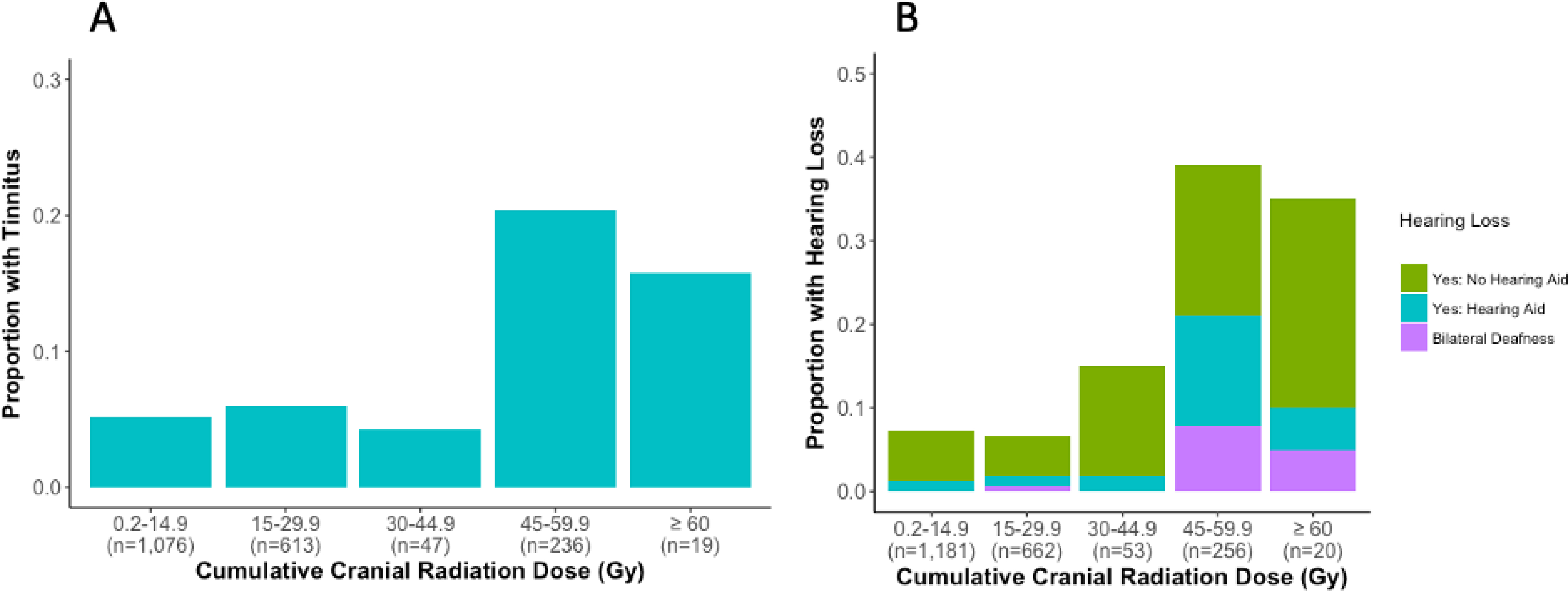
The overall proportion of pediatric cancer survivors with A) tinnitus (p=4.82×10⁻¹⁴) or B) hearing loss (p<2×10⁻¹⁶) based on maximum cranial radiation dose is provided. Statistical significance is based on logistic regression, and sample sizes for each group are indicated within each panel on the x-axis.
Table 2.
Association Between CRT-Related Tinnitus or Hearing Loss and Selected Characteristics of Childhood Cancer Survivors.
| Characteristic | Phenotype | n | Number of Controls Considered (Percent with Characteristic) | Number of Cases Considered (Percent with Characteristic) | OR (95% CI) | p | Age-Adjusted OR (95% CI) | Age-Adjusted p |
|---|---|---|---|---|---|---|---|---|
| Persistent Dizziness or Vertigo | Tinnitus | 1,953 | 1,815 (3.2%) | 138 (21.0%) | 8.06 (4.91, 13.01) | < 2×10 −16 | 7.91 (4.81,12.78) | < 2×10 −16 |
| Hearing Loss | 2,156 | 1,894 (4.5%) | 262 (13.4%) | 3.24 (2.11, 4.88) | 3.13×10 −8 | 3.22 (2.10, 4.85) | 3.71×10 −8 | |
| Prescribed Antidepressant | Tinnitus | 1,875 | 1,741 (4.8%) | 134 (9.7%) | 2.15 (1.11, 3.83) | 0.01 | 2.05 (1.06, 3.68) | 0.02 |
| Hearing Loss | 2,064 | 1,811 (4.9%) | 253 (8.7%) | 1.84 (1.11, 2.94) | 0.01 | 1.82 (1.10, 2.91) | 0.02 | |
| Prescribed Antihypertensive Medication | Tinnitus | 1,959 | 1,818 (22.8%) | 141 (34.8%) | 1.80 (1.24, 2.58) | 0.002 | 1.66 (1.14, 2.41) | 0.008 |
| Hearing Loss | 2,163 | 1,903 (23.3%) | (260) 28.5% | 1.31 (0.97, 1.74) | 0.07 | 1.27 (0.94, 1.71) | 0.11 |
Age-adjustment reflects the use of age at last observation as a covariate in the logistic regression model. Bold indicates p ≤ 0.05.
Patients who received CRT, but did not receive cisplatin or carboplatin were included in the CRT-related tinnitus and hearing loss cohorts.
CRT-related tinnitus was associated with hearing loss (age-adjusted OR=18, 95% CI: 12.3–26.5, p<2×10⁻¹⁶; Supplemental Figure 3A). Both CRT-related tinnitus and hearing loss were associated with persistent dizziness or vertigo (tinnitus: age-adjusted OR=7.9, 95% CI: 4.8–12.8, p<2×10⁻¹⁶; hearing loss: age-adjusted OR=3.2, 95% CI: 2.1–4.9, p=3.7×10⁻⁸; Table 2). Tinnitus and hearing loss cases also reported higher antidepressant use (tinnitus: age-adjusted OR=2.1, 95% CI: 1.1–3.7, p=0.02; hearing loss: age-adjusted OR=1.8, 95% CI: 1.1–2.9, p=0.02; Table 2). Self-reported health was significantly lower in tinnitus and hearing loss cases than controls (tinnitus: age-adjusted OR=1.7, 95% CI: 1.4–2.1, p=1.5×10⁻⁶; hearing loss: age-adjusted OR=1.5, 95% CI: 1.3–1.7, p=1.7×10⁻⁶; Supplemental Figure 3B and C).
Genome-Wide Association Studies
GWAS of CRT-related tinnitus identified a prominent signal in chromosome 1 in which 33 SNPs met genome-wide significance (p<5×10⁻⁸; Supplemental Table 3). The most significant SNP was rs203248 (OR=8.7, 95% CI: 1.5–47.9, p=1.5×10⁻⁹; Figure 2A and B). This SNP is intronic to DCAF6, which encodes for a ligand-dependent coactivator of nuclear receptors. rs203248 is in perfect LD with nearly all SNPs meeting genome-wide significance (R2=1.0, p<0.0001; Supplemental Figure 4A). One SNP in perfect LD is rs73024126 (OR=7.8, 95% CI: 1.4–43.4, p=1.6×10⁻⁸) that has a relatively high CADD score (12.1) that places it within the top 10% of deleterious mutations, and may regulate the binding of transcription factors to DCAF6 (RegulomeDB score: 2b - likely to affect binding; Supplemental Figure 4B). In addition, rs433173 and rs430565 are eQTLs for DCAF6, rs369914 and rs370952 are eQTLs for DCAF6, TBX19, MPC2 and GPR161, and rs73030214 is an eQTL for TBX19 and is in LD with rs203248 (r2=0.91; p<0.0001; Supplemental Figure 4B). Gene-based association analysis identified DCAF6 and NAV2 as nearly genome-wide significant (p=8.6×10⁻⁶ and p=1.6×10⁻⁵; Supplemental Figure 5A and 5B; Supplemental Table 4). NAV2 encodes for a member of the neuron navigator gene family.
Figure 2. Genome-Wide Association Studies of Radiation-Associated Ototoxicity in Pediatric Cancer Survivors.
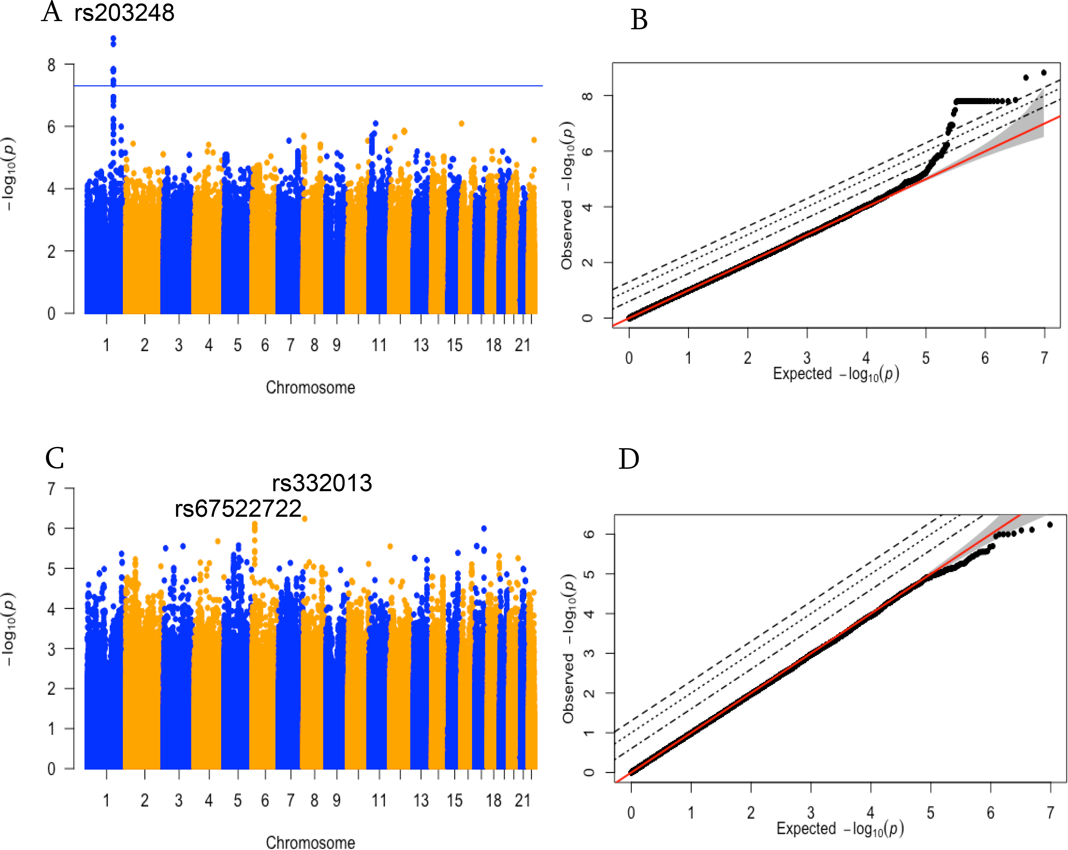
A) Manhattan plot of GWAS results for CRT-related tinnitus reveals a prominent signal in chromosome 1 exceeding genome-wide significance (p<5×10⁻⁸), with the most significant SNP being rs203248 (p=1.50×10⁻⁹). B) Quantile-Quantile plot of GWAS results for CRT-related tinnitus. C) Manhattan plot of GWAS results for CRT-related hearing loss reveals rs332013 in chromosome 8 (p=5.79×10⁻⁷) and rs67522722 (p=7.78×10⁻⁷) in chromosome 6 as nearly genome-wide significant. D) Quantile-Quantile plot of GWAS results for CRT-related hearing loss. Covariates in both GWAS include maximum cranial radiation dose, age at last observation, and 20 European genetic principal components accounting for population substructure.
GWAS of CRT-related hearing loss identified rs332013 in chromosome 8 as near genome-wide significant as defined by p<8×10⁻⁷ (OR=0.6, 95% CI: 0.4–0.9, p=5.8×10⁻⁷; Figure 2C and D; Supplemental Table 5). This SNP is intronic to ERI1, encoding for an RNA exonuclease. rs332013 has a high CADD score (17.5), indicative of a deleterious mutation, and is an eQTL for multiple genes, including ERI1, MFHAS1, and SGK223 (Supplemental Figure 6A and 6B). Three SNPs in LD with rs332013 (rs6991294, rs1077951, and rs1077950; R2 = 0.7, p<0.0001; Supplemental Figure 6A) are also eQTLs for the same genes (Supplemental Figure 6B). The next most significant SNP is rs67522722 (OR=2.5, 95% CI: 1.1–5.4, p=7.8×10⁻⁷) in chromosome 6, which is intronic to ATXN1, a gene that encodes for ataxin-1 that regulates various aspects of protein production. rs67522722 is in perfect LD with four SNPs (R2 = 1.0, p<0.0001; Supplemental Figure 6C), including rs34675197 (OR=2.5, 95% CI: 1.1–5.3, p=1.0×10⁻⁶), which has a relatively high CADD score (11.5), and appears to regulate binding of transcription factors to ATXN1 (RegulomeDB score: 2b - likely to affect binding; Supplemental Figure 6D). Gene-based association analysis identified no genome-wide significant genes (Supplemental Figure 5; Supplemental Table 6).
We further evaluated the biological significance of these findings by determining whether the expression level of genes associated with top SNPs from both GWAS correlated with radiosensitivity in CNS tumor cell lines in silico. Both NAV2 and MPC2 expression were significantly correlated with radiosensitivity (NAV2: Spearman Rho=0.5, p=0.003; R2=0.2, p=0.01; MPC2: Spearman Rho=0.4, p=0.04; R2=0.08, p=0.1; Figure 3), indicative of a protective function against CRT-related damage. No other examined gene showed a significant association with radiosensitivity in these cell lines (Supplemental Table 7). The positive association between NAV2 or MPC2 expression and radiosensitivity also appeared to be specific to cancer cell lines of CNS origin (Supplemental Table 8).
Figure 3. Scatter Plots of Radiosensitivity as a Function of Normalized NAV2 or MPC2 Expression.

Scatter plots of radiosensitivity as a function of normalized gene expression are provided for A) NAV2 (ρ=0.48, p=0.003; R2=0.18, p=0.01) and B) MPC2 (ρ=0.35, p=0.04; R2=0.08, p=0.10). Radiosensitivity, measured as the area under the survival curve derived from a linear-quadratic model to fit 9-day viability assay data, for all 36 CNS tumor cell lines, was obtained from the RadioGx package in R, and normalized gene expression data were downloaded from the Cancer Cell Line Encyclopedia. Correlation was assessed nonparametrically using the Spearman rank method, as well as by linear regression.
To examine whether any top genetic variants associated with CRT-related tinnitus or hearing loss were confounded by primary brain cancer diagnosis, we performed a separate GWAS of brain cancer in CCSS patients. Among 4,435 pediatric cancer survivors (cases: 614; controls: 3,821), we performed a logistic regression-based GWAS using the same 20 principal components of European ancestry as covariates. None of the top ototoxicity SNPs had a statistically significant association with childhood brain cancer (Supplemental Table 9), validating that the SNPs were associated with CRT-related tinnitus or hearing loss independent of brain cancer.
Replication Analysis in SJLIFE and UKBiobank
We examined whether the above SNPs reaching genome-wide or near genome-wide significance were significant in an independent cohort of cancer survivors (tinnitus analysis: n=952; hearing loss analysis: n=331) (Table 3). None of the top GWAS-identified SNPs in DCAF6, or NAV2 were significantly associated with CRT-related tinnitus. However, SNPs intronic to ATXN1 (rs67522722 and rs34675197; p=0.03) were significantly associated with hearing loss, and had the same direction of effect as the discovery cohort. Three SNPs intronic to ERI1 were marginally associated with hearing loss (rs6991294, rs1077951, and rs1077950; p=0.08), while one SNP (rs332013; p = 0.23) was not significant.
Table 3.
Evaluation of Top SNPs in CRT-Related Tinnitus and Hearing Loss GWAS in SJLIFE.
| SNP | Gene | MAF (Cases) | MAF (Controls) | GWAS OR (95% CI) | GWAS p | SJLIFE OR (95% CI) | SJLIFE p |
|---|---|---|---|---|---|---|---|
| CRT-Related Tinnitus | |||||||
| rs203248 | DCAF6 | 0.05 | 0.008 | 8.67 (1.52, 47.94) | 1.50×10⁻⁹ | 0.77 (0.09, 6.50) | 0.81 |
| rs73024126 | DCAF6 | 0.05 | 0.008 | 7.81 (1.43, 43.38) | 1.59×10⁻⁸ | 0.77 (0.09, 6.50) | 0.81 |
| rs430565 | DCAF6 | 0.05 | 0.01 | 7.04 (1.40, 35.16) | 1.72×10⁻⁸ | 1.38 (0.29, 6.65) | 0.69 |
| rs369914 | DCAF6 | 0.05 | 0.009 | 7.03 (1.38, 35.87) | 4.55×10⁻⁸ | 1.38 (0.29, 6.65) | 0.69 |
| rs370952 | DCAF6 | 0.05 | 0.01 | 5.15 (1.19, 22.42) | 1.78×10⁻⁶ | 1.20 (0.33, 4.33) | 0.78 |
| rs433173 | DCAF6 | 0.05 | 0.01 | 4.87 (1.16, 20.29) | 3.34×10⁻⁶ | 1.20 (0.33, 4.33) | 0.78 |
| rs7106624 | NAV2 | 0.45 | 0.31 | 1.86 (1.07, 3.22) | 1.99×10⁻⁶ | 1.15 (0.83, 1.58) | 0.40 |
| CRT-Related Hearing Loss | |||||||
| rs332013 | ERI1 | 0.28 | 0.38 | 0.57 (0.35, 0.93) | 5.79×10⁻⁷ | 1.26 (0.87, 1.82) | 0.23 |
| rs6991294 | ERI1 | 0.23 | 0.30 | 0.63 (0.39, 1.01) | 2.12×10⁻⁴ | 1.42 (0.95, 2.12) | 0.08 |
| rs1077951 | ERI1 | 0.23 | 0.30 | 0.62 (0.38, 1.01) | 1.47×10⁻⁴ | 1.42 (0.95, 2.12) | 0.08 |
| rs1077950 | ERI1 | 0.23 | 0.30 | 0.62 (0.38, 1.01) | 1.51×10⁻⁴ | 1.42 (0.95, 2.12) | 0.08 |
| rs67522722 | ATXN1 | 0.09 | 0.04 | 2.47 (1.13, 5.37) | 7.78×10⁻⁷ | 2.64 (1.12, 6.20) | 0.03 |
| rs34675197 | ATXN1 | 0.09 | 0.04 | 2.45 (1.11, 5.31) | 1.01×10⁻⁶ | 2.64 (1.12, 6.20) | 0.03 |
Significance values were calculated using logistic regression with the same covariates from GWAS. P-values ≤ 0.05 are highlighted in bold.
The sample sizes for the discovery cohorts of CRT-related tinnitus and hearing loss (CCSS) were 1,991 and 2,198 patients, while the sample sizes for the replication cohorts of CRT-related tinnitus and hearing loss (SJLIFE) were 952 and 331 patients.
We also examined whether the above SNPs were significant in an independent cohort of de novo hearing loss and tinnitus using publicly available data through the UK Biobank (n = 8,148 individuals)38. None of the CRT-induced tinnitus GWAS SNPs were significantly associated with de novo tinnitus-related phenotypes (Supplemental Table 10). Although no SNPs in ERI1 were associated with hearing loss related phenotypes in UK Biobank, SNPs intronic to ATXN1 (rs67522722 and rs34675197) were significantly associated with hearing difficulty/problems (p=3.6× 10⁻⁴) and hearing difficulty/problems with background noise (p=0.03) and was marginally significant with hearing difficulty/problems: I am completely deaf (p=0.09; Supplemental Table 11).
Discussion
The current study marks an advance in understanding genetic and clinical factors associated with CRT-related hearing loss/tinnitus in children. The incidence of tinnitus and hearing loss was significantly associated with cumulative CRT dose, with 4.7 and 2.3 times more survivors with tinnitus and hearing loss, respectively, among survivors treated with 45–59.9 Gy compared to 30–44.9 Gy CRT dose, consistent with previous literature on hearing loss in children5 and adults41. Independent of age at last observation and maximum CRT dose, males were 1.7 and 1.3 times more likely than females to experience tinnitus and hearing loss, respectively. Survivors with CRT-related tinnitus are more likely to experience hearing loss, persistent dizziness/vertigo, poorer self-reported health, and greater use of antidepressants, similar to results found in studies of young adult cancer survivors treated with cisplatin18. This indicates deleterious effects of tinnitus on quality of life regardless of etiology. GWAS of CRT-related tinnitus identified rs203248, intronic to DCAF6, as genome-wide significant in the CCSS discovery cohort only; while GWAS of CRT-related hearing loss identified rs67522722 as near genome-wide significant in the CCSS discovery cohort that replicated in the SJLIFE cohort and a cohort of de novo hearing loss.
Consistent with our finding that males were more likely to experience CRT-related ototoxicity, males are also more likely to experience de novo tinnitus and hearing loss in the general population42–44. This association may be explained by differences in environmental factors such as noise exposure, different hypertension rates, smoking rates, and biological sex differences including a growing body of literature demonstrating a role for estrogen in modulating the sex differences in hearing 44, 45.
The GWAS of CRT-related hearing loss identified rs67522722 as near genome-wide significant. This SNP is intronic to ATXN1, and is in perfect LD with rs34675197, which appears to regulate the binding of transcription factors to ATXN1. Further, rs67522722 was significantly associated with CRT-related hearing loss in SJLIFE (p=0.03), and had the same direction of effect as well as with a different etiology of hearing loss (de novo) as determined by positive responses to the questions regarding hearing difficulty/problems (p=3.6× 10⁻⁴) and hearing difficulty/problems with background noise (p=0.03) in the UKBiobank38. Spinocerebellar ataxia type 1 is an inherited neurodegenerative disease associated with a gain of function mutation in ataxin-1 that contributes to cerebellar and brain stem degeneration. Recently, mechanistic studies have validated the importance of ATXN1-CIC complexes in the pathophysiology of the neurodegenerative disease46, 47, highlighting the importance of ataxin-1 in neuronal maintenance. Atxn1 (mouse homolog) is expressed in mouse cochlear inner and outer hair cells as identified in the gEAR database48.
The GWAS of CRT-related tinnitus identified a prominent signal in chromosome 1 led by rs203248, intronic to DCAF6, a gene that encodes for a ligand-dependent coactivator of nuclear receptors, including glucocorticoid receptor and androgen receptor. Recent studies have confirmed the importance of glucocorticoid receptor in the maintenance of normal hearing49–51 and Dcaf6 (mouse homolog) is also expressed in mouse cochlear inner and outer hair cells as identified in the gEAR database48. SNPs intronic to DCAF6 were not replicated in SJLIFE. In addition to DCAF6, gene expression of NAV2 and MPC2 were positively correlated with radiosensitivity in CNS tumor cell lines in silico, with NAV2 also identified as near genome-wide significant in the gene-based analysis. MPC2 encodes for mitochondrial pyruvate carrier 2, a protein vital for neuron survival due to their reliance on glucose and pyruvate metabolism to generate ATP52. Importantly, MPC function has also been linked to radiosensitivity, as inhibiting the carrier increases oxygen availability, markedly sensitizing SiHa xenografts in nude mice to radiation53. Our in silico analysis in CNS tumor cell lines is in accord with these in vivo data, as lower MPC2 expression was associated with increased radiosensitivity.
Approximately 29% of patients in the CCSS cohort were given prophylactic CRT for ALL; yet recent evidence indicates that prophylactic CRT is unnecessary in ALL, as only 2 of ~1,100 children not given CRT died due to CNS relapse54, 55. Although this suggests the overall proportion of pediatric cancer survivors exposed to CRT will likely decrease over time, prophylactic CRT for ALL was routine clinical practice up until recently56. Fortunately, the expectation is that fewer pediatric patients will experience ototoxicity as the number of patients with prophylactic CRT decreases.
Major strengths of our study include the longitudinal nature of the CCSS data collection that enabled the first genome-wide analysis of CRT-associated ototoxicity and replication of ATXN1 variants approaching genome-wide significance in two independent cohorts. Due to the dearth of information regarding genetic susceptibility to ototoxicity and its associated pathophysiology, our study provides novel information on a treatment-related toxicity that is frequently encountered in the clinical setting among pediatric and adult-onset cancer survivors. Inherent limitations of our study include the lack of collection of several factors associated with ototoxicity, including the usage of aminoglycosides or other ototoxic supportive care agents, the specific location of brain tumors, and brain surgery status. Although the presence of brain tumors could be a confounder, we demonstrated that SNPs associated with tinnitus/hearing loss were not associated with brain tumors. Furthermore, the radiation exposure variable used in the analysis was not based upon direct estimation of radiation dose to the cochlea. In addition, the hearing loss phenotype differed in the CCSS discovery set (self-report) and SJLIFE cohort (Chang score based on audiometry). Although the optimal assessment for hearing loss is pure-tone audiometry57, evaluation of patient-reported symptoms are increasingly recognized as valid26 and time/cost-effective58. However, subjective perceptions of hearing loss differ from audiometrically-defined hearing loss in the general population with younger individuals tending to overestimate and older participants tending to underestimate their hearing impairment59–61, and underestimation in cisplatin-treated testicular cancer survivors24, 62.
In summary, survivors of childhood cancer receiving CRT are at increased risk for developing hearing loss and tinnitus with males at greater risk for both than females. Those who develop these toxicities are more likely to use antidepressants and report poorer overall health. Untreated hearing loss, including resulting from CRT has been linked to many health conditions including cognitive decline and dementia12, 63, 64. Importantly, midlife hearing loss is the single largest modifiable risk factor for dementia63. Given the detrimental effects of tinnitus/hearing loss on neurological and behavioral development, patients and their families should be educated with regard to potential non-genetic risk factors and comorbidities associated with treatment-related ototoxicity prior to therapy initiation and during long-term follow-up. Health care providers can improve management of survivors of childhood cancer by informing patients of associated comorbidities (dizziness, vertigo) after completion of cranial radiation therapy. Genome-wide association studies reveal genetic variants in ATXN1, a gene associated with spinocerebellar ataxia type 1, to be significantly associated with CRT-related hearing loss; further functional studies to uncover potential mechanisms of its relationship with radiation-associated ototoxicity are warranted.
Supplementary Material
Acknowledgments:
This work was supported by the National Cancer Institute (CA55727, G.T. Armstrong, Principal Investigator) and CA195547 (M.M. Hudson and L. L. Robison, Principal Investigators). Support to St. Jude Children’s Research Hospital also provided by the Cancer Center Support (CORE) grant (CA21765, C. Roberts, Principal Investigator) and the American Lebanese-Syrian Associated Charities (ALSAC). The work was also supported by grant R01 CA157823 (L.B. Travis, Principal Investigator), the University of Chicago Comprehensive Cancer Center Women’s Board (M.E. Dolan). The Genotype-Tissue Expression (GTEx) Project was supported by the Common Fund of the Office of the Director of the National Institutes of Health, and by the NCI, NHGRI, NHLBI, NIDA, NIMH, and NINDS. The data used for the analyses described in this manuscript were obtained from the GTEx Portal on 11/25/19. The dbGaP Study Accession for CCSS is phs001327.v1.p1
Footnotes
Conflict of interest: None.
Data availability: Summary statistics for both SNP based GWAS of CRT-related hearing loss and tinnitus as well as MAGMA based gene analysis of CRT-related hearing loss and tinnitus can be found at https://ccss.stjude.org/tools-and-documents/summary-statistics-for-published-analyses.html
References
- 1.Surveillance, Epidemiology, and End Results (SEER) Program released April 2019.www.seer.cancer.gov.
- 2.Armstrong GT, Chen Y, Yasui Y, et al. Reduction in Late Mortality among 5-Year Survivors of Childhood Cancer. N Engl J Med. 2016;374: 833–842. [DOI] [PMC free article] [PubMed] [Google Scholar]
- 3.Gibson TM, Mostoufi-Moab S, Stratton KL, et al. Temporal patterns in the risk of chronic health conditions in survivors of childhood cancer diagnosed 1970–99: a report from the Childhood Cancer Survivor Study cohort. Lancet Oncol. 2018;19: 1590–1601. [DOI] [PMC free article] [PubMed] [Google Scholar]
- 4.Bhakta N, Liu Q, Ness KK, et al. The cumulative burden of surviving childhood cancer: an initial report from the St Jude Lifetime Cohort Study (SJLIFE). Lancet. 2017;390: 2569–2582. [DOI] [PMC free article] [PubMed] [Google Scholar]
- 5.Whelan K, Stratton K, Kawashima T, et al. Auditory complications in childhood cancer survivors: a report from the childhood cancer survivor study. Pediatr Blood Cancer. 2011;57: 126–134. [DOI] [PMC free article] [PubMed] [Google Scholar]
- 6.Meijer AJM, Clemens E, Hoetink AE, van Grotel M, van den Heuvel-Eibrink MM. Tinnitus during and after childhood cancer: A systematic review. Crit Rev Oncol Hematol. 2019;135: 1–7. [DOI] [PubMed] [Google Scholar]
- 7.Landier W Ototoxicity and cancer therapy. Cancer. 2016;122: 1647–1658. [DOI] [PubMed] [Google Scholar]
- 8.Han BI, Lee HW, Kim TY, Lim JS, Shin KS. Tinnitus: characteristics, causes, mechanisms, and treatments. J Clin Neurol. 2009;5: 11–19. [DOI] [PMC free article] [PubMed] [Google Scholar]
- 9.Grewal S, Merchant T, Reymond R, McInerney M, Hodge C, Shearer P. Auditory late effects of childhood cancer therapy: a report from the Children’s Oncology Group. Pediatrics. 2010;125: e938–950. [DOI] [PMC free article] [PubMed] [Google Scholar]
- 10.Bartnik G, Stepien A, Raj-Koziak D, Fabijanska A, Niedzialek I, Skarzynski H. Troublesome tinnitus in children: epidemiology, audiological profile, and preliminary results of treatment. Int J Pediatr. 2012;2012: 945356. [DOI] [PMC free article] [PubMed] [Google Scholar]
- 11.Olivier TW, Bass JK, Ashford JM, et al. Cognitive Implications of Ototoxicity in Pediatric Patients With Embryonal Brain Tumors. J Clin Oncol. 2019;37: 1566–1575. [DOI] [PMC free article] [PubMed] [Google Scholar]
- 12.Bass JK, Liu W, Banerjee P, et al. Association of Hearing Impairment With Neurocognition in Survivors of Childhood Cancer. JAMA Oncol. 2020;6: 1363–1371. [DOI] [PMC free article] [PubMed] [Google Scholar]
- 13.Packer RJ, Gurney JG, Punyko JA, et al. Long-term neurologic and neurosensory sequelae in adult survivors of a childhood brain tumor: childhood cancer survivor study. J Clin Oncol. 2003;21: 3255–3261. [DOI] [PubMed] [Google Scholar]
- 14.King AA, Seidel K, Di C, et al. Long-term neurologic health and psychosocial function of adult survivors of childhood medulloblastoma/PNET: a report from the Childhood Cancer Survivor Study. Neuro Oncol. 2017;19: 689–698. [DOI] [PMC free article] [PubMed] [Google Scholar]
- 15.Wells EM, Ullrich NJ, Seidel K, et al. Longitudinal assessment of late-onset neurologic conditions in survivors of childhood central nervous system tumors: a Childhood Cancer Survivor Study report. Neuro Oncol. 2018;20: 132–142. [DOI] [PMC free article] [PubMed] [Google Scholar]
- 16.Xu H, Robinson GW, Huang J, et al. Common variants in ACYP2 influence susceptibility to cisplatin-induced hearing loss. Nat Genet. 2015;47: 263–266. [DOI] [PMC free article] [PubMed] [Google Scholar]
- 17.Wheeler HE, Gamazon ER, Frisina RD, et al. Variants in WFS1 and Other Mendelian Deafness Genes Are Associated with Cisplatin-Associated Ototoxicity. Clin Cancer Res. 2017;23: 3325–3333. [DOI] [PMC free article] [PubMed] [Google Scholar]
- 18.El Charif O, Mapes B, Trendowski MR, et al. Clinical and Genome-wide Analysis of Cisplatin-induced Tinnitus Implicates Novel Ototoxic Mechanisms. Clin Cancer Res. 2019;25: 4104–4116. [DOI] [PMC free article] [PubMed] [Google Scholar]
- 19.Langer T, Clemens E, Broer L, et al. Usefulness of current candidate genetic markers to identify childhood cancer patients at risk for platinum-induced ototoxicity: Results of the European PanCareLIFE cohort study. Eur J Cancer. 2020;138: 212–224. [DOI] [PubMed] [Google Scholar]
- 20.Robison LL, Armstrong GT, Boice JD, et al. The Childhood Cancer Survivor Study: a National Cancer Institute-supported resource for outcome and intervention research. J Clin Oncol. 2009;27: 2308–2318. [DOI] [PMC free article] [PubMed] [Google Scholar]
- 21.Howell RM, Smith SA, Weathers RE, Kry SF, Stovall M. Adaptations to a Generalized Radiation Dose Reconstruction Methodology for Use in Epidemiologic Studies: An Update from the MD Anderson Late Effect Group. Radiat Res. 2019;192: 169–188. [DOI] [PMC free article] [PubMed] [Google Scholar]
- 22.Anderson CA, Pettersson FH, Clarke GM, Cardon LR, Morris AP, Zondervan KT. Data quality control in genetic case-control association studies. Nat Protoc. 2010;5: 1564–1573. [DOI] [PMC free article] [PubMed] [Google Scholar]
- 23.Sapkota Y, Turcotte LM, Ehrhardt MJ, et al. Genome-Wide Association Study in Irradiated Childhood Cancer Survivors Identifies HTR2A for Subsequent Basal Cell Carcinoma. J Invest Dermatol. 2019;139: 2042–2045 e2048. [DOI] [PMC free article] [PubMed] [Google Scholar]
- 24.Frisina RD, Wheeler HE, Fossa SD, et al. Comprehensive Audiometric Analysis of Hearing Impairment and Tinnitus After Cisplatin-Based Chemotherapy in Survivors of Adult-Onset Cancer. J Clin Oncol. 2016;34: 2712–2720. [DOI] [PMC free article] [PubMed] [Google Scholar]
- 25.Hua C, Bass JK, Khan R, Kun LE, Merchant TE. Hearing loss after radiotherapy for pediatric brain tumors: effect of cochlear dose. Int J Radiat Oncol Biol Phys. 2008;72: 892–899. [DOI] [PubMed] [Google Scholar]
- 26.Bass JK, Hua CH, Huang J, et al. Hearing Loss in Patients Who Received Cranial Radiation Therapy for Childhood Cancer. J Clin Oncol. 2016;34: 1248–1255. [DOI] [PMC free article] [PubMed] [Google Scholar]
- 27.Purcell S, Neale B, Todd-Brown K, et al. PLINK: a tool set for whole-genome association and population-based linkage analyses. Am J Hum Genet. 2007;81: 559–575. [DOI] [PMC free article] [PubMed] [Google Scholar]
- 28.Chang CC, Chow CC, Tellier LC, Vattikuti S, Purcell SM, Lee JJ. Second-generation PLINK: rising to the challenge of larger and richer datasets. Gigascience. 2015;4: 7. [DOI] [PMC free article] [PubMed] [Google Scholar]
- 29.Morton LM, Sampson JN, Armstrong GT, et al. Genome-Wide Association Study to Identify Susceptibility Loci That Modify Radiation-Related Risk for Breast Cancer After Childhood Cancer. J Natl Cancer Inst. 2017;109. [DOI] [PMC free article] [PubMed] [Google Scholar]
- 30.Watanabe K, Taskesen E, van Bochoven A, Posthuma D. Functional mapping and annotation of genetic associations with FUMA. Nat Commun. 2017;8: 1826. [DOI] [PMC free article] [PubMed] [Google Scholar]
- 31.de Leeuw CA, Mooij JM, Heskes T, Posthuma D. MAGMA: generalized gene-set analysis of GWAS data. PLoS Comput Biol. 2015;11: e1004219. [DOI] [PMC free article] [PubMed] [Google Scholar]
- 32.Consortium GT. The Genotype-Tissue Expression (GTEx) project. Nat Genet. 2013;45: 580–585. [DOI] [PMC free article] [PubMed] [Google Scholar]
- 33.Psych EC, Akbarian S, Liu C, et al. The PsychENCODE project. Nat Neurosci. 2015;18: 1707–1712. [DOI] [PMC free article] [PubMed] [Google Scholar]
- 34.Boyle AP, Hong EL, Hariharan M, et al. Annotation of functional variation in personal genomes using RegulomeDB. Genome Res. 2012;22: 1790–1797. [DOI] [PMC free article] [PubMed] [Google Scholar]
- 35.Kircher M, Witten DM, Jain P, O’Roak BJ, Cooper GM, Shendure J. A general framework for estimating the relative pathogenicity of human genetic variants. Nat Genet. 2014;46: 310–315. [DOI] [PMC free article] [PubMed] [Google Scholar]
- 36.Rentzsch P, Witten D, Cooper GM, Shendure J, Kircher M. CADD: predicting the deleteriousness of variants throughout the human genome. Nucleic Acids Res. 2019;47: D886–D894. [DOI] [PMC free article] [PubMed] [Google Scholar]
- 37.Brinkman TM, Bass JK, Li Z, et al. Treatment-induced hearing loss and adult social outcomes in survivors of childhood CNS and non-CNS solid tumors: Results from the St. Jude Lifetime Cohort Study. Cancer. 2015;121: 4053–4061. [DOI] [PMC free article] [PubMed] [Google Scholar]
- 38.Elliott LT, Sharp K, Alfaro-Almagro F, et al. Genome-wide association studies of brain imaging phenotypes in UK Biobank. Nature. 2018;562: 210–216. [DOI] [PMC free article] [PubMed] [Google Scholar]
- 39.Barretina J, Caponigro G, Stransky N, et al. The Cancer Cell Line Encyclopedia enables predictive modelling of anticancer drug sensitivity. Nature. 2012;483: 603–607. [DOI] [PMC free article] [PubMed] [Google Scholar]
- 40.Manem VS, Lambie M, Smith I, et al. Modeling Cellular Response in Large-Scale Radiogenomic Databases to Advance Precision Radiotherapy. Cancer Res. 2019;79: 6227–6237. [DOI] [PMC free article] [PubMed] [Google Scholar]
- 41.Mujica-Mota M, Waissbluth S, Daniel SJ. Characteristics of radiation-induced sensorineural hearing loss in head and neck cancer: a systematic review. Head Neck. 2013;35: 1662–1668. [DOI] [PubMed] [Google Scholar]
- 42.Shargorodsky J, Curhan GC, Farwell WR. Prevalence and characteristics of tinnitus among US adults. Am J Med. 2010;123: 711–718. [DOI] [PubMed] [Google Scholar]
- 43.Bhatt JM, Lin HW, Bhattacharyya N. Prevalence, Severity, Exposures, and Treatment Patterns of Tinnitus in the United States. JAMA Otolaryngol Head Neck Surg. 2016;142: 959–965. [DOI] [PMC free article] [PubMed] [Google Scholar]
- 44.Agrawal Y, Platz EA, Niparko JK. Prevalence of hearing loss and differences by demographic characteristics among US adults: data from the National Health and Nutrition Examination Survey, 1999–2004. Arch Intern Med. 2008;168: 1522–1530. [DOI] [PubMed] [Google Scholar]
- 45.Shuster BZ, Depireux DA, Mong JA, Hertzano R. Sex differences in hearing: Probing the role of estrogen signaling. J Acoust Soc Am. 2019;145: 3656. [DOI] [PMC free article] [PubMed] [Google Scholar]
- 46.Rousseaux MWC, Tschumperlin T, Lu HC, et al. ATXN1-CIC Complex Is the Primary Driver of Cerebellar Pathology in Spinocerebellar Ataxia Type 1 through a Gain-of-Function Mechanism. Neuron. 2018;97: 1235–1243 e1235. [DOI] [PMC free article] [PubMed] [Google Scholar]
- 47.Perez Ortiz JM, Mollema N, Toker N, et al. Reduction of protein kinase A-mediated phosphorylation of ATXN1-S776 in Purkinje cells delays onset of Ataxia in a SCA1 mouse model. Neurobiol Dis. 2018;116: 93–105. [DOI] [PMC free article] [PubMed] [Google Scholar]
- 48.Orvis J GB, Kancherla J et al. gEAR: gene Expression Analysis Resource portal for community-driven, multi-omic data exploration. bioRxiv 2020;2020.08.28.272039; doi: 10.1101/2020.08.28.272039. [DOI] [PMC free article] [PubMed] [Google Scholar]
- 49.Hebert S, Lupien SJ. The sound of stress: blunted cortisol reactivity to psychosocial stress in tinnitus sufferers. Neurosci Lett. 2007;411: 138–142. [DOI] [PubMed] [Google Scholar]
- 50.Simoens VL, Hebert S. Cortisol suppression and hearing thresholds in tinnitus after low-dose dexamethasone challenge. BMC Ear Nose Throat Disord. 2012;12: 4. [DOI] [PMC free article] [PubMed] [Google Scholar]
- 51.Hayes SH, Manohar S, Majumdar A, Allman BL, Salvi R. Noise-induced hearing loss alters hippocampal glucocorticoid receptor expression in rats. Hear Res. 2019;379: 43–51. [DOI] [PMC free article] [PubMed] [Google Scholar]
- 52.McCommis KS, Finck BN. Mitochondrial pyruvate transport: a historical perspective and future research directions. Biochem J. 2015;466: 443–454. [DOI] [PMC free article] [PubMed] [Google Scholar]
- 53.Corbet C, Bastien E, Draoui N, et al. Interruption of lactate uptake by inhibiting mitochondrial pyruvate transport unravels direct antitumor and radiosensitizing effects. Nat Commun. 2018;9: 1208. [DOI] [PMC free article] [PubMed] [Google Scholar]
- 54.Pui CH, Campana D, Pei D, et al. Treating childhood acute lymphoblastic leukemia without cranial irradiation. N Engl J Med. 2009;360: 2730–2741. [DOI] [PMC free article] [PubMed] [Google Scholar]
- 55.Jeha S, Pei D, Choi J, et al. Improved CNS Control of Childhood Acute Lymphoblastic Leukemia Without Cranial Irradiation: St Jude Total Therapy Study 16. J Clin Oncol. 2019;37: 3377–3391. [DOI] [PMC free article] [PubMed] [Google Scholar]
- 56.Vora A, Andreano A, Pui CH, et al. Influence of Cranial Radiotherapy on Outcome in Children With Acute Lymphoblastic Leukemia Treated With Contemporary Therapy. J Clin Oncol. 2016;34: 919–926. [DOI] [PMC free article] [PubMed] [Google Scholar]
- 57.Baiduc RR, Poling GL, Hong O, Dhar S. Clinical measures of auditory function: the cochlea and beyond. Dis Mon. 2013;59: 147–156. [DOI] [PMC free article] [PubMed] [Google Scholar]
- 58.Oldenburg J, Fossa SD, Dahl AA. Scale for chemotherapy-induced long-term neurotoxicity (SCIN): psychometrics, validation, and findings in a large sample of testicular cancer survivors. Qual Life Res. 2006;15: 791–800. [DOI] [PubMed] [Google Scholar]
- 59.Hong O, Ronis DL, Antonakos CL. Validity of self-rated hearing compared with audiometric measurement among construction workers. Nurs Res. 2011;60: 326–332. [DOI] [PubMed] [Google Scholar]
- 60.Kamil RJ, Genther DJ, Lin FR. Factors associated with the accuracy of subjective assessments of hearing impairment. Ear Hear. 2015;36: 164–167. [DOI] [PMC free article] [PubMed] [Google Scholar]
- 61.Kirk KM, McGuire A, Nasveld PE, Treloar SA. Comparison of self-reported and audiometrically-measured hearing loss in the Australian Defence Force. Int J Audiol. 2012;51: 294–298. [DOI] [PubMed] [Google Scholar]
- 62.Bokemeyer C, Berger CC, Hartmann JT, et al. Analysis of risk factors for cisplatin-induced ototoxicity in patients with testicular cancer. Br J Cancer. 1998;77: 1355–1362. [DOI] [PMC free article] [PubMed] [Google Scholar]
- 63.Livingston G, Sommerlad A, Orgeta V, et al. Dementia prevention, intervention, and care. Lancet. 2017;390: 2673–2734. [DOI] [PubMed] [Google Scholar]
- 64.Golub JS, Brickman AM, Ciarleglio AJ, Schupf N, Luchsinger JA. Association of Subclinical Hearing Loss With Cognitive Performance. JAMA Otolaryngol Head Neck Surg. 2019. [DOI] [PMC free article] [PubMed] [Google Scholar]
Associated Data
This section collects any data citations, data availability statements, or supplementary materials included in this article.