Abstract
A female patient diagnosed of infiltrative breast carcinoma using tru-cut biopsy underwent 18flourine-fluorodeoxyglucose (18F-FDG) positron emission tomography/computed tomography (PET/CT) for staging. The tumor was located in the superior external quadrant of the right breast, and did not exhibit pathological uptake in 18F-FDG PET/CT. Later, gallium-68 (68Ga) fibroblast activation protein-specific inhibitor (FAPI)-04 PET/CT imaging was performed and the primary tumor showed intense radiotracer accumulation. This presumes that 68Ga-FAPI PET/CT imaging is superior to 18F-FDG imaging in detecting the primary tumor in breast cancer, thereby suggesting the replacement of FAPI by 18F-FDG in breast-cancer staging in the future.
Keywords: 68Ga-FAPI, 18F-FDG, PET/CT, breast cancer
Abstract
Tru-cut biyopsi sonucu infiltratif meme karsinomu gelen bir kadın hastaya evreleme amacıyla 18fluoride-florodeoksiglikoz (18F-FDG) pozitron emisyon tomografi/bilgisayarlı tomografi (PET/BT) görüntüleme yapıldı. Sağ meme üst dış kadranda yer alan tümör, 18F-FDG PET/BT’de patolojik aktivite tutulumu göstermedi. Daha sonra hastaya galyum-68 (68Ga)-fibroblast aktivasyon protein spesifik inhibitör (FAPI)-04 PET/BT görüntüleme yapıldı ve primer tümör yoğun radyofarmasötik tutulumu gösterdi. Bu olgu 68Ga-FAPI PET/BT görüntülemenin meme kanserinde primer tümörleri tespit etmede 18F-FDG görüntülemeden üstün olabileceğini göstermiştir ve gelecekte meme kanseri evrelemesinde 18F-FDG’nin yerini FAPI’nın alabileceğini düşündürmektedir.
Figure 1.

A tru-cut biopsy performed on a 48-year-old woman, whose breast ultrasound revealed a Breast Imaging Reporting and Data System-4B lesion measuring 20×9 mm in the right breast at the 10 o’clock position. The histologic diagnosis was infiltrative breast carcinoma and tumor cells were in the form of single-cell infiltration in focal areas (hematoxylin and eosin ×20 and ×40) without myoepithelium in the collagenous stroma (estrogen receptor is strongly positive in 95% of the cells, progesterone moderately positive in 5% of the cells, and CerbB2 is negative).
Figure 2.

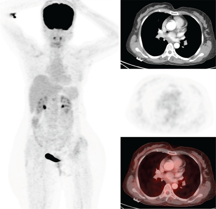
18Flourine-fluorodeoxyglucose (18F-FDG) positron emission tomography/computed tomography (PET/CT) performed for breast-cancer staging. However, the primary tumor in the superior external quadrant of the right breast did not exhibit pathological uptake in the maximum intensity projection (MIP) and axial CT/PET-fusion images.
Figure 3.

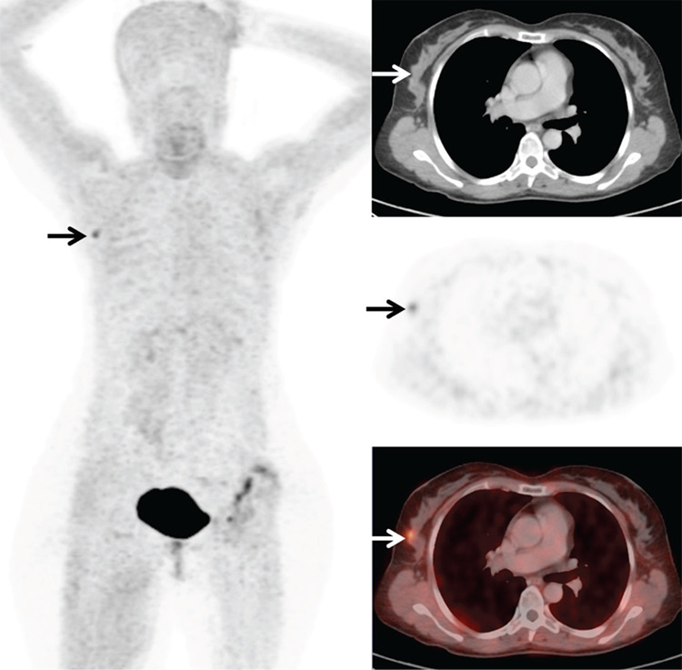
Gallium-68 (68Ga)-fibroblast activation protein-specific inhibitor (FAPI)-04 PET/CT revealing intense radiotracer accumulation in the primary tumor distinguishing it from the MIP (a) image. The axial view of 68Ga-FAPI-04 PET/CT (CT, PET, and fusion images, respectively) demonstrated intense radiotracer uptake in a lesion in the superior external quadrant of the right breast, of about 1.5 cm in size with a maximum standardized uptake value (SUVmax) of 5.3 (arrows). 68Ga-FAPI is a recently introduced imaging agent targeting fibroblast activation protein that is highly expressed in various tumors (1,2). Recent case reports reveal that the mean SUVmax of breast cancers and metastases were found to be high (3,4,5). A recent study showed that 68Ga-FAPI-04 PET/CT is superior to 18F-FDG PET/CT in detecting primary tumors in patients with breast cancer by demonstrating its high sensitivity, high SUVmax, and high tumor-to-background ratio (6). 68Ga-FAPI-04 PET/CT is also superior to 18F-FDG PET/CT in detecting lymph node, hepatic, bone, and cerebral metastases owing to its lower background activity and higher uptake in subcentimetric lesions (6). Thus, our case depicts that 68Ga-FAPI-04 PET/CT should be considered in cases with 18F-FDG-negative breast cancer.
Footnotes
Ethics
Informed Consent: Written informed consent was obtained from the patient.
Peer-review: Externally peer-reviewed.
Authorship Contributions
Surgical and Medical Practices: H.K., C.G., H.E., C.C., Concept: H.K., C.G., H.E., C.C., Design: H.K., C.G., H.E., C.C., Data Collection or Processing: H.K., C.G., H.E., C.C., Analysis or Interpretation: H.K., C.G., H.E., C.C., Literature Search: H.K., C.G., H.E., C.C., Writing: H.K., C.G., H.E., C.C.
Conflict of Interest: No conflict of interest was declared by the authors.
Financial Disclosure: The authors declared that this study received no financial support.
References
- 1.Kratochwil C, Flechsig P, Lindner T, Abderrahim L, Altmann A, Mier W, Adeberg S, Rathke H, Röhrich M, Winter H, Plinkert PK, Marme F, Lang M, Kauczor HU, Jäger D, Debus J, Haberkorn U, Giesel FL. 68Ga-FAPI PET/CT: Tracer Uptake in 28 Different Kinds of Cancer. J Nucl Med. 2019;60:801–805. doi: 10.2967/jnumed.119.227967. [DOI] [PMC free article] [PubMed] [Google Scholar]
- 2.Chen H, Pang Y, Wu J, Zhao L, Hao B, Wu J, Wei J, Wu S, Zhao L, Luo Z, Lin X, Xie C, Sun L, Lin Q, Wu H. Comparison of [68Ga]Ga-DOTA-FAPI-04 and [18F] FDG PET/CT for the diagnosis of primary and metastatic lesions in patients with various types of cancer. Eur J Nucl Med Mol Imaging. 2020;47:1820–1832. doi: 10.1007/s00259-020-04769-z. [DOI] [PubMed] [Google Scholar]
- 3.Pang Y, Zhao L, Chen H. 68Ga-FAPI Outperforms 18F-FDG PET/CT in Identifying Bone Metastasis and Peritoneal Carcinomatosis in a Patient With Metastatic Breast Cancer. Clin Nucl Med. 2020;45:913–915. doi: 10.1097/RLU.0000000000003263. [DOI] [PubMed] [Google Scholar]
- 4.Can C, Gündoğan C, Güzel Y, Kaplan İ, Kömek H. 68Ga-FAPI Uptake of Thyroiditis in a Patient With Breast Cancer. Clin Nucl Med. 2021;46:683–685. doi: 10.1097/RLU.0000000000003637. [DOI] [PubMed] [Google Scholar]
- 5.Gündoğan C, Güzel Y, Can C, Alabalik U, Kömek H. False-Positive 68Ga-Fibroblast Activation Protein-Specific Inhibitor Uptake of Benign Lymphoid Tissue in a Patient With Breast Cancer. Clin Nucl Med. 2021;46:e433–e435. doi: 10.1097/RLU.0000000000003594. [DOI] [PubMed] [Google Scholar]
- 6.Kömek H, Can C, Güzel Y, Oruç Z, Gündoğan C, Yildirim ÖA, Kaplan İ, Erdur E, Yıldırım MS, Çakabay B. 68Ga-FAPI-04 PET/CT, a new step in breast cancer imaging: a comparative pilot study with the 18F-FDG PET/CT. Ann Nucl Med. 2021;35:744–752. doi: 10.1007/s12149-021-01616-5. [DOI] [PubMed] [Google Scholar]


