Abstract
Background
Posterior wall fracture is the most common type of acetabular fracture, the traditional open reduction and fixation through the Kocher–Langenbeck approach required a large incision and extensive muscle and soft tissue dissection, resulting in more blood loss, more complications and delayed recovery after the operation. Hip arthroscopy has been widely used in clinical practice but rarely reported in acetabular fractures.
Case Presentation
We present the case of a 14‐year‐old boy with acetabular posterior wall fracture who was treated with hip arthroscopy reduction and fixation using anchors. He began to walk with partial weight‐bearing assisted by double crutches, and returned to school with crutches at 3 days after surgery. Although hip arthroscopy is technically more demanding, it’s an optimal choice for selected patients of acetabular fracture with the advantages of less invasive and faster postoperative recovery.
Keywords: Acetabulum, Arthroscopic surgery, Fracture fixation, Hip fractures
We present the case of a 14‐year‐old boy with acetabular posterior wall fracture who was treated with hip arthroscopy reduction and fixation using anchors. Hip arthroscopy is an optimal choice for selected patients of acetabular fracture with the advantages of less invasive and faster postoperative recovery.
Introduction
As the most common type of acetabular fracture, the posterior wall fracture is considered to be the simplest and easily treatable fracture. More than 80% of nonoperative patients resulted in unsatisfactory clinical outcomes 1 , therefore, open reduction and fixation has become the primary choice for the treatment of acetabular posterior wall fractures. In view of the anatomical characteristics of the acetabulum, an open surgery is accompanied by greater invasive and blood loss. Especially, it is less cost effective in the relatively simple cases that cannot be fixed with plates and screws, it is necessary to explore a minimally invasive reduction and fixation method for such cases. Hip arthroscopy has become the main surgical procedure for diagnosis and treatment of some hip diseases such as femoroacetabular impingement, Labrum injury and so on 2 . Here, we performed a case of adolescent acetabular posterior wall fracture treated by hip arthroscopy and report a successful outcome.
Case Presentation
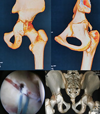
A 14‐year‐old boy unfortunately suffered a traffic accident in a car that resulted pain and limited mobility in his right hip joint. He was initially admitted to the county hospital and computed tomography (CT) showed a fracture of the right posterior acetabular wall. The patient was transferred to our hospital 8 days after the injury. The preoperative 3‐dimensional (3D) CT showed the fracture of the right posterior acetabular wall (Fig. 1A) and the major osseous fragment displaced significantly (Fig. 1B). An operative reduction of the fracture is essential to restore the instability of the hip. In order to achieve enhanced recovery after surgery and the patient can go back to school as soon as possible, hip arthroscopic reduction and fixation was performed.
Fig. 1.
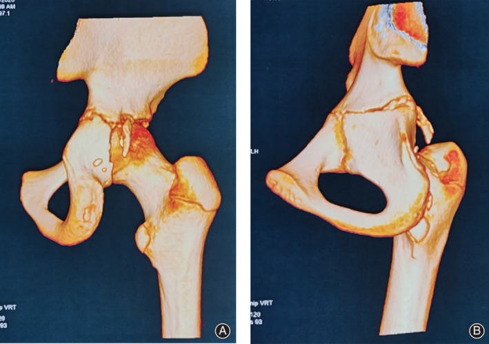
(A)Preoperative posterior view of 3D‐CT showed the fracture of the acetabular posterior wall. (B) Preoperative posterior oblique view of 3D‐CT showed the displaced of the major osseous fragment.
Surgical Technique
Patient was in supine position on a fracture table with the right limb in traction after general anesthesia, and a C‐arm was used for fluoroscopic guidance to establish the anterolateral portal. Anterior and posterolateral portals were made under direct visualization. After hematoma evacuation by water lavage and synovectomy, the major osseous fragment of the acetabular posterior wall was found (Fig. 2) and used shaver handpiece to push it reduction. Two 2.9 mm bio‐absorbable anchors (OSTEORAPTOR Suture Anchor, Smith & Nephew, Andover, MA) were inserted into the superior and inferior of the fracture surface of acetabular posterior wall adjoined the major osseous fragment. The sutures of the anchors embraced the osseous fragment and tied it up to fix (Fig. 3). Finally, a 2.8 mm anchor (TWINFIX Ti anchor; Smith & Nephew, Andover, MA) was used to through the center of the fragment and inserted into the posterior wall, the sutures of this anchor and the bio‐absorbable anchors were tied together to enhanced fixation. A diagram (Fig. 4) showed the surgical procedure.
Fig. 2.
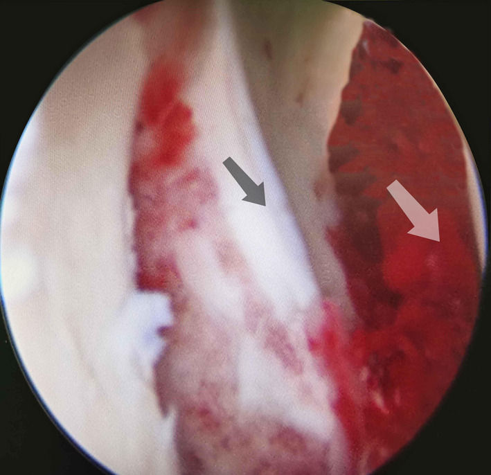
The arthroscopic view showed the major osseous fragment (black arrow) of the acetabular posterior wall and residual hemarthrosis (white arrow).
Fig. 3.
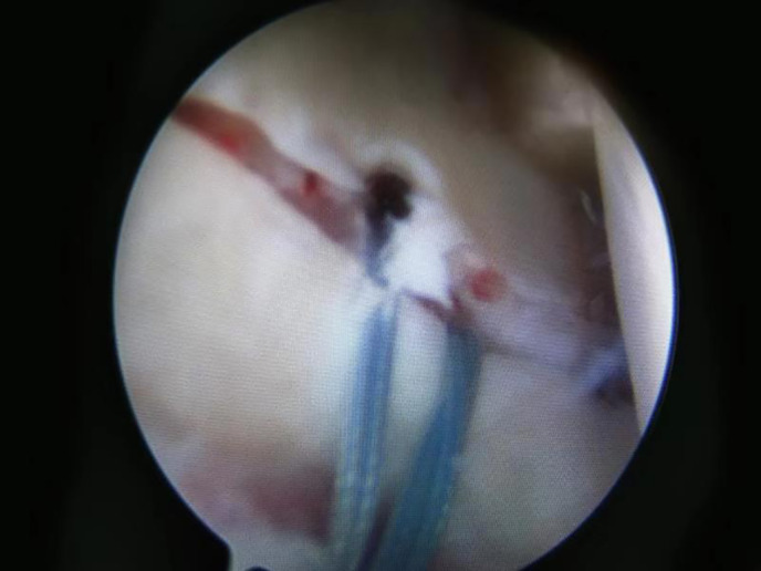
The arthroscopic view showed the major osseous fragment was replaced and fixed by anchors.
Fig. 4.

Surgical procedure diagrams: (A) Placed two bio‐absorbable anchors in the superior and inferior of the fracture surface of acetabular posterior wall adjoined the major osseous fragment. (B) Reduced the osseous fragment and tied it up by the sutures of the anchors. (C) Placed a Ti anchor into the posterior wall through the center of the fragment and tied its sutures together with the sutures of bio‐absorbable anchors.
A postoperative 3D‐CT was performed and showed anatomical reduction of the major osseous fragment (Fig. 5). The next day after the surgery, the patient began to walk with partial weight‐bearing assisted by double crutches and returned to school with crutches at 3 days after surgery. At 3 months follow up postoperatively, anteroposterior X‐ray of pelvis showed bone union of the acetabular posterior wall (Fig. 6), the patient was pain free and recovered full activities.
Fig. 5.
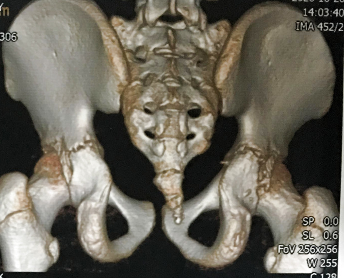
Postoperative 3D‐CT demonstrated anatomical reduction of the major osseous fragment of the posterior wall.
Fig. 6.
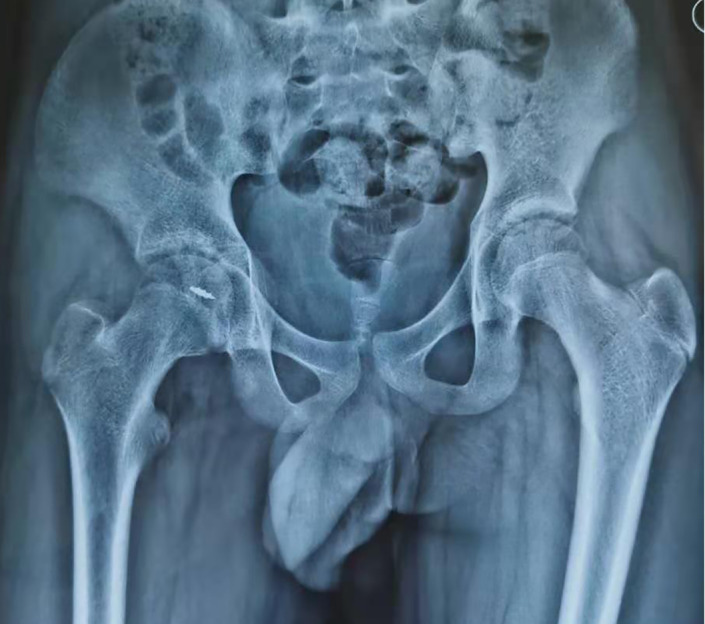
Postoperative 3 months anteroposterior X‐ray of pelvis showed bone union of the acetabular posterior wall.
Discussion
Bed rest and traction were main conservative treatment methods for acetabular posterior wall fractures that were not significantly displaced and do not affect the stability of the hip joint. Complications of bed rest include thromboembolism, pneumonia, bedsore and Joint stiffness. Most acetabular posterior wall fractures require open reduction and internal fixation, which required wide dissection and led to a prolonged recovery. In this case, the adolescent patient wanted fastest recovery in order to return to school, but the posterior wall fracture was significantly displaced and requires surgical reduction and fixation, compared to hip arthroscopy, open reduction and internal fixation was a viable option but not the optimal option.
As a representative of minimally invasive surgery, arthroscopic technology has been successfully used in the treatment of joint diseases and intra‐articular fractures. For the diagnosis and treatment of femoroacetabular impingement, pigmented villonodular synovitis, septic arthritis and synovial chondromatosis, hip arthroscopy is a gold‐standard procedure 2 . The application of hip arthroscopy in trauma is still less reported. A systematic review by Niroopan et al. in 2016 showed the hip arthroscopy was an effective and safe approach in trauma, 32 studies (25 case reports and seven case series) reported a total of 144 hip traumatic patients underwent hip arthroscopy for six indications: eight patients for bullet extraction, six for femoral head fixation, 82 for loose body removal, six for acetabular fracture fixation, 20 for labral intervention, and 23 for ligamentum teres debridement. Successful surgery was achieved in 96% of these patients 3 (Table 1).
TABLE 1.
Hip arthroscopic management of acetabular fracture reported in the literature
| Author | Year | Study design | No.of hips | Main injury | Surgical methods |
|---|---|---|---|---|---|
| Yamamoto et al. 4 | 2003 | Case series | 11 hips(1 acetabular fracture) | Weight‐bearing region fracture | Reduction and percutaneous pinning were performed under arthroscopic observation |
| Yang et al. 5 | 2010 | Case report | 2 |
Case 1: transverse fracture Case 2: anterior column fracture |
Fixed by percutaneous screw and arthroscopy as an assisting tool for intra‐articular observation |
| Park et al. 6 | 2013 | Case report | 1 | Posterior wall fractures, femoral head fractures, dislocation | The avulsed torn labrum was reattached with 2 anchors through the midanterior portal. Osteochondral fragments were curetted and removed. |
| Kim et al. 7 | 2014 | Case report | 2 |
Case 1: posterior wall fracture Case 2: anterior column fracture |
Fixed by cannulated screws under direct arthroscopic visualization |
| Stabile et al. 8 | 2014 | Case report | 1 | Posterior wall fracture, dislocation, bucket‐handle labral tear | Bucket‐handle labral tear had an attached osseous fragment that was reduced under direct visualization with the use of a switching stick and fixed by anchors |
| Park et al. 9 | 2016 | Case report | 2 | Posterior wall fracture | Cannulated screws to fixate the fragment under direct arthroscopic visualization |
| Gürınar et al. 10 | 2019 | Case report | 1 | Posterior wall fracture | Arthroscopic reduction and fixation using a cannulated screw |
To our knowledge, there are no cases series of hip arthroscopic treatment of acetabular fractures were reported so far, only several case reports. In 2003, Yamamoto et al. first described the usefulness of hip arthroscopic in hip trauma and reported 11 hips from 10 cases, and one acetabular weight‐bearing region fracture was treated by reduction and percutaneous pinning fixation under arthroscopic observation 4 . Yang et al. described the technique of percutaneous screw fixation of acetabular fractures using arthroscopy as an assisting tool for intra‐articular observation and report two cases resulted satisfied outcomes 5 . A transverse fracture of the acetabulum and another anterior column fracture of the acetabulum were fixed percutaneously using 6.5‐mm‐diameter cannulated screws under fluoroscopic guidance and arthroscopy was used for direct visualization of the fracture site until satisfactory compression 5 . Similarly, Kim et al. reported two patients in 2014, one posterior acetabular wall fracture and one acetabular anterior column fracture, fixed by percutaneously 4.0‐mm‐diameter and 3.5‐mm‐diameter cannulated screws under direct arthroscopic visualization 7 . In addition, Park et al. 9 reported two and Gürpınar et al. 10 reported one acetabular posterior wall fractures were treated with the similar surgical technique.
For the acetabular wall fracture fragment too little to fix by cannulated screws, hip arthroscopic debridement to remove osseous fragment or/and osteosynthesis using anchors to fix the fragment was another effective method 6 , 8 . For this adolescent patient, the little osseous fragments were removed by lavage to prevent traumatic arthritis, and the primary fragment was fixed by three anchors. The disadvantages of this method are the fixed effect is not as good as screw fixing, and the surgical procedure is more difficult. But the advantage is the whole process is under hip arthroscopy observation and without more fluoroscopic guidance for percutaneous screws.
Kim et al. stated that the indications of hip arthroscopic surgery for acetabular fractures is narrow and can be made only for those cases with minimal and moderate displaced 7 . Based on experience, we believe that the indications of osteosynthesis for acetabular fracture under hip arthroscopy were also limited. This osteosynthesis only suitable for the simple acetabular wall fracture with small fragment which easy to reduce through the arthroscopic portals. Indications of hip arthroscopy with percutaneous screw fixation was more than osteosynthesis and can be used for more types of acetabular wall fracture and acetabular anterior or posterior column fractures do not required open reduction 5 , 7 .With the improvement of arthroscopic instruments for fixation of reduced acetabulum and surgical technique, arthroscopic minimally invasive surgery may be extended to more complex acetabular fracture.
Hip arthroscopy for reduction and fixation of acetabular fracture offers the advantage of superior visualization and reduction of the articular surface, and additional benefits from joint lavage and debridement can help to remove the loose osteochondral fragments, which is thought to cause future osteoarthritis 4 . Although hip arthroscopy is technically more demanding, it is an optimal choice for selected acetabular fractures with the advantages of less invasive and faster postoperative recovery.
Acknowledgment
The author(s) disclosed receipt of the following financial support for the research, authorship, and/or publication of this article: this work is funded by Yunnan Province Talents Program (2018HB001) and The National Key Research and Development Program of China (2017YFC1103904).
Disclosure: The author(s) declared no potential conflicts of interest with respect to the research, authorship, and/or publication of this article.
Contributor Information
Bing Yang, Email: yangbin@foxmail.com.
Hong‐bo Tan, Email: tantoo@163.com.
References
- 1. Baumgaertner MR. Fractures of the posterior wall of the acetabulum. J Am Acad Orthop Surg, 1999, 7: 54–65. [DOI] [PubMed] [Google Scholar]
- 2. Jamil M, Dandachli W, Noordin S, Witt J. Hip arthroscopy: indications, outcomes and complications. Int J Surg, 2018, 54: 341–344. [DOI] [PubMed] [Google Scholar]
- 3. Niroopan G, de Sa D, Macdonald A, Burrow S, Larson CM, Ayeni OR. Hip arthroscopy in trauma: a systematic review of indications, efficacy, and complications. Art Ther, 2016, 32: 692–703. [DOI] [PubMed] [Google Scholar]
- 4. Yamamoto Y, Ide T, Ono T, Hamada Y. Usefulness of arthroscopic surgery in hip trauma cases. Art Ther, 2003, 19: 269–273. [DOI] [PubMed] [Google Scholar]
- 5. Yang JH, Chouhan DK, Oh KJ. Percutaneous screw fixation of acetabular fractures: applicability of hip arthroscopy. Art Ther, 2010, 26: 1556–1561. [DOI] [PubMed] [Google Scholar]
- 6. Park MS, Yoon SJ, Choi SM. Hip arthroscopic management for femoral head fractures and posterior acetabular wall fractures (Pipkin type IV). Arthrosc Tech, 2013, 2: e221–e225. [DOI] [PMC free article] [PubMed] [Google Scholar]
- 7. Kim H, Baek J, Park S, Ha Y. Arthroscopic reduction and internal fixation of acetabular fractures. Knee Surg Sports Traumatol Arthrosc, 2014, 22: 867–870. [DOI] [PubMed] [Google Scholar]
- 8. Stabile KJ, Neumann JA, Mannava S, Howse EA, Stubbs AJ. Arthroscopic treatment of bucket‐handle labral tear and acetabular fracture. Arthrosc Tech, 2014, 3: e283–e287. [DOI] [PMC free article] [PubMed] [Google Scholar]
- 9. Park JY, Chung WC, Kim CK, Huh SH, Kim SJ, Jung BH. Arthroscopic reduction and transportal screw fixation of acetabular posterior wall fracture: technical note. Hip Pelvis, 2016, 28: 120–126. [DOI] [PMC free article] [PubMed] [Google Scholar]
- 10. Gürpınar T, Polat B, Kanay E, Polat A, öztürkmen Y. Arthroscopic fixation of a posterior acetabular wall fracture: a case report. Cureus, 2019, 11: e6264. [DOI] [PMC free article] [PubMed] [Google Scholar]


