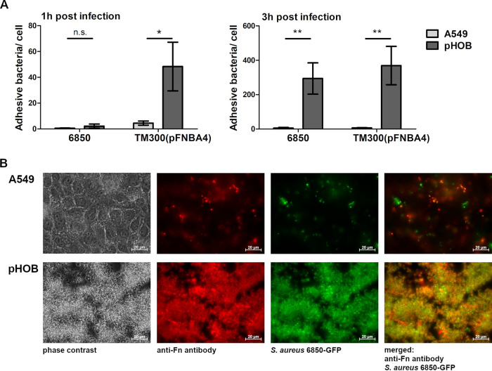
FIG 5.
Staphylococci adhere to a large proportion to pHOB. (A) Binding of S. aureus 6850 or S. carnosus TM300(pFNBA4) to A549 and pHOB cells after 1 and 3 h of infection (MOI 50). To determine the amount of adhesive bacteria, the quantified amount of bacteria from an experimental setup with lysostaphin (only intracellular bacteria) was subtracted from an experimental setup without lysostaphin (adhesive and intracellular bacteria). The number of host cells was determined, and the number of viable bacteria was assessed by lysing of host cells and plate counting. Data represent the means ± SD from 3 independent experiments. *, P < 0.05; **, P ≤ 0.01, unpaired t test. (B) Representative fluorescence microscopy images of A549 and pHOB cells infected with S. aureus 6850-GFP (MOI 50) for 3 h, with a washing step but no lysostaphin step; therefore, no distinction between extra- and intracellular bacteria is possible. Images of phase contrast, anti-Fn staining (red), S. aureus 6850-GFP signal (green), and overlay (anti-Fn and S. aureus GFP-signal) are presented.

