Abstract
Protein phosphatase 1, comprising the regulatory subunit Reg1 and the catalytic subunit Glc7, has a role in glucose repression in Saccharomyces cerevisiae. Previous studies showed that Reg1 regulates the Snf1 protein kinase in response to glucose. Here, we explore the functional relationships between Reg1, Glc7, and Snf1. We show that different sequences of Reg1 interact with Glc7 and Snf1. We use a mutant Reg1 altered in the Glc7-binding motif to demonstrate that Reg1 facilitates the return of the activated Snf1 kinase complex to the autoinhibited state by targeting Glc7 to the complex. Genetic evidence indicated that the catalytic activity of Snf1 negatively regulates its interaction with Reg1. We show that Reg1 is phosphorylated in response to glucose limitation and that this phosphorylation requires Snf1; moreover, Reg1 is dephosphorylated by Glc7 when glucose is added. Finally, we show that hexokinase PII (Hxk2) has a role in regulating the phosphorylation state of Reg1, which may account for the effect of Hxk2 on Snf1 function. These findings suggest that the phosphorylation of Reg1 by Snf1 is required for the release of Reg1-Glc7 from the kinase complex and also stimulates the activity of Glc7 in promoting closure of the complex.
The yeast Saccharomyces cerevisiae regulates metabolism, gene expression, and growth in response to carbon source availability. When glucose is abundant, the expression of a large number of genes, including those involved in the utilization of alternative carbon sources, gluconeogenesis, respiration, and peroxisomal functions, is repressed at the level of transcription by a process known as glucose repression. This process has been extensively studied both biochemically and genetically (for recent reviews, see references 4, 16, and 26). These studies show that both the Snf1 (Cat1, Ccr1) protein kinase and the Reg1 (Hex2, Srn1)-Glc7 (Cid1) protein phosphatase play a crucial role in glucose repression.
Snf1 is a serine-threonine protein kinase that regulates transcription both by inhibiting transcription repressors (e.g., Mig1) and by stimulating transcription activators (e.g., Cat8 and Sip4) (see reference 4 for a review). The Snf1 kinase is found in complexes containing the activating subunit Snf4 (Cat3) and members of the Sip1/Sip2/Gal83 family (25). Glucose regulates the activity of the kinase (55, 56) and regulates the interaction between Snf1 and Snf4 within the Snf1 kinase complex (24). In glucose-grown cells, the Snf1 kinase complex exists predominantly in an inactive autoinhibited conformation in which the catalytic domain of Snf1 binds to the regulatory domain of the protein (24). When glucose is limiting, the catalytic domain is released and the activating subunit Snf4 binds to the regulatory domain, which leads to an active, open conformation of the complex (24). Activation requires phosphorylation and an essential, conserved threonine in the activation loop of Snf1, which is phosphorylated during the activation of other kinases (11, 30, 55, 56).
Another major component of the glucose repression pathway is the Reg1-Glc7 protein phosphatase complex. GLC7 is an essential gene that encodes the catalytic subunit of protein phosphatase type 1 (PP1) (48). It is involved in glucose repression and also in the regulation of different processes including glycogen metabolism, translation, sporulation, chromosome segregation, and cell cycle progression (3, 12, 14, 22, 38, 46, 48, 54). As is the case for its mammalian counterpart, the Glc7 protein phosphatase participates in the regulation of these processes via the binding of specific regulatory subunits that target the phosphatase to the corresponding substrates (15, 21, 33, 49, 50, 57). Reg1 is one of these regulatory subunits, which targets Glc7 to substrates involved in the glucose repression pathway and other processes (15, 23, 34, 36, 49, 51). Genetic evidence indicates that Reg1 is required for glucose repression, and Reg1 is physically associated with Glc7, as judged by their strong interaction in the two-hybrid system and their coimmunoprecipitation from cell extracts (49).
The Reg1-Glc7 phosphatase complex has a role in regulating the Snf1 kinase complex. Reg1 (but not Glc7) interacts with the Snf1 kinase in the two-hybrid system (30). This interaction is glucose regulated, increasing when glucose is limiting. Moreover, in a reg1Δ mutant, the Snf1 complex remains in an open conformation even in the presence of glucose. It was proposed that in response to glucose, Glc7, targeted by Reg1, dephosphorylates Snf1 or another component of the Snf1 complex and thereby facilitates its conformational change from its active state to the autoinhibited form (30). Reg1 interacts very strongly with a kinase-dead Snf1 mutant, Snf1K84R, which has arginine substituted for the invariant lysine in the ATP-binding site; moreover, this interaction is not inhibited by glucose. These findings suggest that Snf1 negatively regulates its own interaction with Reg1.
Another component of the glucose repression pathway is hexokinase PII (Hxk2), a glycolytic enzyme that in addition to phosphorylating glucose is involved in regulating glucose repression (9, 10, 16, 31, 32). This enzyme is located in both the nucleus and the cytoplasm, consistent with dual roles in signaling and catalysis (20, 39), but its actual role in glucose repression is still poorly understood. When glucose is limiting, Hxk2 is a phosphoprotein (27, 52), and the Reg1-Glc7 phosphatase complex dephosphorylates Hxk2 in response to glucose (1, 40).
In this study, we explore the functional and physical relationships between Reg1, Glc7, and Snf1. We show that it is the recruitment of Glc7 by Reg1 that promotes the conformational change of the Snf1 complex in response to glucose. We also show that Reg1 is phosphorylated in a Snf1-dependent manner in response to glucose limitation and that Reg1 is then dephosphorylated, dependent on Glc7, when glucose is added back. Finally, we address the relationship of Hxk2 to the Reg1-Glc7 and Snf1 complexes and show that Hxk2 has a role in regulating the phosphorylation of Reg1.
MATERIALS AND METHODS
Strains and genetic methods.
S. cerevisiae strains used in this study were FY250 (MATα his3 leu2 trp1 ura3 SUC2; gift from F. Winston, Harvard Medical School, Boston, Mass.), FY250 snf1Δ (FY250 containing snf1Δ10), MCY3278 (FY250 containing reg1Δ::URA3), MCY3000 (MATa lys2 his3 trp1 ura3 leu2 glc7-T152K), CTY10-5d (MATa ade2 his3 leu2 trp1 gal4 gal80 URA3::lexAop-lacZ; gift from R. Sternglanz, State University of New York, Stony Brook), and CTY10-5d hxk2Δ and CTY10-5d reg1Δ (CTY10-5d derivatives containing hxk2Δ::URA3 or reg1Δ::URA3). To construct strains FY250 hxk2Δ and MCY3000 hxk2Δ, an SpeI-XhoI fragment of pSB21 (see below) was used to introduce hxk2Δ::TRP1 by gene disruption (42), and the absence of hexokinase PII was confirmed by Western blotting with an anti-Hxk2 antibody (α-Hxk2).
Standard methods for genetic analysis and transformation were used. Yeast cultures were grown in synthetic complete (SC) medium lacking appropriate supplements to maintain selection for plasmids (41).
Oligonucleotides.
The oligonucleotides used were HXK2-1 (AAATGGATCCATTTAGGTCC), HXK2-2 (GATCATAGAATTCATGTTCAC), HXK2-5 (GCGGGGATCCAGAGCTCCACATTGG), HXK2-6 (TACGGGATCCTTATATAAGCATCTTTTACTAC), REG1-1 (TCAAGAATTCTAGCAAATTACTTCG), REG1-2 (TAATCTCGAGGATAATCCCATGGAATTG), and REG1-7 (CATCCCTCGAGTGTGAAGCTGGATATCG). Restriction sites are underlined.
Plasmids.
To construct the disruption plasmid pSB21, in which TRP1 replaces codons 15 to 415 of HXK2, oligonucleotides HXK2-5 and HXK2-6 were used to amplify by PCR the HXK2 sequence from nucleotide −602 to 281 nucleotides after the stop codon, using genomic DNA from strain FY250 as the template. The fragment was digested with BamHI and subcloned into pSK93 (see below) to yield pSK-HXK2(5-6), and then a SmaI/SalI fragment from the latter was subcloned into pRS305 (44), yielding pRS-HXK2(5-6). A SmaI/PstI TRP1 fragment from plasmid YDp-W (2) was used to replace a NcoI/PstI fragment from pRS-HXK2(5-6), generating pSB21.
To construct GAD-Hxk2, oligonucleotides HXK2-1 and HXK2-2 were used to amplify by PCR the HXK2 gene from genomic DNA of FY250. The amplified fragment was digested with BamHI and EcoRI and subcloned into pACTII (29).
To construct GAD-Reg1 fusions, oligonucleotide pairs REG1-1–REG1-2 and REG1-1–REG1-7 were used to amplify by PCR a region of REG1 from codon 3 to codons 447 and 485, respectively, using pRJ65 (49) as the template. The amplified fragments were digested with EcoRI and XhoI and subcloned into pACTII (29) to give pSB31 (pACTII-Reg13-447) and pSB32 (pACTII-Reg13-485), respectively. pSB50 contains an EcoRI/EcoRV fragment from pSB32 subcloned into pRS303 (44). pSB51 contains a NcoI/SalI fragment from pHW-2 (LexA-Reg1401-760 [see below]) subcloned into pSB50. pSB52 (pACTII-Reg13-760) contains an EcoRI fragment from pSB51 in pACTII. A NcoI/SalI fragment from pRJ65 (49) was subcloned into pSB50 to yield pSB54. An EcoRI/XhoI fragment from the latter was subcloned into pACTII to yield pSB55 (pACTII-Reg13-1014). GAD-Reg1424-760 (pBF374), GAD-Reg1424-1014 (pBF414), and GAD-Reg1760-1014 (pBF394) in vector pACT were a gift from Frank Li and Mark Johnston (Washington University School of Medicine).
Plasmid pSB16 expresses HA-Reg1, which has the hemagglutinin epitope (HA) tag fused to the N terminus, and was constructed by subcloning an EcoRI/SalI fragment containing the REG1 coding region from pRJ65 (49) into pWS93 (45). A derivative of pWS93 carrying TRP1 as the selectable marker (pSK93, a gift from Sergei Kuchin) was used similarly to construct pSB17. HA-Reg1 fully complemented a reg1Δ mutation and was more stable than the LexA-Reg1 fusion protein previously described (49). An EcoRI/SalI fragment from plasmid pLexA-Reg1F468R (1) was subcloned into pSK93 to obtain pSB44 (HA-Reg1F468R).
To construct pSB53 (HA-Reg11-443), we subcloned an EcoRI/NcoI fragment from pRJ65 (49) into pEG202 (17) to give pSB27, and an EcoRI/SalI fragment from the latter was then subcloned into pWS93.
Other plasmids used in this study were pVP16 (53), pRJ79 (VP16-Snf1 [30]), pRJ80 (VP16-Snf1K84R [30]), pLexA(202+PL) (43), pRJ55 (LexA-Snf1 [24]), pNI12 (Snf4-GAD [13]), pLexA-Glc7 (49), and pSK120 (HA-Snf1K84R [47]). pHW1 (LexA-Reg11-400), pHW2 (LexA-Reg1401-760), and pHW3 (LexA-Reg1761-1014) in vector pEG202 were a gift from Heather A. Wiatrowski.
Invertase and β-galactosidase assays.
Invertase activity was assayed in whole cells as previously described (24). β-Galactosidase activity was assayed in permeabilized cells and expressed in Miller units as in reference 30. Where indicated, β-galactosidase was assayed using crude protein extracts (5), and activity was expressed as units per milligram of protein.
λ-Phosphatase treatment.
Protein extracts were prepared essentially as described in reference 5. Protein extracts (2 μg) in λ-phosphatase buffer containing 2 mM MnCl2 were treated at 30°C for 30 min with 50 U of λ-phosphatase (New England BioLabs) in the presence or absence of a mixture of phosphatase inhibitors (50 mM EDTA, 50 mM NaF, 100 mM sodium phosphate buffer [pH 8]). The reactions were stopped by adding 1 volume of sample buffer and boiling for 3 min.
Preparation of cell extracts by the fast boiling method.
Cells corresponding to 1 U of A600 were collected by rapid centrifugation (14,000 rpm, 1 min), resuspended in 100 μl of Laemmli sample buffer (28), and boiled for 3 min. Glass beads (0.3 g, 450-μm diameter) were added to the suspension, and then cells were vortexed at full speed for 30 s. The suspension was boiled again 3 min and centrifuged at 14,000 rpm 1 min; 10 μl of the supernatant was subjected to sodium dodecyl sulfate-polyacrylamide gel electrophoresis (SDS-PAGE) and immunoblotting.
Coimmunoprecipitation assays.
Preparation of protein extracts and immunoprecipitation procedures were essentially as described previously (5). The extraction buffer was 50 mM HEPES (pH 7.5)–150 mM NaCl–0.5% Triton X-100–1 mM dithiothreitol–10% glycerol and contained 2 mM phenylmethylsulfonyl fluoride and Complete protease inhibitor cocktail (Boehringer Mannheim). α-Snf1 polyclonal antibody (1 μl) was used in each immunoprecipitation reaction. Precipitates were analyzed by Western blotting using α-HA monoclonal antibodies.
Immunoblot analysis.
Crude extracts were separated by SDS-PAGE using 7% acrylamide gels and analyzed by immunoblotting using α-Snf1 and α-Snf4 polyclonal antibodies and commercial monoclonal α-HA (Boehringer Mannheim) or α-LexA (Clontech). Antibodies were detected by enhanced chemiluminescence (ECL) with ECL or ECL Plus reagents (Amersham).
Analysis of 32P-labeled proteins.
Cultures were grown in synthetic medium selective for plasmid maintenance and then transferred to 50 ml of YEP-PO4 containing 2% glucose (1) for two doublings (optical densities at 600 nm [OD600] of 0.2 to 0.8). Cells were then collected by centrifugation at 200 × g for 10 min and resuspended into 50 ml of YEP-PO4 plus 2% glucose at an OD600 of 0.2. [32P]orthophosphate (2 mCi) was added, and cells were grown to an OD600 of 0.8. The culture was divided in two; cells were harvested by centrifugation, washed in 10 ml of the appropriate medium (YEP-PO4 plus 2% glucose or 2% raffinose), and resuspended into 25 ml of the same medium. [32P]orthophosphate (1 mCi) was added to each culture, which was then grown for 20 min. Cells were harvested by centrifugation and washed. Extracts were prepared by the addition of 0.5 ml of acid-washed glass beads and 1 ml of lysis buffer (50 mM Tris-HCl [pH 8.0], Complete protease inhibitor cocktail, 1 μM microcystin-LR, 150 mM NaCl, 1% Triton X-100). This mixture was vortexed five times for 20 s with 1 min on ice between vortexing. The entire sample was then centrifuged at 200 × g for 2 min, and the supernatant (about 1 ml) was drawn off into a microcentrifuge tube and centrifuged at 12,000 × g for 10 min. For immunoprecipitation, the supernatant was precleared with 20 μl of a slurry of equal parts of lysis buffer and protein G-agarose for 30 min. Immunoprecipitations were done as described previously (19) with 5 μg of α-LexA, using preconjugated α-LexA–agarose (Santa Cruz).
RESULTS
Reg1 coimmunoprecipitates with the Snf1 protein kinase.
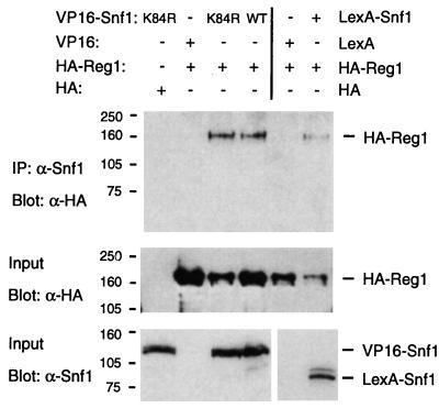
To confirm the interaction between Reg1 and the Snf1 protein kinase detected in the two-hybrid assay (30), we introduced plasmids expressing the functional proteins HA-Reg1 and VP16-Snf1 (30) into a snf1Δ strain. VP16-Snf1 was immunoprecipitated from cell extracts with a polyclonal Snf1 antibody, and the precipitates were analyzed by Western blotting. HA-Reg1 coimmunoprecipitated with VP16-Snf1 and also with the inactive mutant VP16-Snf1K84R (Fig. 1). HA-Reg1 also coimmunoprecipitated with LexA-Snf1, thereby confirming that the VP16 moiety was not responsible.
FIG. 1.
Coimmunoprecipitation of HA-Reg1 with VP16-Snf1 and LexA-Snf1. Protein extracts (250 μg) were prepared from FY250 snf1Δ cells expressing the indicated proteins. VP16 and LexA fusion proteins were immunoprecipitated (IP) with α-Snf1 polyclonal antibody, and the precipitated proteins were separated by SDS-PAGE and immunoblotted with α-HA monoclonal antibody (Blot) (upper panel). Proteins in the input crude extracts (1 μg) were also immunodetected with either α-HA (middle panel) or α-Snf1 (lower panel). Size standards are indicated in kilodaltons.
Different sequences of Reg1 interact with Snf1 and the PP1 catalytic subunit.
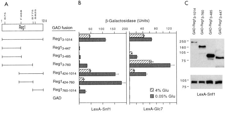
We next fused different regions of Reg1 in frame to the activating domain of Gal4 (GAD) and tested for their interaction with LexA-Snf1 and LexA-Glc7 (Fig. 2). LexA-Snf1 interacted most strongly with GAD-Reg1424-760 and GAD-Reg1424-1014. LexA-Glc7 showed a different interaction profile, interacting most strongly with GAD-Reg13-760. A consensus motif, (R/K)(V/I)XF, for the recognition of regulatory subunits by the catalytic subunit of mammalian PP1 has been identified, and the similar motif (R/K)X(V/I)XF was found in various PP1-binding proteins in yeast (8). Reg1 contains the sequence RHIHF468 (Fig. 2), which is present in all of the GAD-Reg1 proteins that interacted with LexA-Glc7. (GAD-Reg1424-760 also contains the motif, close to the GAD moiety, but no interaction with Glc7 was detected for unknown reasons.) Mutation of this motif dramatically reduces the interaction of Reg1 with Glc7 (1, 7). In contrast, the F468R mutation (1) did not substantially affect the two-hybrid interaction between Reg1 and Snf1. The combination LexA-Reg1F468R plus VP16-Snf1 gave 20 U of β-galactosidase activity after a shift to low glucose, and LexA-Reg1F468R plus VP16-Snf1K84R gave 330 U during growth in 4% glucose (values are averages for four to six transformants of CTY10-5d). These values are the same as those for LexA-Reg1 (see Fig. 7). Thus, distinct Reg1 sequences mediate its interactions with Snf1 and Glc7.
FIG. 2.
Two-hybrid interaction of Snf1 and Glc7 with different sequences of Reg1. (A) Reg1 sequences fused to GAD that were used in the two-hybrid analysis. GAD-Reg1 fusions were used because certain regions of Reg1 show self-activating activity. The positions of F468 and serines in potential Snf1 recognition sites (see text) are indicated. (B) CTY10-5d transformants expressing the GAD-Reg1 proteins and either LexA-Snf1 or LexA-Glc7 were grown to mid-log phase in selective SC–4% glucose medium; cells were then washed with water and shifted to SC–0.05% glucose medium for 3 h; values are average β-galactosidase activities of four to six transformants, and bars show standard deviations. (C) Western blots of proteins from transformants growing in 4% glucose and expressing LexA-Snf1 and the indicated GAD-Reg1 fusion; 10-μl aliquots of cell extracts prepared by the fast boiling method (see Materials and Methods) were immunoblotted with α-HA (top) or α-Snf1 (bottom). The production of GAD-Reg1424-1014, GAD-Reg1424-760, and GAD-Reg1760-1014 could not be assessed because these constructs lack the HA epitope tag; the production of GAD-Reg1 fusion proteins in transformants expressing LexA-Glc7 followed the same trend as transformants expressing LexA-Snf1 (data not shown). Size standards are indicated in kilodaltons.
FIG. 7.
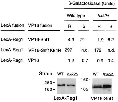
Two-hybrid interaction of Reg1 and Snf1 in an hxk2Δ mutant. Wild-type CTY10-5d cells and an hxk2Δ mutant derivative of CTY10-5d expressing the indicated fusion proteins were treated as described for Fig. 2. Values for cells growing in 4% glucose (R) and cells shifted to 0.05% glucose for 3 h (S) are average β-galactosidase activities of four transformants; standard deviations were lower than 10% of the average values in all cases. A Western blot of wild-type (WT) and hxk2Δ transformants expressing LexA-Reg1 and VP16-Snf1 is shown; 10-μl aliquots of crude extracts (boiling method) were immunodetected with α-LexA (left) or α-Snf1 (right). Levels of VP16-Snf1K84R were also similar in both strains (data not shown). n.d., not determined. Size standards are indicated in kilodaltons.
Reg1 regulates the conformation of the Snf1 kinase complex by targeting Glc7 to the complex.
The interaction between Snf1 and Snf4 within the kinase complex increases in response to glucose limitation; Snf4 binds to the regulatory domain of Snf1 and the autoinhibition of the Snf1 catalytic domain is relieved, leading to an open, active conformation of the kinase complex (24). Reg1 has a role in regulating the interaction of Snf1 and Snf4, by promoting the autoinhibited conformation of the complex (24, 30). Previously, we inferred that these effects of Reg1 result from the targeting of PP1 activity to the kinase complex. The availability of the Reg1F468R mutant allowed a direct test of this model.
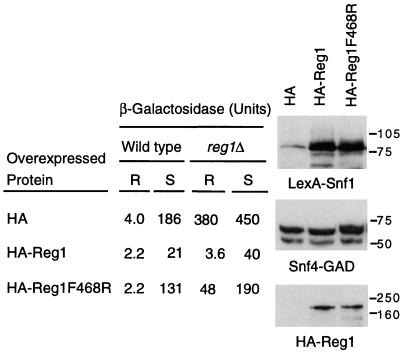
We first analyzed the interaction of Snf1 and Snf4 in wild-type cells that overexpressed either HA-Reg1 or HA-Reg1F468R from the ADH1 promoter. Overexpression of HA-Reg1 caused an almost 10-fold decrease in the interaction between Snf1 and Snf4 in low glucose (Fig. 3). However, when the Reg1F468R form of the protein was overexpressed, no significant decrease was detected. In a reg1Δ mutant, the Snf1-Snf4 interaction increased and was no longer inhibited by glucose (Fig. 3 and reference 24). The overexpression of HA-Reg1 in the mutant complemented the defect and reduced Snf1-Snf4 interaction 100-fold in glucose-grown cells. In contrast, overexpression of HA-Reg1F468R reduced the interaction only eightfold. Western analysis showed similar levels of the fusion proteins in all cases (Fig. 3). These findings indicate that the effects of Reg1 on the conformation of the Snf1 kinase complex occur via the recruitment of the catalytic subunit of PP1, which most likely dephosphorylates Snf1 or another component of the complex.
FIG. 3.
Reg1 and Glc7 regulate the two-hybrid interaction of Snf1 and Snf4 within the kinase complex in response to glucose. Two-hybrid interaction between LexA-Snf1 and Snf4-GAD was measured in wild-type CTY10-5d and reg1Δ mutant transformants expressing the indicated HA-Reg1 fusion proteins. Values for cells growing in 4% glucose (R) and cells shifted to 0.05% glucose for 3 h (S) are average β-galactosidase activities of four to six transformants, with standard deviations lower than 15% in all cases. A Western blot of wild-type transformants growing in 4% glucose and expressing the indicated proteins is shown; 10-μl aliquots of crude extracts (boiling method) were immunodetected either with α-Snf1 (upper panel), α-Snf4 (middle panel), or α-HA (lower panel). Analysis of protein levels at the end of the 3-h shift did not reveal any dramatic changes, and the production of the different fusion proteins in reg1Δ transformants was similar to wild type (data not shown). Size standards are indicated in kilodaltons.
Reg1 is phosphorylated in response to glucose limitation in a Snf1-dependent manner.
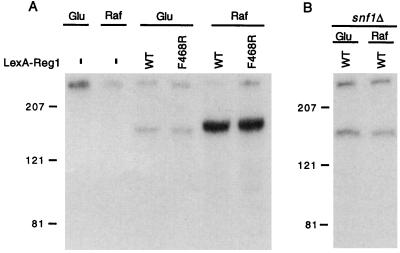
Genetic evidence indicates that the Snf1 catalytic activity negatively regulates the interaction between Snf1 and Reg1: a mutation in the ATP-binding site of Snf1 (Snf1K84R) increases this interaction and relieves inhibition of the interaction by glucose (see Fig. 7 and reference 30). To determine whether the mechanism entails phosphorylation of Reg1, we examined Reg1 for phosphorylation in vivo in response to glucose limitation (Fig. 4A). Cells expressing LexA-Reg1 or LexA-Reg1F468R were labeled with [32P]orthophosphate during growth in 2% glucose and then transferred to fresh medium containing [32P]orthophosphate and either 2% glucose or 2% raffinose for 20 min. Extracts were prepared, and the LexA fusion protein was immunoprecipitated, separated by gel electrophoresis, and detected by autoradiography. Phosphorylation of both the wild-type and mutant LexA-Reg1 proteins increased substantially (10- to 20-fold in three different experiments) during growth in raffinose, that is, in the absence of glucose. In contrast, the majority of the phosphoproteins detected by two-dimensional gel electrophoresis and autoradiography are the same in glucose- and raffinose-grown cells (1). This differential phosphorylation of LexA-Reg1 requires the Snf1 kinase because a shift to 2% raffinose did not cause a substantial increase in phosphorylation in a snf1Δ mutant (Fig. 4B). Similar results were obtained for HA-Reg1 (data not shown).
FIG. 4.
Snf1-dependent phosphorylation of Reg1 in response to glucose limitation. (A) Strain MCY3278 expressed no LexA fusion protein (−), wild-type (WT) LexA-Reg1, or LexA-Reg1F468R. (B) Strain FY250 snf1Δ expressed LexA-Reg1. Cultures were grown in 2% glucose, labeled with [32P]orthophosphate, collected by centrifugation, and resuspended in medium containing [32P]orthophosphate and either 2% glucose (Glu) or 2% raffinose (Raf) for another 20 min as described in Materials and Methods. Extracts were prepared, and proteins were immunoprecipitated with preconjugated α-LexA–agarose. Precipitates were subjected to SDS-PAGE in 6% acrylamide, and gels were dried for autoradiography. Size standards are indicated in kilodaltons.
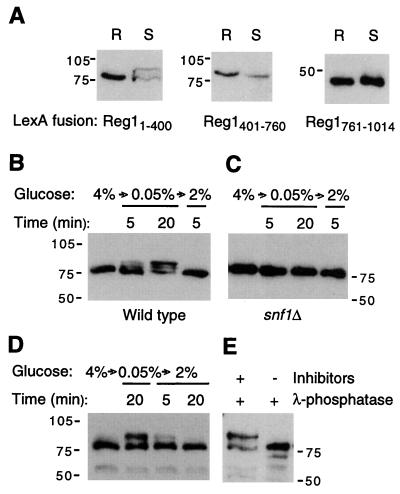
Phosphorylation of Reg1 does not cause an easily detectable change in electrophoretic mobility of the protein because Reg1 is large (it has a calculated molecular mass of 113 kDa but migrates in SDS-PAGE with a mobility of >160 kDa). We therefore constructed truncated forms of the protein fused to LexA to facilitate detection of changes in mobility by immunoblot analysis. We first tested if any of these forms showed a different mobility after shifting the cells from medium containing high (4%) glucose to low (0.05%) glucose. Only the N-terminal part of the protein (LexA-Reg11-400) showed a clear change in mobility after the shift (Fig. 5A). This modification occurred very rapidly, within 5 min (Fig. 5B), and was dependent on the Reg1 sequence because an HA-Reg11-443 derivative was modified similarly (Fig. 5D). This modification did not occur in a snf1Δ mutant, indicating that the Snf1 protein kinase is directly or indirectly responsible (Fig. 5C). This region of Reg1 showed only weak interaction with Snf1 in the two-hybrid assay; however, phosphorylation may require only transient interaction.
FIG. 5.
N terminus of Reg1 is phosphorylated in low glucose in a Snf1-dependent manner. (A) FY250 cells expressing different regions of Reg1 fused to LexA were grown in selective SC–4% glucose medium (R). When they reached the mid-log phase, the cells were washed with water and shifted to SC–0.05% glucose medium for 3 h (S). Crude extracts (10 μl) prepared by the fast boiling method were subjected to immunoblot analysis with α-LexA. (B) FY250 cells expressing LexA-Reg11-400 were grown in selective SC–4% glucose medium and then were shifted to SC–0.05% glucose medium for 5 min or 20 min; after this time, 2% glucose was added to the medium, and cells were collected after 5 min. Crude extracts prepared by the boiling method (10 μl) were analyzed by immunoblotting with α-LexA. (C) FY250 snf1Δ cells expressing LexA-Reg11-400 were treated and analyzed as for panel B. (D) FY250 cells expressing HA-Reg11-443 were treated as for panel B except that an additional sample was taken 20 min after the addition of glucose; proteins in the crude extracts were immunodetected with α-HA. (E) glc7-T152K cells expressing HA-Reg11-443 were grown in selective SC–4% glucose medium and shifted to 0.05% glucose for 20 min; 2-μg aliquots of protein extract were treated with λ-phosphatase (50 U) in the presence or absence of phosphatase inhibitors (see Materials and Methods); HA-Reg11-443 was immunodetected with α-HA. Size standards are indicated in kilodaltons.
To confirm that the modification detected here is indeed phosphorylation, we used λ-phosphatase to treat protein extracts from cells expressing HA-Reg11-443 and shifted to low glucose for 20 min. We used glc7-T152K mutant cells to prevent loss of the modified form of HA-Reg11-443 due to action of the endogenous phosphatase (see below). The modified form disappeared during the treatment with λ-phosphatase unless phosphatase inhibitors were included in the reaction mixture, indicating that the modification was due to phosphorylation (Fig. 5E).
Since Reg1 was phosphorylated in a Snf1-dependent manner, we searched the sequence for possible Snf1 phosphorylation sites. Three sequences matched the proposed consensus sequence ΦXRXXSXXXΦ, where Φ is a hydrophobic residue (M, V, L, I, or F) (6): LKRTRS75MGLL, LGKSGS775TNSL and LKRNSS825SGNF (consensus residues are underlined [Fig. 2]). The serine residues in these sites were substituted with alanine by site-directed mutagenesis. All of the mutated forms were able to restore regulated invertase synthesis in a reg1Δ mutant (data not shown). Moreover, the phosphorylation observed in LexA-Reg11-400 did not occur at serine 75, because the mutant LexA-Reg11-443S75A was similarly modified upon removal of glucose (data not shown). Thus, we have no evidence that these serines are physiologically important; however, the consensus sequence for Snf1 phosphorylation is not yet completely defined (18).
Reg1 is dephosphorylated by Glc7 in response to glucose.
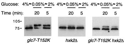
The phosphorylation of LexA-Reg11-400 and HA-Reg11-443 was reversible: if glucose was added back to cells after a shift to low glucose, the modified form rapidly disappeared (Fig. 5B and D). To determine whether Glc7 is responsible for this dephosphorylation, we used the mutant allele glc7-T152K, which partially relieves glucose repression but does not interfere with the function of Glc7 in other pathways (48, 49). The mutant was defective in dephosphorylating LexA-Reg11-400 after the addition of glucose, and the modified form was still present after 5 min (Fig. 6). However, the modified form was not prominent in cells growing exponentially in 4% glucose, presumably because the mutant phosphatase is still partially active.
FIG. 6.
PP1 and hexokinase PII are involved in regulating the phosphorylation state of Reg1. Strains with the indicated genotypes (see Materials and Methods) were transformed with a plasmid expressing LexA-Reg11-400. Cells were grown and proteins were analyzed and immunodetected with α-LexA as for Fig. 5B. Size standards are indicated in kilodaltons.
Hexokinase PII has a role in regulating the phosphorylation of Reg1.
The hexokinase PII, encoded by HXK2, both phosphorylates glucose and regulates glucose repression (16). The hxk2Δ and reg1Δ mutations are similar in several respects: both relieve glucose repression of many of the same genes, including SUC, MAL, GAL, HXT2, and HXT4, but do not relieve glucose repression of gluconeogenic genes (16); both affect glucose induction of HXT1 expression (37); both cause Snf1 and Snf4 to exhibit two-hybrid interaction in cells growing in high glucose (24); and snf1 is epistatic to both (35). In addition, the Reg1-Glc7 phosphatase complex dephosphorylates Hxk2 in response to glucose (1, 40). We therefore considered a role for Hxk2 in regulating the phosphorylation of Reg1.
We first examined LexA-Reg11-400 in a hxk2Δ mutant, expecting to observe phosphorylation even in glucose-grown cells because the Snf1 kinase is active in glucose in this mutant. Instead, little modification was detected, even after a shift to low glucose (Fig. 6). This result was confirmed using HA-Reg11-443 (data not shown).
One possibility is that the interaction between Reg1 and Snf1 is severely impaired in a hxk2Δ mutant. However, the two-hybrid interaction of LexA-Reg1 with VP16-Snf1 or VP16-Snf1K84R was reduced only two- to threefold in the mutant (Fig. 7). In addition, the interaction between LexA-Reg1 and GAD-Glc7 was normal in the hxk2Δ mutant (data not shown).
Alternatively, the absence of phosphorylation of Reg1 in the hxk2Δ mutant could be due to an elevated Glc7 activity, which would mask the phosphorylation by Snf1. We therefore analyzed the phosphorylation of LexA-Reg11-400 in a double glc7-T152K hxk2Δ mutant (Fig. 6). The phosphorylated species was observed in cells shifted to low glucose for 20 min and was still detectable 5 min after the addition of glucose. The amount of the modified species was lower than in the parental glc7-T152K mutant strain (Fig. 6), consistent with the effect of hxk2Δ in a wild-type background. These results indicate that in a hxk2Δ mutant, Reg1 is phosphorylated in response to low glucose but is rapidly dephosphorylated by Glc7. Although these data do not exclude the possibility that Hxk2 stimulates the phosphorylation of Reg1, a simpler model is that Hxk2 interferes with its dephosphorylation by Glc7.
These results imply that Hxk2 interacts with Snf1 and/or Reg1-Glc7. We detected a very weak two-hybrid interaction between LexA-Snf1 and GAD-Hxk2 in derepressed cells (blue reaction in filter assays and a twofold increase in β-galactosidase activity relative to negative controls) but detected no interaction between LexA-Reg1 or LexA-Glc7 and GAD-Hxk2. One possibility is that Hxk2 interacts with Reg1-Glc7 that is bound to Snf1. To test this idea, we overexpressed Snf1K84R, which interacts strongly with Reg1 (Fig. 7 and reference 30). In the presence of high levels of Snf1K84R, interaction between LexA-Reg1 and GAD-Hxk2 was indeed detected (Table 1).
TABLE 1.
Two-hybrid interaction between Reg1 and Hxk2 in cells overexpressing Snf1K84R
| LexA fusion | GAD fusion | Overexpressed protein | β-Galactosidase (U/mg of protein)a
|
|
|---|---|---|---|---|
| Glu | Raf | |||
| LexA-Reg1 | GAD-Hxk2 | HA | 0.2 | 0.1 |
| LexA-Reg1 | GAD-Hxk2 | HA-Snf1K84R | 3.4 | 7.0 |
| LexA-Reg1 | GAD | HA-Snf1K84R | 0.2 | 0.1 |
CTY10-5d transformants expressing the indicated proteins were grown to mid-log phase in either selective SC–4% glucose (Glu) or SC–2% raffinose plus 0.05% glucose (Raf) medium. β-Galactosidase was measured in cell extracts. Values are average β-galactosidase activities of four to six transformants; standard deviations were lower than 10% of the average value.
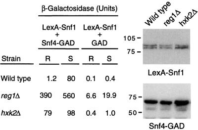
Further evidence consistent with a functional relationship between Hxk2 and Reg1-Glc7 was that overexpressing Reg1 suppressed the defect in glucose repression of invertase synthesis in a hxk2Δ mutant. Glucose-grown hxk2Δ cells carrying the empty vector pWS93 expressed 67 U of invertase activity, whereas cells overexpressing HA-Reg1 (plasmid pSB16) expressed only 12 U, an 82% reduction (values are the average of at least two transformants). Finally, both reg1Δ and hxk2Δ caused a constitutive two-hybrid interaction between Snf1 and Snf4 in cells growing in 4% glucose (Fig. 8), in agreement with previous results (24). These findings are consistent with the view that the phosphorylation of Reg1, which is reduced in an hxk2Δ mutant, stimulates the ability of Reg1-Glc7 to promote the return of the open, activated Snf1 complex to a closed, autoinhibited state.
FIG. 8.
Two-hybrid interaction of Snf1 and Snf4 in reg1Δ and hxk2Δ mutants. Wild-type CTY10-5d cells and reg1Δ and hxk2Δ derivatives of CTY10-5d were transformed with plasmids expressing LexA-Snf1 and Snf4-GAD or GAD alone. Cells were grown and treated as described in the legend of Fig. 2. Values for cells growing in 4% glucose (R) and cells shifted to 0.05% glucose for 3 h (S) are average β-galactosidase activities of four transformants; standard deviations were lower than 10% of the average values in all cases. A Western blot of the wild-type and reg1Δ and hxk2Δ transformants expressing the indicated fusion proteins is shown; 10-μl aliquots of crude extracts (boiling method) were immunodetected with α-Snf1 (top) or α-Snf4 (bottom). Size standards are indicated in kilodaltons.
DISCUSSION
We have examined the regulatory interactions between the Reg1-Glc7 form of PP1 and the Snf1 kinase complex. Previous work indicated that phosphorylation and dephosphorylation of Snf1 are important in the regulation of its activity in response to the glucose signal. Phosphorylation is required for activation of the kinase, and the conserved activation-loop threonine is required for Snf1 activity and for the positive regulatory interaction between Snf1 and Snf4 within the kinase complex (11, 30, 55, 56). Conversely, the Reg1-Glc7 protein phosphatase also regulates the kinase complex (24, 30).
We have examined the physical and functional interactions of Reg1 with Glc7 and Snf1. We show that different sequences of Reg1 interact with these two proteins. The binding of Glc7 is mediated by the RHIHF468 motif, which matches the consensus motif (R/K)X(V/I)XF found in other regulatory subunits of PP1 (1, 7, 8). The F468R mutation greatly reduces the binding of Reg1 to Glc7 (1, 7) but does not affect its binding to Snf1. We were therefore able to use the Reg1F468R form to demonstrate that the effects of Reg1 on the conformation of the Snf1 kinase complex require the recruitment of the catalytic subunit Glc7. Thus, these effects are mediated by PP1 activity. Glc7 presumably dephosphorylates Snf1, or possibly another component of the kinase complex, to promote the autoinhibited conformation of the complex.
The Snf1 kinase negatively regulates its own interaction with Reg1, as the kinase-dead Snf1K84R protein interacts strongly with Reg1, even in the presence of glucose (30). We present evidence that the mechanism entails phosphorylation of Reg1. The N-terminal region of Reg1 (Reg11-400) is rapidly phosphorylated when cells are shifted to low glucose, and this phosphorylation is dependent on Snf1 activity. Moreover, we show that dephosphorylation occurs soon after the addition of glucose to the medium and that a mutation in Glc7 (glc7-T152K) affects this process.
The phosphorylation state of Reg1 is also affected by hexokinase PII, a protein that not only phosphorylates glucose in the glycolytic pathway but also regulates glucose repression by an unknown mechanism (16). We found that LexA-Reg11-400 is not phosphorylated in a hxk2Δ mutant. It is possible that Hxk2 contributes to efficient binding of Reg1 to Snf1 in low glucose, but the two-hybrid interaction of the full-length proteins (LexA-Reg1 and VP16-Snf1) was reduced only two- to threefold in a hxk2Δ mutant. Hxk2 could also stimulate the phosphorylation of Reg1 by Snf1 by some other mechanism. Alternatively, Hxk2 may interfere with the dephosphorylation of Reg1 by Glc7. Consistent with this view, phosphorylation of LexA-Reg11-400 was detected in the double glc7-T152K hxk2Δ mutant, which has impaired PP1 function. Regardless of the precise mechanism, these data suggest that the effects of Hxk2 on glucose repression are mediated by Reg1-Glc7. Previous genetic evidence supported a functional relationship between Hxk2 and Reg1-Glc7, and we have presented additional evidence, namely, that overexpressing Reg1 suppresses the defect in glucose repression of invertase synthesis of a hxk2Δ mutant. Finally, we detected a two-hybrid interaction between LexA-Reg1 and GAD-Hxk2 in glucose-limited cells when, in addition to the overexpression of the two fusion proteins, Snf1K84R was also overexpressed.
What is the role of the phosphorylation of Reg1? Analysis of the hxk2Δ mutant suggests that the phosphorylation of Reg1 stimulates the ability of Reg1-Glc7 to promote the return of the open, activated Snf1 complex to a closed, autoinhibited state. In this mutant, Reg1 is not phosphorylated, and the interaction between Snf1 and Snf4 within the Snf1 complex is constitutive and occurs even in cells growing in high glucose (reference 24 and this study). Phosphorylation is also required for release of Reg1 from the kinase complex, as Reg1 remains tightly bound to the kinase-dead SnfK84R protein in glucose-grown cells (30). Finally, the N-terminal 200 residues of Reg1 play a role in glucose repression of SUC2 (7), and it is possible that the N-terminal phosphorylation of Reg1 modulates this function.
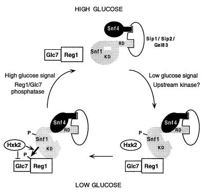
We propose the following model for the role of the Reg1-Glc7 phosphatase complex in regulation of the Snf1 complex (Fig. 9). Reg1-Glc7 binds to Snf1 when the kinase is activated by phosphorylation, which occurs at a much higher rate in glucose-limited cells than in glucose-grown cells. Reg1 is then phosphorylated by Snf1, and Hxk2 either stimulates the binding and/or phosphorylation of Reg1 or inhibits the dephosphorylation of Reg1 by Glc7. In response to a glucose signal, Reg1-Glc7 facilitates the transition back to the autoinhibited state, presumably by dephosphorylating Snf1, and Reg1-Glc7 is released from its association with the kinase complex. The phosphorylation of Reg1 by Snf1 appears to stimulate the activity of Glc7 in promoting closure of the complex, and this phosphorylation is also required for release of Reg1-Glc7 from the kinase complex. Glc7 then dephosphorylates Reg1. The glucose signal most likely inhibits the initial phosphorylation of Snf1 but may also activate Reg1/Glc7 function. The complexity of these regulatory interactions suggests that the Snf1 kinase activity is finely tuned in response to glucose.
FIG. 9.
Model for regulation of the Snf1 kinase complex in response to glucose. Cells growing in high glucose maintain the Snf1 complex predominantly in an autoinhibited conformation in which the regulatory domain (RD) binds to the catalytic kinase domain (KD). Low levels of glucose favor phosphorylation of the Snf1 kinase, possibly by an upstream kinase. The catalytic domain is released from autoinhibition and Snf4 binds to the regulatory domain, leading to an open and active conformation of the complex. The Reg1-Glc7 phosphatase complex then binds to Snf1, and Reg1 is phosphorylated by Snf1. Hxk2 either stimulates the binding and/or phosphorylation of Reg1 or inhibits the dephosphorylation of Reg1 by Glc7. Reg1-Glc7 facilitates the transition back to the autoinhibited state, presumably by dephosphorylating Snf1. The phosphorylation of Reg1 appears to stimulate closure of the complex. Reg1-Glc7 is released from its association with the kinase complex, and this release also requires phosphorylation of Reg1 by Snf1. Glc7 then dephosphorylates Reg1. In glucose-grown reg1, glc7-T152K, or hxk2 mutant cells, the Snf1 kinase, once activated, becomes trapped in the activated state. Each kinase complex contains one of the related proteins Sip1, Sip2, and Gal83.
ACKNOWLEDGMENTS
We thank Sergei Kuchin, Heather A. Wiatrowski, and Frank Li for plasmids and Peter Sherwood and Olivier Vincent for useful discussion.
This work was supported by Public Health Service grant GM34095 from the National Institutes of Health to M.C. P.S. was supported by an FPU program fellowship from the Spanish Ministry of Education and Culture.
REFERENCES
- 1.Alms G R, Sanz P, Carlson M, Haystead T A. Reg1p targets protein phosphatase 1 to dephosphorylate hexokinase PII in Saccharomyces cerevisiae: characterizing the effects of a phosphatase subunit on the yeast proteome. EMBO J. 1999;18:4157–4168. doi: 10.1093/emboj/18.15.4157. [DOI] [PMC free article] [PubMed] [Google Scholar]
- 2.Berben G, Dumont J, Gilliquet V, Bolle P A, Hilger F. The YDp plasmids: a uniform set of vectors bearing versatile gene disruption cassettes for Saccharomyces cerevisiae. Yeast. 1991;7:475–477. doi: 10.1002/yea.320070506. [DOI] [PubMed] [Google Scholar]
- 3.Cannon J, Pringle J R, Fiechter A, Khalil M. Characterization of glycogen-deficient glc mutants of Saccharomyces cerevisiae. Genetics. 1994;136:485–503. doi: 10.1093/genetics/136.2.485. [DOI] [PMC free article] [PubMed] [Google Scholar]
- 4.Carlson M. Glucose repression in yeast. Curr Opin Microbiol. 1999;2:202–207. doi: 10.1016/S1369-5274(99)80035-6. [DOI] [PubMed] [Google Scholar]
- 5.Celenza J L, Carlson M. Mutational analysis of the Saccharomyces cerevisiae SNF1 protein kinase and evidence for functional interaction with the SNF4 protein. Mol Cell Biol. 1989;9:5034–5044. doi: 10.1128/mcb.9.11.5034. [DOI] [PMC free article] [PubMed] [Google Scholar]
- 6.Dale S, Wilson W, Edelman A, Hardie D G. Similar substrate recognition motifs for mammalian AMP-activated protein kinase, higher plant HMG-CoA reductase kinase-A, yeast SNF1, and mammalian calmodulin-dependent protein kinase I. FEBS Lett. 1995;361:191–195. doi: 10.1016/0014-5793(95)00172-6. [DOI] [PubMed] [Google Scholar]
- 7.Dombek K M, Voronkova V, Raney A, Young E T. Functional analysis of the yeast Glc7-binding protein Reg1 identifies a PP1-binding motif as essential for repression of ADH2 expression. Mol Cell Biol. 1999;19:6029–6040. doi: 10.1128/mcb.19.9.6029. [DOI] [PMC free article] [PubMed] [Google Scholar]
- 8.Egloff M P, Johnson D F, Moorhead G, Cohen P T W, Cohen P, Barford D. Structural basis for the recognition of regulatory subunits by the catalytic subunit of protein phosphatase 1. EMBO J. 1997;16:1876–1887. doi: 10.1093/emboj/16.8.1876. [DOI] [PMC free article] [PubMed] [Google Scholar]
- 9.Entian K-D. Genetic and biochemical evidence for hexokinase PII as a key enzyme involved in carbon catabolite repression in yeast. Mol Gen Genet. 1980;178:633–637. doi: 10.1007/BF00337871. [DOI] [PubMed] [Google Scholar]
- 10.Entian K-D, Mecke D. Genetic evidence for a role of hexokinase isozyme PII in carbon catabolite repression in Saccharomyces cerevisiae. J Biol Chem. 1982;257:870–874. [PubMed] [Google Scholar]
- 11.Estruch F, Treitel M A, Yang X, Carlson M. N-terminal mutations modulate yeast SNF1 protein kinase function. Genetics. 1992;132:639–650. doi: 10.1093/genetics/132.3.639. [DOI] [PMC free article] [PubMed] [Google Scholar]
- 12.Feng Z H, Wilson S E, Peng Z Y, Schlender K K, Reimann E M, Trumbly R J. The yeast GLC7 gene required for glycogen accumulation encodes a type 1 protein phosphatase. J Biol Chem. 1991;266:23796–23801. [PubMed] [Google Scholar]
- 13.Fields S, Song O. A novel genetic system to detect protein-protein interactions. Nature (London) 1989;340:245–246. doi: 10.1038/340245a0. [DOI] [PubMed] [Google Scholar]
- 14.Francisco L, Wang W, Chan C S M. Type 1 protein phosphatase acts in opposition to Ipl1 protein kinase in regulating yeast chromosome segregation. Mol Cell Biol. 1994;14:4731–4740. doi: 10.1128/mcb.14.7.4731. [DOI] [PMC free article] [PubMed] [Google Scholar]
- 15.Frederick D L, Tatchell K. The REG2 gene of Saccharomyces cerevisiae encodes a type 1 protein phosphatase-binding protein that functions with Reg1p and the Snf1 protein kinase to regulate growth. Mol Cell Biol. 1996;16:2922–2931. doi: 10.1128/mcb.16.6.2922. [DOI] [PMC free article] [PubMed] [Google Scholar]
- 16.Gancedo J M. Yeast carbon catabolite repression. Microbiol Mol Biol Rev. 1998;62:334–361. doi: 10.1128/mmbr.62.2.334-361.1998. [DOI] [PMC free article] [PubMed] [Google Scholar]
- 17.Golemis E A, Serbriiskii I, Gyuris J, Brent R. Interaction trap/two-hybrid system to identify interacting proteins. In: Ausubel F M, Brent R, Kingston R E, Moore D D, Seidman J G, Smith J A, Struhl K, editors. Current protocols in molecular biology. 3, 20.1. New York, N.Y: John Wiley & Sons; 1997. [Google Scholar]
- 18.Hardie D G, Carling D, Carlson M. The AMP-activated/SNF1 protein kinase subfamily: metabolic sensors of the eukaryotic cell? Annu Rev Biochem. 1998;67:821–855. doi: 10.1146/annurev.biochem.67.1.821. [DOI] [PubMed] [Google Scholar]
- 19.Harlow E, Lane D. Antibodies: a laboratory manual. Cold Spring Harbor, N.Y: Cold Spring Harbor Laboratory; 1988. [Google Scholar]
- 20.Herrero P, Martinez-Campa C, Moreno F. The hexokinase 2 protein participates in regulatory DNA-protein complexes necessary for glucose repression of the SUC2 gene in Saccharomyces cerevisiae. FEBS Lett. 1998;434:71–76. doi: 10.1016/s0014-5793(98)00872-2. [DOI] [PubMed] [Google Scholar]
- 21.Hisamoto N, Frederick D L, Sugimoto K, Tatchell K, Matsumoto K. The EGP1 gene may be a positive regulator of protein phosphatase type 1 in the growth control of Saccharomyces cerevisiae. Mol Cell Biol. 1995;15:3767–3776. doi: 10.1128/mcb.15.7.3767. [DOI] [PMC free article] [PubMed] [Google Scholar]
- 22.Hisamoto N, Sugimoto K, Matsumoto K. The Glc7 type 1 protein phosphatase of Saccharomyces cerevisiae is required for cell cycle progression in G2/M. Mol Cell Biol. 1994;14:3158–3165. doi: 10.1128/mcb.14.5.3158. [DOI] [PMC free article] [PubMed] [Google Scholar]
- 23.Huang D, Chun K T, Goebl M G, Roach P J. Genetic interactions between REG1/HEX2 and GLC7, the gene encoding the protein phosphatase type 1 catalytic subunit in Saccharomyces cerevisiae. Genetics. 1996;143:119–127. doi: 10.1093/genetics/143.1.119. [DOI] [PMC free article] [PubMed] [Google Scholar]
- 24.Jiang R, Carlson M. Glucose regulates protein interactions within the yeast SNF1 protein kinase complex. Genes Dev. 1996;10:3105–3115. doi: 10.1101/gad.10.24.3105. [DOI] [PubMed] [Google Scholar]
- 25.Jiang R, Carlson M. The Snf1 protein kinase and its activating subunit, Snf4, interact with distinct domains of the Sip1/Sip2/Gal83 component in the kinase complex. Mol Cell Biol. 1997;17:2099–2106. doi: 10.1128/mcb.17.4.2099. [DOI] [PMC free article] [PubMed] [Google Scholar]
- 26.Johnston M. Feasting, fasting and fermenting. Trends Genet. 1999;15:29–33. doi: 10.1016/s0168-9525(98)01637-0. [DOI] [PubMed] [Google Scholar]
- 27.Kriegel T M, Rush J, Vojtek A B, Clifton D, Fraenkel D G. In vivo phosphorylation site of hexokinase 2 in Saccharomyces cerevisiae. Biochemistry. 1994;33:148–152. doi: 10.1021/bi00167a019. [DOI] [PubMed] [Google Scholar]
- 28.Laemmli U K. Cleavage of structural proteins during the assembly of the head of bacteriophage T4. Nature (London) 1970;227:680–685. doi: 10.1038/227680a0. [DOI] [PubMed] [Google Scholar]
- 29.Legrain P, Dokhelar M-C, Transy C. Detection of protein-protein interactions using different vectors in the two-hybrid system. Nucleic Acids Res. 1994;22:3241–3242. doi: 10.1093/nar/22.15.3241. [DOI] [PMC free article] [PubMed] [Google Scholar]
- 30.Ludin K, Jiang R, Carlson M. Glucose-regulated interaction of a regulatory subunit of protein phosphatase 1 with the Snf1 protein kinase in Saccharomyces cerevisiae. Proc Natl Acad Sci USA. 1998;95:6245–6250. doi: 10.1073/pnas.95.11.6245. [DOI] [PMC free article] [PubMed] [Google Scholar]
- 31.Ma H, Bloom L M, Walsh C T, Botstein D. The residual enzymatic phosphorylation activity of hexokinase II mutants is correlated with glucose repression in Saccharomyces cerevisiae. Mol Cell Biol. 1989;9:5643–5649. doi: 10.1128/mcb.9.12.5643. [DOI] [PMC free article] [PubMed] [Google Scholar]
- 32.Ma H, Botstein D. Effects of null mutations in the hexokinase genes of Saccharomyces cerevisiae on catabolite repression. Mol Cell Biol. 1986;6:4046–4052. doi: 10.1128/mcb.6.11.4046. [DOI] [PMC free article] [PubMed] [Google Scholar]
- 33.MacKelvie S H, Andrews P D, Stark M J R. The Saccharomyces cerevisiae gene SDS22 encodes a potential regulator of the mitotic function of yeast type 1 protein phosphatase. Mol Cell Biol. 1995;15:3777–3785. doi: 10.1128/mcb.15.7.3777. [DOI] [PMC free article] [PubMed] [Google Scholar]
- 34.Matsumoto K, Yoshimatsu T, Oshima Y. Recessive mutations conferring resistance to carbon catabolite repression of galactokinase synthesis in Saccharomyces cerevisiae. J Bacteriol. 1983;153:1405–1414. doi: 10.1128/jb.153.3.1405-1414.1983. [DOI] [PMC free article] [PubMed] [Google Scholar]
- 35.Neigeborn L, Carlson M. Mutations causing constitutive invertase synthesis in yeast: genetic interactions with snf mutations. Genetics. 1987;115:247–253. doi: 10.1093/genetics/115.2.247. [DOI] [PMC free article] [PubMed] [Google Scholar]
- 36.Niederacher D, Entian K D. Characterization of Hex2 protein, a negative regulatory element necessary for glucose repression in yeast. Eur J Biochem. 1991;200:311–319. doi: 10.1111/j.1432-1033.1991.tb16187.x. [DOI] [PubMed] [Google Scholar]
- 37.Ozcan S, Johnston Three different regulatory mechanisms enable yeast hexose transporter (HXT) genes to be induced by different levels of glucose. Mol Cell Biol. 1995;15:1564–1572. doi: 10.1128/mcb.15.3.1564. [DOI] [PMC free article] [PubMed] [Google Scholar]
- 38.Ramaswamy N T, Li L, Khalil M, Cannon J F. Regulation of yeast glycogen metabolism and sporulation by Glc7p protein phosphatase. Genetics. 1998;149:57–72. doi: 10.1093/genetics/149.1.57. [DOI] [PMC free article] [PubMed] [Google Scholar]
- 39.Randez-Gil F, Herrero P, Sanz P, Prieto J A, Moreno F. Hexokinase PII has a double cytosolic-nuclear localisation in Saccharomyces cerevisiae. FEBS Lett. 1998;425:475–478. doi: 10.1016/s0014-5793(98)00289-0. [DOI] [PubMed] [Google Scholar]
- 40.Randez-Gil F, Sanz P, Entian K-D, Prieto J A. Carbon source-dependent phosphorylation of hexokinase PII and its role in the glucose-signaling response in yeast. Mol Cell Biol. 1998;18:2940–2948. doi: 10.1128/mcb.18.5.2940. [DOI] [PMC free article] [PubMed] [Google Scholar]
- 41.Rose M D, Winston F, Hieter P. Methods in yeast genetics, a laboratory course manual. Cold Spring Harbor, N.Y: Cold Spring Harbor Laboratory Press; 1990. [Google Scholar]
- 42.Rothstein R J. One-step gene disruption in yeast. Methods Enzymol. 1983;101C:202–210. doi: 10.1016/0076-6879(83)01015-0. [DOI] [PubMed] [Google Scholar]
- 43.Ruden D M, Ma J, Li Y, Wood K, Ptashne M. Generating yeast transcriptional activators containing no yeast protein sequences. Nature (London) 1991;350:250–252. doi: 10.1038/350250a0. [DOI] [PubMed] [Google Scholar]
- 44.Sikorski R S, Hieter P. A system of shuttle vectors and yeast host strains designed for efficient manipulation of DNA in Saccharomyces cerevisiae. Genetics. 1989;122:19–27. doi: 10.1093/genetics/122.1.19. [DOI] [PMC free article] [PubMed] [Google Scholar]
- 45.Song W, Carlson M. Srb/mediator proteins interact functionally and physically with transcriptional repressor Sfl1. EMBO J. 1998;17:5757–5765. doi: 10.1093/emboj/17.19.5757. [DOI] [PMC free article] [PubMed] [Google Scholar]
- 46.Stark M J. Yeast protein serine/threonine phosphatases: multiple roles and diverse regulation. Yeast. 1996;12:1647–1675. doi: 10.1002/(SICI)1097-0061(199612)12:16%3C1647::AID-YEA71%3E3.0.CO;2-Q. [DOI] [PubMed] [Google Scholar]
- 47.Treitel M A, Kuchin S, Carlson M. Snf1 protein kinase regulates phosphorylation of the Mig1 repressor in Saccharomyces cerevisiae. Mol Cell Biol. 1998;18:6273–6280. doi: 10.1128/mcb.18.11.6273. [DOI] [PMC free article] [PubMed] [Google Scholar]
- 48.Tu J, Carlson M. The GLC7 type 1 protein phosphatase is required for glucose repression in Saccharomyces cerevisiae. Mol Cell Biol. 1994;14:6789–6796. doi: 10.1128/mcb.14.10.6789. [DOI] [PMC free article] [PubMed] [Google Scholar]
- 49.Tu J, Carlson M. REG1 binds to protein phosphatase type 1 and regulates glucose repression in Saccharomyces cerevisiae. EMBO J. 1995;14:5939–5946. doi: 10.1002/j.1460-2075.1995.tb00282.x. [DOI] [PMC free article] [PubMed] [Google Scholar]
- 50.Tu J, Song W, Carlson M. Protein phosphatase type 1 interacts with proteins required for meiosis and other cellular processes in Saccharomyces cerevisiae. Mol Cell Biol. 1996;16:4199–4206. doi: 10.1128/mcb.16.8.4199. [DOI] [PMC free article] [PubMed] [Google Scholar]
- 51.Tung K-S, Norbeck L L, Nolan S L, Atkinson N S, Hopper A K. SRN1, a yeast gene involved in RNA processing, is identical to HEX2/REG1, a negative regulator in glucose repression. Mol Cell Biol. 1992;12:2673–2680. doi: 10.1128/mcb.12.6.2673. [DOI] [PMC free article] [PubMed] [Google Scholar]
- 52.Vojtek A B, Fraenkel D G. Phosphorylation of yeast hexokinases. Eur J Biochem. 1990;190:371–375. doi: 10.1111/j.1432-1033.1990.tb15585.x. [DOI] [PubMed] [Google Scholar]
- 53.Vojtek A B, Hollenberg S M, Cooper J A. Mammalian Ras interacts directly with the serine/threonine kinase Raf. Cell. 1993;74:205–214. doi: 10.1016/0092-8674(93)90307-c. [DOI] [PubMed] [Google Scholar]
- 54.Wek R C, Cannon J F, Dever T E, Hinnebusch A G. Truncated protein phosphatase GLC7 restores translational activation of GCN4 expression in yeast mutants defective for the eIF-2α kinase GCN2. Mol Cell Biol. 1992;12:5700–5710. doi: 10.1128/mcb.12.12.5700. [DOI] [PMC free article] [PubMed] [Google Scholar]
- 55.Wilson W A, Hawley S A, Hardie D G. Glucose repression/derepression in budding yeast: SNF1 protein kinase is activated by phosphorylation under derepressing conditions, and this correlates with a high AMP:ATP ratio. Curr Biol. 1996;6:1426–1434. doi: 10.1016/s0960-9822(96)00747-6. [DOI] [PubMed] [Google Scholar]
- 56.Woods A, Munday M R, Scott J, Yang X, Carlson M, Carling D. Yeast SNF1 is functionally related to mammalian AMP-activated protein kinase and regulates acetyl-CoA carboxylase in vivo. J Biol Chem. 1994;269:19509–19516. [PubMed] [Google Scholar]
- 57.Zhang S, Guha S, Volkert F C. The Saccharomyces SHP1 gene, which encodes a regulator of phosphoprotein phosphatase 1 with differential effects on glycogen metabolism, meiotic differentiation and mitotic cell cycle progression. Mol Cell Biol. 1995;15:2037–2050. doi: 10.1128/mcb.15.4.2037. [DOI] [PMC free article] [PubMed] [Google Scholar]


