PLOS ONE logoLink to PLOS ONE
. 2021 Oct 21;16(10):e0250681. doi: 10.1371/journal.pone.0250681

Clinical efficacy of prebiotics and glycosaminoglycans versus placebo In dogs with food responsive enteropathy receiving a hydrolyzed diet: A pilot study

Barbara Glanemann 1, Yeon-Jung Seo 2, Simon L Priestnall 3, Oliver A Garden 4, Logan Kilburn 5, Mariana Rossoni-Serao 5, Sergi Segarra 6, Jonathan P Mochel 2, Karin Allenspach 7,*
Editor: Mathilde Body-Malapel8
PMCID: PMC8530283  PMID: 34673776

Abstract

Induction of remission is easily achieved with dietary treatment in dogs diagnosed with Food Responsive Chronic Diarrhea (FRD). Administration of prebiotics and glycosaminoglycans (GAGs) may improve epithelial cell integrity and therefore be useful as adjunct treatment. This study evaluated whether the relapse rate of FRD dogs that are switched back to a normal diet can be influenced using supplemental treatment with prebiotics and GAGs. A randomized, controlled clinical trial (RCCT) was performed in dogs diagnosed with FRD. Dogs were diagnosed based on clinical exclusion diagnosis, endoscopic biopsies showing predominantly lymphoplasmacytic infiltration, and response to dietary treatment. Dogs were randomized to be fed a combination of prebiotics and GAGs (group 1) or placebo (group 2) in addition to a hydrolyzed diet. At week 10, a second endoscopy was performed and dogs were switched back to normal diet. Relapse rate was monitored every 2 weeks after that until week 18. Statistical analysis was performed for each outcome (Canine Chronic Enteropathy Clinical Activity Index (CCECAI), clinicopathological data, endoscopic scoring, mWSAVA histological scoring index (mWSAVA), and number of relapses following switch to normal diet) using a linear mixed effects model for group comparison. Time, group, and their interactions were included as a fixed effect, whereas each dog was treated as a random effect. Of the 35 dogs enrolled into the clinical trial, 10 in each group reached the point of second endoscopy. A total of 13 dogs (n = 8 in group 1 and n = 5 in group 2) reached the trial endpoint of 18 weeks. After switching back to normal diet, none of the dogs in either group relapsed. No significant differences were found over time or between groups for CCECAI, endoscopy scoring and histological scoring. Although there was a clinical worsening in the placebo group after switching back to the original diet, this was not statistically significant (CCECAI p = 0.58). Post-hoc power calculation revealed that 63 dogs per group would have been needed to detect statistically significant differences in CIBDAI between treatment groups. Standard dietary treatment induced rapid clinical response in all cases, however, additional supplementation with prebiotics and GAGs did not significantly improve clinical outcome within 4 months after switching back to normal diet. Since there are very few RCCT published in CE in dogs, this pilot study provides important power analyses for planning of further studies.

Introduction

Canine chronic enteropathies (CE) are a group of inflammatory intestinal diseases that are usually defined by response to treatment as food-responsive disease (FRD), antibiotic-responsive disease (ARD) and (idiopathic) inflammatory bowel disease (IBD), with the latter also being termed steroid-responsive disease (SRD) by most authors [1]. FRD is defined as chronic diarrhea that responds quickly (within 10–14 days) to feeding a novel protein or hydrolyzed diet exclusively. If they respond, the recommendation for these dogs is to keep them on the diet for 10–12 weeks, before switching them back to their normal diet [1, 2]. While induction of remission is easily achieved with dietary treatment in dogs with FRD, relapses can occur when the dogs are switched back to their normal diet after the trial elimination diet [2, 3]. Glycosaminoglycans (GAGs) have previously been reported to have beneficial effects on intestinal epithelial cells. In particular, increased expression of tight junction proteins, such as zonulin-1, have been observed in mice as well as human colon cancer cell lines after treatment with GAGs [4, 5]. This effect is mediated through binding of GAGs to CD44- and TLR4-receptors on the surface of intestinal epithelial cells, which results in increased intestinal stem cell proliferation, and therefore enhances intestinal regeneration [6]. The effect of GAGs on enhancing the barrier function of the intestinal epithelium, has been shown to protect animals from translocation of pathogenic bacteria and LPS into the peripheral blood [6]. In addition, GAGs have been shown to have a beneficial effect on the composition of the microbiome, mainly by reducing the number of pro-inflammatory proteobacteria, which also results in increased fecal butyrate concentrations [7, 8]. Finally, in a recent multi-center study of FRD in dogs, GAGs in combination with prebiotics had a beneficial effect on serum markers of oxidative stress over a 18-week treatment period [9]. The aim of the work presented here was therefore to assess whether the relapse rate of FRD dogs that are switched back to a normal diet can be influenced using supplemental treatment with prebiotics and GAGs. Furthermore, the effect of additional prebiotic/GAG treatment on clinical severity, biochemical parameters, endoscopic lesion scoring and histology score was evaluated.

It was assumed that dogs with FRD present with a homogeneous phenotype, mild to moderate clinical disease, and less confounding factors (like concurrent treatment) compared to other forms of canine CE and that this might make comparison between treatment groups easier and more meaningful. In addition, correlations between routine laboratory parameters, clinical severity as assessed by the canine chronic enteropathy clinical activity index (CCECAI [1] and CIBDAI [9]), histological scores as assessed by modified WSAVA (mWSAVA) scoring index [10], profiles were investigated with the goal of identifying differences between treatment groups. Since GAGs have previously been shown to be able to modify SCFA concentrations in the feces [7], we also evaluated (SCFA) concentrations in the feces of the dogs in this study.

Materials and methods

Conduction of the prospective double-blinded placebo-controlled clinical trial

Ethical approvals and products used

The clinical trial was performed after approval of the Ethics and Welfare Committee of the RVC and adhering to ASPA standards (ASPA project licence number 70/7393). Written owner consent was obtained for all study participants. The test substance consisted of 2500mg resistant starch, 300mg prebiotics (β-glucans and mannanoligosacharides (MOS)), 200mg chondroitin sulphate, 20mg glycosaminoglycans, 560mg bentonite, 400mg flavourings (hydrolysed; of pork and poultry origin) and 20mg iron oxide (treatment group 1). The placebo product consisted of 660mg bentonite, 400mg flavourings (hydrolysed; of pork and poultry origin), and 20mg iron oxide, with no active ingredients. Both the product and placebo were formulated in identical powder (treatment group 2).

Inclusion and exclusion criteria

Cases eligible for inclusion were those with a chronic history (≥ 3 weeks) of vomiting and/ or diarrhea ± weight loss, in which the diagnosis of chronic enteropathy was confirmed based on the following: no other cause for the clinical signs identified on routine hematology, serum biochemistry (including values of trypsin-like immunoreactivity (TLI) and canine pancreatic lipase (cPLI) within the reference ranges), ACTH-stimulation test and abdominal imaging (survey radiographs and/ or abdominal ultrasound). In addition, histopathological findings of intestinal mucosal biopsies had to be consistent with chronic enteropathy (lymphoplasmacytic, eosinophilic or mixed inflammatory infiltration with or without architectural changes). Exclusion criteria were the presence of concurrent diseases or treatment with antimicrobials and/or steroidal or non-steroidal anti-inflammatory drugs in the 7 days prior to presentation. In addition, lactating or pregnant bitches, dogs < 6 months of age and dogs < 5 kg body weight were not included in the study.

Study design

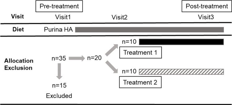
Dogs recruited for the clinical trial were seen at four separate visits: visit 1 (recruitment, inclusion and dispensing of trial capsules once histopathological results of intestinal biopsies were available approximately 10 days later), visit 2 (14 ±3 days after starting the trial medication), visit 3 (70+/- 2 days after starting the trial medication), and visit 7 (126 ±3 days after starting the trial medication; see also Fig 1).

Fig 1. Study design.

Fig 1

A total of thirty-five dogs were enrolled and fifteen dogs were excluded. Each box represents the period when the dogs were on Purina HA diet (grey), Treatment 1 with GAG (black), and Treatment 2 with placebo (upward diagonal).

The procedures performed at each visit can be seen in Table 1.

Table 1. Outline of procedures performed at each visit.
Visit Number Physical examination (general and IBD) CCECAI Blood sample Urine sample Faecal sample (Diagnostic) Ultra-sound Endo-scopy
Visit 1
Visit 2
Visit 3
Visit 7

After confirmation of the diagnosis, patients were randomized to receive either the product or placebo in addition to the hydrolyzed diet (Purina H/A, St. Louis, MO, USA). Patients were withdrawn from the study if their CCECAI at visit 2 or 3 increased more than 30% from the previous visit, if they suffered from an adverse event associated with the study medication or if owners could not adhere to the daily medication routine and/ or attend scheduled study visits.

Randomization and blinding

The patients were discharged after visist 1 from the hospital once they had recovered sufficiently from anaesthesia. The owners were then told to feed the diet exclusively whilst awaiting biopsy results. Owners were contacted approximately one-week post discharge to assess response to diet. Dogs who were diet-responsive and met all other inclusion criteria were enrolled and owners told to start study medication. Dogs who had not responded to the hypoallergenic diet were fed Purina HA for a further week, reassessed and then enrolled or excluded from the study. If the dog was not enrolled onto the study, its treatment allocation was added to the end of the randomized treatment allocation protocol (RTAP) and the treatment re-allocated. Recruitment continued until twenty dogs had been enrolled with 10 dogs in each group. Both the dogs’ owners and evaluators (including clinicians and those analyzing samples and tissues) had no knowledge of the treatment group assignments. The study clinical trial nursing staff acted as dispensers and were not blinded. Randomization was performed using block randomization with an allocation ratio of one-to-one, a random number generator in excel and a block size of 4. No patients required un-blinding during the course of the study.

Assessment of clinical severity

Clinical severity was assessed using the CCECAI or CIBDAI at all 4 visits as described previously [1].

Assessment of relapse rate after switching back to normal diet

After visit 3, all dogs were switched back to their normal diet by gradually mixing the hydrolyzed diet with the normal diet over 1 week. The owners were called weekly between visit 3 and visit 7 in order to assess clinical signs (CIBDAI) via questionnaire. Patients were considered to have relapsed if their CCECAI at visit 4 through 7 increased more than 30% from the previous visit [11].

Endoscopy and endoscopic scoring of lesions

Gastroduodenoscopy, ileoscopy, and colonoscopy were performed according to standard operating procedures using the available video-endoscopes appropriate for the respective dog’s size. Macroscopic scoring of endoscopic findings was recorded using the World Small Animal Veterinary Association (WSAVA) approved endoscopic examination report [12]. Multiple biopsies (15–20) were taken with a single-use endoscopic biopsy forceps from both the duodenum, ileum and colon and 10–15 biopsies from each site were transferred immediately into 2% neutral-buffered formalin.

Histopathology

Initial histopathological assessment was performed at the diagnostic pathology department of the RVC as part of the routine diagnostic procedure. However, after finalizing the study, all slides were reviewed by one of the authors (SLP), when mWSAVA standardized classification of all slides was performed [10, 12]. At this stage, SLP was blinded to the original diagnosis, the visit number and treatment group of the animals. mWSAVA scores [10] were recorded as total scores (summation of scores for duodenum, ileum and colon; mWSAVATotal) per dog and time point, as well as site-specific mWSAVA sub-scores for fundic and antral stomach scores (fundus: SFSUBTOTAL; antrum: SASUBTOTAL), duodenum (DSUBTOTAL), ileum (ISUBTOTAL), and colon (CSUBTOTAL), per dog and time point.

Analysis of fecal Short Chain Fatty Acid (SCFA) concentrations

Fecal SCFA concentrations were determined using gas chromatography (3800 Varian GC, Agilent Technologies, Santa Clara, CA). For each sample, 2.5 mL of distilled water was added to one gram of thawed feces in a conical tube and vortexed. Samples were centrifuged at 4°C for 10 min at 2,000 × g for supernatant removal. One mL of supernatant was then transferred into a 1.5 mL centrifuge tube and mixed with 0.2 mL of metaphosphoric acid for deproteinizing and 0.1 mL of isocaproic acid as an internal standard (48.3 mM; Sigma-Aldrich, Saint Louis, MO). A standard curve was generated using five concentrations of acetate, butyrate, propionate, valerate, isovalerate, and isobutyrate (Sigma-Aldrich, Saint Louis, MO). The tubes were then centrifuged at 12,000 × g at 4°C for 25 min. Aliquots of the supernatant (1 mL) from standard and fecal samples were transferred to 1.5 mL GC vials and duplicate injections of 100 uL were used by the GC for analysis. A flame ionization detector was used with an oven temperature of 60–200°C. The Nukol capillary column (15 m x 0.25 mm x 0.25 μm; Sigma-Aldrich‎, Bellefonte, PA) was operated with highly purified He, as the carrier gas, at 1 mL/min. Concentrations of acetate, butyrate, propionate, valerate, isovalerate, and isobutyrate were calculated using the ratio of the peak area of each compound to the internal standard and standard curve regression. Molar proportions of SCFA (%) were calculated as the individual SCFA / total SCFA concentration × 100.

Statistical analyses

Statistical analysis was performed using R software version 3.5.1 (R Foundation for Statistical Computing, Vienna, Austria). Normality was tested for each variable using Shapiro-Wilk for all parametric approaches. When the assumption did not hold, the nonparametric test (Mann-Whitney) was used for the variables measured only at one (visit = 1) or two time points (visits = 1,3) to compare differences between two groups. With variables involving multiple time points (more than two), each outcome was analyzed using a linear mixed effects model for group comparison. For each analysis, time, group, and their interactions were included as a fixed effect, whereas each dog was treated as a random effect. To test the correlation between variables of interest, spearman’s rank correlation coefficients and the corresponding p-values were calculated. For all statistical analyses, a p-value < 0.05 was considered significant.

Results

All raw data pertaining to clinical data of the study can be found in S2 File.

Animals

Of the 35 dogs enrolled into the clinical trial, 10 in each group reached the point of second endoscopy (visit 3). A total of 13 dogs (n = 8 in the group 1 and n = 5 in group 2) reached the trial endpoint of 18 weeks (visit 7). The demographics of the dogs are shown in Table 2. There were no differences in median age (group 1: 64.40 months ± 40.10 (57.13 ± 36.92); group 2: 42.40 months ± 22.43 (35.00 ± 14.53); p = 0.7) breed, or sex between the treatment groups.

Table 2. Demographics of dogs included into the study.

Group Age in months (at Visit 1) Gender Breed
1 25 FN Lurcher
1 44 FN Cross Breed
1 56 FE Bearded Collie
1 81 FN Staffordshire Bull Terrier
1 120 FN Cocker Spaniel
1 14 ME Cockapoo
1 29 ME Border Collie
1 53 MN Labrador Retriever
1 91 MN Labrador Retriever
1 131 MN Staffordshire Bull Terrier
2 19 FE Whippet
2 54 FE German Shepherd
2 22 ME Boxer
2 23 ME Border Collie
2 23 MN French Bulldog
2 36 ME Collie X
2 36 MN Labradoodle
2 58 MN Lhasa Apso
2 73 MN Staffordshire Bull Terrier
2 80 ME German Shepherd

Six/35 dogs did not respond to the dietary treatment and were not included in the trial. Of the remaining 29 dogs, 9 dogs discontinued the study for the following reasons: Five dogs were withdrawn because of worsening of clinical signs (one dog prior to visit 2, and 4 dogs prior to visit 3). In addition, one dog was withdrawn because the dog refused to take the test compound, and one dog was withdrawn prior to visit 3 because it required treatment with antibiotics for a skin condition. Two further dogs were excluded from the trial: One dog was diagnosed with adenocarcinoma after enrollment, and one dog was withdrawn because of owner non-compliance.

Clinical severity and biochemical parameters

All dogs responded quickly and sustained to dietary treatment for 10 weeks. There were no significant differences in CCECAI or CIBDAI between treatment groups from visit 1 to 7 (mean ± SD CCECAI for group 1: visit 1: 4.90 ± 1.52, visit 3: 2.25 ± 2.19, visit 7: 1.38 ± 0.74; for group 2: visit 1: 6.00 ± 2.94, visit 3: 2.2 ± 1.1, visit 7: 1.8 ± 2.49; mean ±SD CIBDAI for group 1: visit 1: 5.00 1.63, visit 3: 2.25 ±2.19, visit 7: 1.12 ± 035; for group 2: visit 1: 5.6 ±2.99, visit 3: 2.00 ± 1.41, visit 7: 1.8 ±2.49). Although there was a slight clinical worsening in the placebo group after switching back to the original diet (visit 3 to visit 7), this was not statistically significant (CCECAI p = 0.79; CIBDAI p = 0.65) (see Fig 2).

Fig 2. Group comparison of average (±sd) CCECAI and CIBDAI over time.

Fig 2

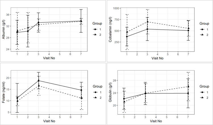
None of the dogs had serum albumin, globulin, cobalamin and folate concentrations outside of the references ranges at any time point throughout the study. Serum TLI and PLI were only measured once at visit 1 and were all within reference ranges. Mean serum albumin and globulin concentrations increased from visit 1 to visit 3 and then again to visit 7, although this was not statistically significant (mean ± SD serum albumin concentrations for group 1: visit 1: 29 ± 4.19, visit 3: 33.25 ± 3.35, visit 7: 33.79 ± 3.98; group 2: visit 1: 30.32 ±6.41, visit 3: 32.52 ±1.98, visit 7: 33.66 ±1.02; mean ± SD serum globulin concentration for group 1: visit 1: 22.12 ± 3.39, visit 3: 23. 8±3.58, visit 7: 23.86 ±4.66; for group 2: visit 1: 21.13 ±3.41, visit 3: 23.8 ±3.58, visit 7: 23.86 ±4.66; for group 2: visit 1: 21.12 ±3.41, visit 3: 23.9 ±3.04, visit 7: 26.02 ±4.55). Serum folate concentration increased from visit 1 to visit 3, and then decreased again slightly at visit 7 (serum folate concentration ± SD for group 1: visit 1: 11.22 ± 6.35, visit 3: 18.79 ± 3.62, visit 7: 14.64 ± 3.43; for group 2: visit 1: 9.85 ±2.07, visit 3: 16.54 ±4.19, visit 7: 11.16 ±5.03). Furthermore, there were no group effects on average for serum albumin (p = 0.26), cobalamin (p = 0.71), folate (p = 0.35), and globulin concentration over time (p = 0.86) (See Fig 3).

Fig 3. Group comparison of average (±sd) for serum concentrations of albumin, cobalamin, folate and globulin over time.

Fig 3

Endoscopy scores

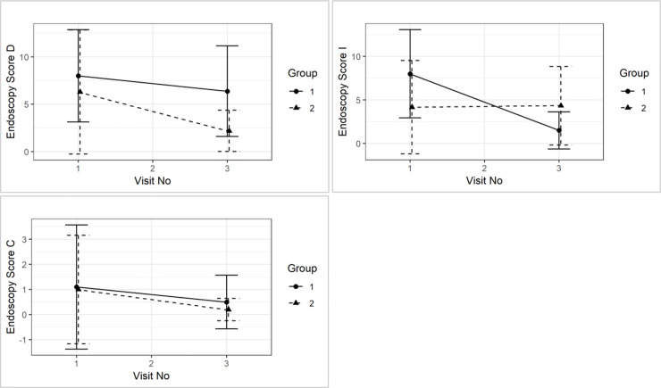
Endoscopy scores in the duodenum and ileum were higher than those in the colon at diagnosis (Endoscopy score D Group 1: Mean = 8.0, Median = 9.0 at visit 1; Mean = 6.4, Median = 5.0 at visit 3; Group 2: Mean = 6.3, Median = 3.5 at visit 1; Mean = 2.2, Median = 3.0 at visit 3. Endoscopy score I group 1: Mean = 8.0, Median Mean = 1.1, Median = 0.0 at visit 1; Mean = 0.5, Median = 0.0 at visit 3; Mean = 1.0, Median = 0.0 at visit 1; Mean = 0.2, Median = 0.0 at visit 3).

Although all scores reduced numerically from visit 1 to visit 3, this was not statistically significant over time. There were also no significant time and group interactions in endoscopy scores for the duodenum (p = 0.38), ileum (p = 0.71), and colon (p = 0.75) (see Fig 4).

Fig 4. Group comparison of mean (±sd) endoscopy score for duodenum (D), ileum (I), and colon (C) between visit 1 and visit 3 and between treatment groups.

Fig 4

Modified WSAVA histology scores

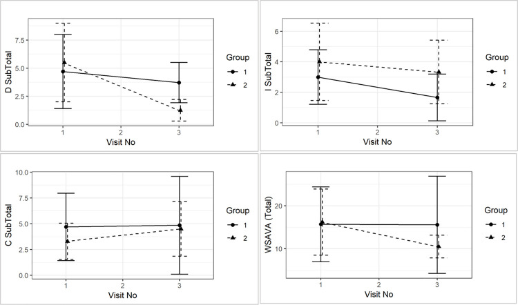
There were no differences between mWSAVATotal, nor DSUBTOTAL, ISUBTOTAL and CSUBTOTAL at the time of diagnosis. Similarly, no statistically significant differences between groups were detected in mWSAVATotal (p = 0.40), nor DSUBTOTAL (p = 0.16), ISUBTOTAL (p = 0.92) and CSUBTOTAL (p = 0.47) over time (see Fig 5).

Fig 5. Comparison of mWSAVATotal (p = 0.8), nor DSUBTOTAL (p = 0.2), ISUBTOTAL (p = 01.0) and CSUBTOTAL (p = 0.6) between visit 1 and visit 3 and between treatment groups.

Fig 5

Correlation analyses

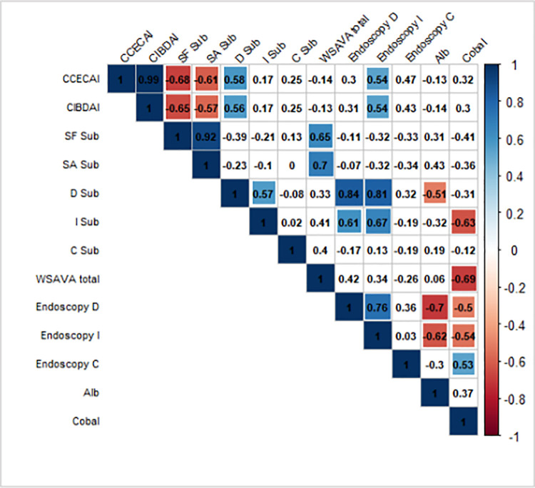
A graphical display of a correlation matrix is given in Fig 6 for the variables CCECAI, CIBDAI, mWSAVA SF, SA, as well as mWSAVA DSUBTOTAL,, ISUBTOTAL, and CSUBTOTAL scores. Modified mWSAVATotal, as well as endoscopic scores for duodenum, ileum and colon, and serum albumin and cobalamin concentrations.

Fig 6. Pearson correlation coefficients (combining all the visits and groups 1 and 2) using corrplot package in R.

Fig 6

Positive correlations are displayed in blue color and negative correlations are in red. Additional colors (corresponding to their correlation values) are added in the plot when the test for linear association between paired variables results in p-value < 0.05 (p-values are not shown in the plot). CCECAI = Canine Chronic Enteropathy Clinical Activity Index; CIBAI = Canine IBD Activity Index; SF sub = Stomach fundic mWSAVA subscore; SA = Stomach antral mWSAVA subscore; D sub = Duodenum subscore mWSAVA; I sub = Ileum subscore mWSAVA; C sub = Colon subscore WSAVA; mWSAVA total = combined mWSAVA subscores for all intestinal sites; Endoscopy D = Endoscopy subscore duodenum; Endoscopy I = Endoscopy subscore Iluem; Endoscopy C = Endoscopy subscore Colon; Alb = serum albumin ocnentration; Cobal = serum cobalamin concentration.

There were significant positive correlations between mWSAVA DSUBTOTAL and mWSAVA ISUBTOTAL scores, as well as between duodenal and ileal endoscopy scores. Furthermore, there was a significant positive correlation found between serum albumin and serum cobalamin concentrations within individual dogs.

Post hoc power analysis

A post-hoc sample size calculation was performed based on the slope of CCECAI and CIBDAI scores between groups using visits 2 and 7, in ordert o detrmine the sample size for detecting a difference in relapse rates between the groups after switching back to normal diet. Based on CCECAI scores, the sample size required to detect a significant difference between groups would have been 238 per group for each time point. Based on CIBDAI scores, the sample size required would have been 63 per group (for each time point).

Fecal SCFA concentrations

Fecal SCFA concentrations were determined at visits 1, 2, 3 and 7 in 14 of the dogs, 9 of which dogs were in group 1 and 5 of which were in group 2. No statistically significant differences were found in fecal concentrations of acetate, butyrate, propionate, valerate, isovalerate, and isobutyrate fecal concentrations over time and between treatment groups (see Fig 7).

Fig 7. Fecal SCFA (acetate, butyrate, propionate, valerate, isovalerate, and isobutyrate) concentrations over time and between groups.

Fig 7

Raw data from the fecal SCFA analysis can be found in S1 File.

Discussion

The objective of the current study was to test the in-vivo clinical effects of the oral administration of GAGs and prebiotics as a supplemental treatment to hydrolyzed diet in dogs with FRD on the relapse rate of FRD dogs after switching back to normal diet.

Most dogs were medium sized breeds with a mean age of around 4.4 years, which is concordant with other recent studies on FRD dogs [2]. The severity of clinical signs was mild to moderate and reduced significantly at visit 2 and 3, regardless of product or placebo treatment, which confirms the diagnosis of FRD. After switching back to the normal diet at visit 3 and until visit 7, none of the dogs experienced any clinical relapse in either group. Therefore, a meaningful assessment of whether the adjunct treatment had an effect on the relapse rate in these dogs cannot be made from this sample population.

No changes in biochemical parameters were noted between the groups over time, nor for endoscopic scores or histological scores, which is consistent with previous reports in FRD dogs [1, 1315]. We did find a strong correlation between serum albumin and serum cobalamin concentration in the individual dogs, which also concurs with previous publications [1]. Furthermore, endoscopic and histological scores did not correlate with clinical scores in the dogs over time, which is a fact that has also been noted in previous studies [1, 9, 16].

However, we found strong positive correlations between duodenal and ileal endoscopic scores, as well as mWSAVA sub-scores for duodenum and ileum. This is a novel finding in our study and may represent the notion that FRD dogs present with diffuse small intestinal inflammation with similar severity in the duodenum and ileum. In a recent study, it was shown that sub-scores of mWSAVA for each intestinal site (duodenum, ileum and colon) correlate better with clinical activity of disease than the total modified mWSAVA score including sub-scores of all sites [10]. Furthermore, endoscopic subscores for the duodenum and ileum in this study were higher than those for the colon at the time of diagnosis. This may represent the fact that most dogs in this study presented with predominantly small intestinal signs, and/or may be due to the fact that endoscopy scores in the small intestine seem to correlate better with clinical activity scores [1, 15].

Fecal SCFA did not show any significant changes over time or between groups in this study. It is possible that the effect of the diet was substantial and therefore, a minor effect of the supplement might have been missed.

Unfortunately, even though a substantial number of potential cases for the clinical trial were screened (n = 35), only 20 dogs (10 of each group) were enrolled, and only 13 finished the study. This resulted in the study being underpowered, with statistically significant differences in CIBDAI scores between groups expected at a minimum number of n = 63 per group. Nevertheless, this study represents one of the few published double-blinded RCCT and will be informative for the design of future clinical trials in canine FRD.

Conclusion

Standard dietary treatment induced rapid clinical response in all cases. Because the study was underpowered, it was not possible to determine whether or not supplementation with prebiotic and GAG had an additional effect on clinical outcomes or frequency of relapses after switching back to normal diet.

Supporting information

S1 File. This file contains all clinical raw data fort he trial.

(PDF)

S2 File. This file contains all statistical output data.

(PDF)

S3 File. This file contains all statistical output for the analyses of fecal Short Chain Fatty Acids concentrations.

(PDF)

Acknowledgments

The authors would like to thank the owners of the dogs participating in this study for taking part; as well as the help of the staff of the Queen Mother Hospital for Animals (QMHA) and the Clinical Investigation Centre of the Royal Veterinary College (RVC) for their support in recruiting cases and acquiring all necessary samples.

The work presented in this manuscript has been performed at the Royal Veterinary College, London, UK.

This paper was presented as an abstract at SEVC 2019, Spain.

Data Availability

All relevant data are within the manuscript and its Supporting Information files.

Funding Statement

This clinical trial study was funded by Bioiberica S.A.U., Barcelona, Spain.

References

  • 1.Allenspach K, Wieland B, Grone A, Gaschen F. Chronic enteropathies in dogs: evaluation of risk factors for negative outcome. J Vet Intern Med. 2007. Aug;21:700–8. doi: 10.1892/0891-6640(2007)21[700:ceideo]2.0.co;2 [DOI] [PubMed] [Google Scholar]
  • 2.Allenspach K. Long-term outcome in dogs with chronic enteropathies: 203 cases. Veterinary Record, in press. 2016; [DOI] [PubMed]
  • 3.Volkmann M, Steiner JM, Fosgate GT, Zentek J, Hartmann S, Kohn B. Chronic Diarrhea in Dogs—Retrospective Study in 136 Cases. J Vet Intern Med. 2017. Jul;31(4):1043–55. doi: 10.1111/jvim.14739 [DOI] [PMC free article] [PubMed] [Google Scholar]
  • 4.Ambrosini YM, Neuber S, Borcherding D, Seo Y-J, Segarra S, Glanemann B, et al. Treatment With Hydrolyzed Diet Supplemented With Prebiotics and Glycosaminoglycans Alters Lipid Metabolism in Canine Inflammatory Bowel Disease. Front Vet Sci. 2020;7:451. doi: 10.3389/fvets.2020.00451 [DOI] [PMC free article] [PubMed] [Google Scholar]
  • 5.Segarra S, Martinez-Subiela S, Cerda-Cuellar M, Martinez-Puig D, Munoz-Prieto A, Rodriguez-Franco F, et al. Oral chondroitin sulfate and prebiotics for the treatment of canine Inflammatory Bowel Disease: a randomized, controlled clinical trial. BMC Vet Res. 2016;12:49. doi: 10.1186/s12917-016-0676-x [DOI] [PMC free article] [PubMed] [Google Scholar]
  • 6.Coppa GV, Facinelli B, Magi G, Marini E, Zampini L, Mantovani V, et al. Human milk glycosaminoglycans inhibit in vitro the adhesion of Escherichia coli and Salmonella fyris to human intestinal cells. Pediatr Res. 2016. Apr;79(4):603–7. doi: 10.1038/pr.2015.262 [DOI] [PubMed] [Google Scholar]
  • 7.Liu F, Zhang N, Li Z, Wang X, Shi H, Xue C, et al. Chondroitin sulfate disaccharides modified the structure and function of the murine gut microbiome under healthy and stressed conditions. Sci Rep. 2017. 28;7(1):6783. doi: 10.1038/s41598-017-05860-6 [DOI] [PMC free article] [PubMed] [Google Scholar]
  • 8.Riehl TE, Santhanam S, Foster L, Ciorba M, Stenson WF. CD44 and TLR4 mediate hyaluronic acid regulation of Lgr5+ stem cell proliferation, crypt fission, and intestinal growth in postnatal and adult mice. Am J Physiol Gastrointest Liver Physiol. 2015. Dec 1;309(11):G874–887. doi: 10.1152/ajpgi.00123.2015 [DOI] [PMC free article] [PubMed] [Google Scholar]
  • 9.Jergens AE, Schreiner CA, Frank DE, Niyo Y, Ahrens FE, Eckersall PD, et al. A scoring index for disease activity in canine inflammatory bowel disease. JVetInternMed. 2003;17:291–7. doi: 10.1111/j.1939-1676.2003.tb02450.x [DOI] [PubMed] [Google Scholar]
  • 10.Allenspach KA, Mochel JP, Du Y, Priestnall SL, Moore F, Slayter M, et al. Correlating Gastrointestinal Histopathologic Changes to Clinical Disease Activity in Dogs With Idiopathic Inflammatory Bowel Disease. Vet Pathol. 2019;56(3):435–43. doi: 10.1177/0300985818813090 [DOI] [PubMed] [Google Scholar]
  • 11.White R, Atherly T, Guard B, Rossi G, Wang C, Mosher C, et al. Randomized, controlled trial evaluating the effect of multi-strain probiotic on the mucosal microbiota in canine idiopathic inflammatory bowel disease. Gut Microbes. 2017. 03;8(5):451–66. doi: 10.1080/19490976.2017.1334754 [DOI] [PMC free article] [PubMed] [Google Scholar]
  • 12.Day MJ, Bilzer T, Mansell J, Wilcock B, Hall EJ, Jergens A, et al. Histopathological standards for the diagnosis of gastrointestinal inflammation in endoscopic biopsy samples from the dog and cat: a report from the World Small Animal Veterinary Association Gastrointestinal Standardization Group. J Comp Pathol. 2008. Apr;138 Suppl 1:S1–43. [DOI] [PubMed] [Google Scholar]
  • 13.Schreiner NM, Gaschen F, Grone A, Sauter SN, Allenspach K. Clinical signs, histology, and CD3-positive cells before and after treatment of dogs with chronic enteropathies. J Vet Intern Med. 2008. Oct;22:1079–83. doi: 10.1111/j.1939-1676.2008.0153.x [DOI] [PubMed] [Google Scholar]
  • 14.Garcia-Sancho M, Rodriguez-Franco F, Sainz A, Mancho C, Rodriguez A. Evaluation of clinical, macroscopic, and histopathologic response to treatment in nonhypoproteinemic dogs with lymphocytic-plasmacytic enteritis. J Vet Intern Med. 2007. Feb;21:11–7. doi: 10.1892/0891-6640(2007)21[11:eocmah]2.0.co;2 [DOI] [PubMed] [Google Scholar]
  • 15.Procoli F, Motskula PF, Keyte SV, Priestnall S, Allenspach K. Comparison of histopathologic findings in duodenal and ileal endoscopic biopsies in dogs with chronic small intestinal enteropathies. J Vet Intern Med. 2013. Mar;27:268–74. doi: 10.1111/jvim.12041 [DOI] [PubMed] [Google Scholar]
  • 16.Walker D. A comprehensive pathological survey of duodenal biopsies from dogs with diet-responsive chronic inflammatory enteropathy. Journal of Veterinary Internal Medicine. 2013; in press. [DOI] [PubMed] [Google Scholar]

Decision Letter 0

Mathilde Body-Malapel

24 Jun 2021

PONE-D-21-11631

Clinical Efficacy of Prebiotics and Glycosaminoglycans versus Placebo In Dogs with Food Responsive Enteropathy Receiving a Hydrolyzed Diet: A Pilot Study

PLOS ONE

Dear Dr. Allenspach,

Thank you for submitting your manuscript to PLOS ONE. After careful consideration, we feel that it has merit but does not fully meet PLOS ONE’s publication criteria as it currently stands. Therefore, we invite you to submit a revised version of the manuscript that addresses the points raised during the review process.

PLOS ONE welcomes primary research that contributes to the base of scientific knowledge, including negative or null results. The important point is that the data presented in the manuscript must support the conclusions drawn and the authors should avoid overstating their conclusions. We believe that this is the case for your manuscript. Therefore, we do not request you to follow the first major comment of the reviewer 2.

Please submit your revised manuscript  within 2 months. If you will need more time than this to complete your revisions, please reply to this message or contact the journal office at plosone@plos.org. When you're ready to submit your revision, log on to https://www.editorialmanager.com/pone/ and select the 'Submissions Needing Revision' folder to locate your manuscript file.

Please include the following items when submitting your revised manuscript:

  • A rebuttal letter that responds to each point raised by the academic editor and reviewer(s). You should upload this letter as a separate file labeled 'Response to Reviewers'.

  • A marked-up copy of your manuscript that highlights changes made to the original version. You should upload this as a separate file labeled 'Revised Manuscript with Track Changes'.

  • An unmarked version of your revised paper without tracked changes. You should upload this as a separate file labeled 'Manuscript'.

If you would like to make changes to your financial disclosure, please include your updated statement in your cover letter. Guidelines for resubmitting your figure files are available below the reviewer comments at the end of this letter.

If applicable, we recommend that you deposit your laboratory protocols in protocols.io to enhance the reproducibility of your results. Protocols.io assigns your protocol its own identifier (DOI) so that it can be cited independently in the future. For instructions see: http://journals.plos.org/plosone/s/submission-guidelines#loc-laboratory-protocols. Additionally, PLOS ONE offers an option for publishing peer-reviewed Lab Protocol articles, which describe protocols hosted on protocols.io. Read more information on sharing protocols at https://plos.org/protocols?utm_medium=editorial-email&utm_source=authorletters&utm_campaign=protocols.

We look forward to receiving your revised manuscript.

Kind regards,

Mathilde Body-Malapel

Academic Editor

PLOS ONE

Journal Requirements:

When submitting your revision, we need you to address these additional requirements.

1. Please ensure that your manuscript meets PLOS ONE's style requirements, including those for file naming. The PLOS ONE style templates can be found at 

https://journals.plos.org/plosone/s/file?id=wjVg/PLOSOne_formatting_sample_main_body.pdf and 

https://journals.plos.org/plosone/s/file?id=ba62/PLOSOne_formatting_sample_title_authors_affiliations.pdf

2. In your Methods section, please provide additional details regarding participant consent from the owners of the animals. In the ethics statement in the Methods and online submission information, please ensure that you have specified (1) whether consent was informed and (2) what type you obtained (for instance, written or verbal). If the need for consent was waived by the ethics committee, please include this information.

3. Thank you for stating the following in the Acknowledgments Section of your manuscript: 

This clinical trial study was funded by Bioiberica S.A.U., Barcelona, Spain.

We note that you have provided funding information that is not currently declared in your Funding Statement. However, funding information should not appear in the Acknowledgments section or other areas of your manuscript. We will only publish funding information present in the Funding Statement section of the online submission form. 

Please remove any funding-related text from the manuscript and let us know how you would like to update your Funding Statement. Currently, your Funding Statement reads as follows: 

This clinical trial study was funded by Bioiberica S.A.U., Barcelona, Spain.

Please include your amended statements within your cover letter; we will change the online submission form on your behalf.

4. We note that you have stated that you will provide repository information for your data at acceptance. Should your manuscript be accepted for publication, we will hold it until you provide the relevant accession numbers or DOIs necessary to access your data. If you wish to make changes to your Data Availability statement, please describe these changes in your cover letter and we will update your Data Availability statement to reflect the information you provide.

5. Please note that in order to use the direct billing option the corresponding author must be affiliated with the chosen institute. Please either amend your manuscript to change the affiliation or corresponding author, or email us at plosone@plos.org with a request to remove this option.

6. PLOS requires an ORCID iD for the corresponding author in Editorial Manager on papers submitted after December 6th, 2016. Please ensure that you have an ORCID iD and that it is validated in Editorial Manager. To do this, go to ‘Update my Information’ (in the upper left-hand corner of the main menu), and click on the Fetch/Validate link next to the ORCID field. This will take you to the ORCID site and allow you to create a new iD or authenticate a pre-existing iD in Editorial Manager. Please see the following video for instructions on linking an ORCID iD to your Editorial Manager account: https://www.youtube.com/watch?v=_xcclfuvtxQ

Additional Editor Comments:

Please review your reference list to ensure that it is complete and correct. If you have cited papers that have been retracted, please include the rationale for doing so in the manuscript text, or remove these references and replace them with relevant current references. Any changes to the reference list should be mentioned in the rebuttal letter that accompanies your revised manuscript. If you need to cite a retracted article, indicate the article’s retracted status in the References list and also include a citation and full reference for the retraction notice.

[Note: HTML markup is below. Please do not edit.]

Reviewers' comments:

Reviewer's Responses to Questions

Comments to the Author

1. Is the manuscript technically sound, and do the data support the conclusions?

The manuscript must describe a technically sound piece of scientific research with data that supports the conclusions. Experiments must have been conducted rigorously, with appropriate controls, replication, and sample sizes. The conclusions must be drawn appropriately based on the data presented.

Reviewer #1: Yes

Reviewer #2: No

**********

2. Has the statistical analysis been performed appropriately and rigorously?

Reviewer #1: Yes

Reviewer #2: Yes

**********

3. Have the authors made all data underlying the findings in their manuscript fully available?

The PLOS Data policy requires authors to make all data underlying the findings described in their manuscript fully available without restriction, with rare exception (please refer to the Data Availability Statement in the manuscript PDF file). The data should be provided as part of the manuscript or its supporting information, or deposited to a public repository. For example, in addition to summary statistics, the data points behind means, medians and variance measures should be available. If there are restrictions on publicly sharing data—e.g. participant privacy or use of data from a third party—those must be specified.

Reviewer #1: Yes

Reviewer #2: Yes

**********

4. Is the manuscript presented in an intelligible fashion and written in standard English?

PLOS ONE does not copyedit accepted manuscripts, so the language in submitted articles must be clear, correct, and unambiguous. Any typographical or grammatical errors should be corrected at revision, so please note any specific errors here.

Reviewer #1: Yes

Reviewer #2: Yes

**********

5. Review Comments to the Author

Please use the space provided to explain your answers to the questions above. You may also include additional comments for the author, including concerns about dual publication, research ethics, or publication ethics. (Please upload your review as an attachment if it exceeds 20,000 characters)

Reviewer #1: Abstract- was it number of relapses that was evaluated? or was it simply whether or not they relapsed?

Introduction-

-reference missing for "relapses can occur when dogs are switched back to their normal diet after the trial elimination diet - text says (Ref)

-The statement " Therefore, supplemental treatments in addition to the diet may help reduce the number of relapses in these dogs after switching them back to their normal diet" seems out of place. It would make more sense to make this argument AFTER you discuss the benefits of GAGs on intestinal epithelial cells, etc. Suggesting moving this statement to directly after ".. oxidative stress over a 18-week treatment period.."

-... "have been observed in mice as well as human colon cancer cell lines after treatment with GAGs (reference typo - "34, 3).

-End of introduction- I think it would be a good idea to present to the reader at least some brief information as to WHY you wanted to compare fecal short chain fatty acids and the other parameters between groups.

Materials & Methods-

Endoscopy and endoscopic scoring lesions- says multiple biopsies were taken from duodenum and colon, but no mention of ileum?

Page 9- typo - "visit"

-I'm a little confused about enrollment because in the abstract it says 35 dogs were enrolled but here on page 9 it says recruitment continued until twenty dogs had been enrolled, 10 dogs in each group

Results-

Page 13- I would suggest making a flow diagram that outlines- how many dogs were screened, how many dogs initially enrolled, how many dogs completed the full study and include reasons for patient drop out

Page 16- typo-- "order to"

Discussion- well written and not overstated

Figures- adequate and appropriate

Reviewer #2: This pilot study aimed to clarify the effect of supplemental treatment with prebiotics and glycosaminoglycans (GAGs) in the relapse rate of food responsive disease (FRD) in dogs that were switched back to a normal diet in a randomized, controlled clinical trial (RCCT).

Major comments:

1. Because there were no significant differences in clinical outcomes between supplemental treatment with prebiotics and GAGs (group 1) and placebo (group 2), it is impossible to conclude that the effect of prebiotics and GAGs in the relapse rate of FRD. Without conclusions, this study would not provide any scientific significance in this field. To determine the effect of prebiotics and GAGs, this study must be conducted by using a larger sample size, as suggested by the authors.

2. This pilot study did not find any significant effects of prebiotics and GAGs on the relapse rate and fecal SCFA profiles in FRD dogs. Thus, it is unclear whether prebiotics and GAGs really improved intestinal environment in the GI tract of dogs. According to the introduction, GAGs have beneficial effects on intestinal barrier functions, microbiome, and fecal short chain fatty acid (SCFA). The authors, therefore, must analyze expression of tight junction proteins in the intestinal epithelial cells and gut microbiome in dogs that received prebiotics and GAGs or placebo.

3. Page 7, Inclusion and exclusion criteria:

To justify the diagnosis and ensure the evenness of group 1 and 2, please provide hematology and serum biochemistry data including TLI, cPLI, and ACTH-stimulation test at visit 1 and compare them between two groups.

4. Page 10, Assessment of relapse rate after switching back to normal diet: “Patients were considered to have relapsed if their CCECAI at visit 4 through 7 increased more than 30% from the previous visit.”

Please provide the scientific evidence for the relapse definition used in this study.

Minor comments:

1. Page 1: FRD is not the abbreviation of “Food Responsive Chronic Enteropathy”.

2. Page 5: Please check the references in the following sentences:

(a) While induction of remission is easily achieved with dietary treatment in dogs with FRD, relapses can occur when the dogs are switched back to their normal diet after the trial elimination diet (Ref).

(b) The effect of GAGs on enhancing the barrier function of the intestinal epithelium, has been shown to protect animals from translocation of pathogenic bacteria and LPS into the peripheral blood6,7,6,8.

3. The authors cited 15 references, but I can find reference no. more than 15 in the text. Please check the manuscript thoroughly.

4. Page 13-14, Clinical severity and biochemical parameters:

Please describe reference ranges of biochemical parameters in the text.

**********

6. PLOS authors have the option to publish the peer review history of their article (what does this mean?). If published, this will include your full peer review and any attached files.

If you choose “no”, your identity will remain anonymous but your review may still be made public.

Do you want your identity to be public for this peer review? For information about this choice, including consent withdrawal, please see our Privacy Policy.

Reviewer #1: No

Reviewer #2: No

[NOTE: If reviewer comments were submitted as an attachment file, they will be attached to this email and accessible via the submission site. Please log into your account, locate the manuscript record, and check for the action link "View Attachments". If this link does not appear, there are no attachment files.]

While revising your submission, please upload your figure files to the Preflight Analysis and Conversion Engine (PACE) digital diagnostic tool, https://pacev2.apexcovantage.com/. PACE helps ensure that figures meet PLOS requirements. To use PACE, you must first register as a user. Registration is free. Then, login and navigate to the UPLOAD tab, where you will find detailed instructions on how to use the tool. If you encounter any issues or have any questions when using PACE, please email PLOS at figures@plos.org. Please note that Supporting Information files do not need this step.

PLoS One. 2021 Oct 21;16(10):e0250681. doi: 10.1371/journal.pone.0250681.r002

Author response to Decision Letter 0


25 Aug 2021

Rebuttal letter to editor and reviewers of PONE-D-21-11631:

Clinical Efficacy of Prebiotics and Glycosaminoglycans versus Placebo In Dogs with Food Responsive Enteropathy Receiving a Hydrolyzed Diet: A Pilot Study

1. Please ensure that your manuscript meets PLOS ONE's style requirements, including those for file naming. The PLOS ONE style templates can be found at

https://journals.plos.org/plosone/s/file?id=wjVg/PLOSOne_formatting_sample_main_body.pdf and

https://journals.plos.org/plosone/s/file?id=ba62/PLOSOne_formatting_sample_title_authors_affiliations.pdf

Many thanks for this comment- this has been done.

2. In your Methods section, please provide additional details regarding participant consent from the owners of the animals. In the ethics statement in the Methods and online submission information, please ensure that you have specified (1) whether consent was informed and (2) what type you obtained (for instance, written or verbal). If the need for consent was waived by the ethics committee, please include this information.

Many thanks for this comment- this has been added to the section: Line 133: Written owner consent was obtained for all study participants.

3. Thank you for stating the following in the Acknowledgments Section of your manuscript:

This clinical trial study was funded by Bioiberica S.A.U., Barcelona, Spain.

We note that you have provided funding information that is not currently declared in your Funding Statement. However, funding information should not appear in the Acknowledgments section or other areas of your manuscript. We will only publish funding information present in the Funding Statement section of the online submission form.

Please remove any funding-related text from the manuscript and let us know how you would like to update your Funding Statement. Currently, your Funding Statement reads as follows:

This clinical trial study was funded by Bioiberica S.A.U., Barcelona, Spain.

Many thanks for this comment. We have taken out the funding statement in the acknowledgement section and have now updated the Funding Statement.

Please include your amended statements within your cover letter; we will change the online submission form on your behalf.

4. We note that you have stated that you will provide repository information for your data at acceptance. Should your manuscript be accepted for publication, we will hold it until you provide the relevant accession numbers or DOIs necessary to access your data. If you wish to make changes to your Data Availability statement, please describe these changes in your cover letter and we will update your Data Availability statement to reflect the information you provide.

Many thanks for your comments. We have included all raw data and statistics data file as supplemental material.

5. Please note that in order to use the direct billing option the corresponding author must be affiliated with the chosen institute. Please either amend your manuscript to change the affiliation or corresponding author, or email us at plosone@plos.org with a request to remove this option.

6. PLOS requires an ORCID iD for the corresponding author in Editorial Manager on papers submitted after December 6th, 2016. Please ensure that you have an ORCID iD and that it is validated in Editorial Manager. To do this, go to ‘Update my Information’ (in the upper left-hand corner of the main menu), and click on the Fetch/Validate link next to the ORCID field. This will take you to the ORCID site and allow you to create a new iD or authenticate a pre-existing iD in Editorial Manager. Please see the following video for instructions on linking an ORCID iD to your Editorial Manager account: https://www.youtube.com/watch?v=_xcclfuvtxQ

Many thanks for this comment- this has been corrected.

Additional Editor Comments:

Please review your reference list to ensure that it is complete and correct. If you have cited papers that have been retracted, please include the rationale for doing so in the manuscript text, or remove these references and replace them with relevant current references. Any changes to the reference list should be mentioned in the rebuttal letter that accompanies your revised manuscript. If you need to cite a retracted article, indicate the article’s retracted status in the References list and also include a citation and full reference for the retraction notice.

Many thanks for this comment. We have gone through the manuscript and corrected all of the references.

Reviewer #1:

Abstract- was it number of relapses that was evaluated? or was it simply whether or not they relapsed?

Many thanks for this comment. Since none of the dogs in this study relapsed after being switched back to a normal diet, no statistics were performed on this.

Introduction-

-reference missing for "relapses can occur when dogs are switched back to their normal diet after the trial elimination diet - text says (Ref)

Many thanks for this comment – this has been added.

-The statement " Therefore, supplemental treatments in addition to the diet may help reduce the number of relapses in these dogs after switching them back to their normal diet" seems out of place. It would make more sense to make this argument AFTER you discuss the benefits of GAGs on intestinal epithelial cells, etc. Suggesting moving this statement to directly after ".. oxidative stress over a 18-week treatment period.."

Many thanks for this comment- this sentence has now been moved further down in the paragraph:

Line 101: The aim of the work presented here was therefore to assess whether the relapse rate of FRD dogs that are switched back to a normal diet can be influenced using supplemental treatment with prebiotics and GAGs.

-... "have been observed in mice as well as human colon cancer cell lines after treatment with GAGs (reference typo - "34, 3).

Many thanks for this comment- this has been corrected.

-End of introduction- I think it would be a good idea to present to the reader at least some brief information as to WHY you wanted to compare fecal short chain fatty acids and the other parameters between groups.

Many thanks for this comment. We have added the following sentence in line 114: Since GAGs have previously been shown to be able to modify SCFA concentrations in the feces, we also evaluated (SCFA) concentrations in the feces of the dogs in this study.

Materials & Methods-

Endoscopy and endoscopic scoring lesions- says multiple biopsies were taken from duodenum and colon, but no mention of ileum?

Many thanks for this comment- this has been corrected.

Page 9- typo - "visit"

Many thanks for this comment- this has been corrected.

-I'm a little confused about enrollment because in the abstract it says 35 dogs were enrolled but here on page 9 it says recruitment continued until twenty dogs had been enrolled, 10 dogs in each group

Many thanks for this comment. We have included more detail on the recruitment and drop-outs form the study: Line 269:

Six/35 dogs did not respond to the dietary treatment and were not included in the trial. Of the remaining 29 dogs, 9 dogs discontinued the study for the following reasons: Five dogs were withdrawn because of worsening of clinical signs (one dog prior to visit 2, and 4 dogs prior to visit 3). In addition, one dog was withdrawn because the dog refused to take the test compound, and one dog was withdrawn prior to visit 3 because it required treatment with antibiotics for a skin condition. Two further dogs were excluded from the trial: One dog was diagnosed with adenocarcinoma after enrollment, and one dog was withdrawn because of owner non-compliance.

Results-

Page 13- I would suggest making a flow diagram that outlines- how many dogs were screened, how many dogs initially enrolled, how many dogs completed the full study and include reasons for patient drop out

Many thanks for this comment. We have included more detail on the recruitment and drop-outs form the study: Line 269:

Six/35 dogs did not respond to the dietary treatment and were not included in the trial. Of the remaining 29 dogs, 9 dogs discontinued the study for the following reasons: Five dogs were withdrawn because of worsening of clinical signs (one dog prior to visit 2, and 4 dogs prior to visit 3). In addition, one dog was withdrawn because the dog refused to take the test compound, and one dog was withdrawn prior to visit 3 because it required treatment with antibiotics for a skin condition. Two further dogs were excluded from the trial: One dog was diagnosed with adenocarcinoma after enrollment, and one dog was withdrawn because of owner non-compliance.

Page 16- typo-- "order to"

Many thanks for this comment- this has been corrected.

Discussion- well written and not overstated

Figures- adequate and appropriate

Reviewer #2: This pilot study aimed to clarify the effect of supplemental treatment with prebiotics and glycosaminoglycans (GAGs) in the relapse rate of food responsive disease (FRD) in dogs that were switched back to a normal diet in a randomized, controlled clinical trial (RCCT).

Major comments:

1. Because there were no significant differences in clinical outcomes between supplemental treatment with prebiotics and GAGs (group 1) and placebo (group 2), it is impossible to conclude that the effect of prebiotics and GAGs in the relapse rate of FRD. Without conclusions, this study would not provide any scientific significance in this field. To determine the effect of prebiotics and GAGs, this study must be conducted by using a larger sample size, as suggested by the authors.

Many thanks for this comment. As suggested by editors, we have not replied to this comment.

2. This pilot study did not find any significant effects of prebiotics and GAGs on the relapse rate and fecal SCFA profiles in FRD dogs. Thus, it is unclear whether prebiotics and GAGs really improved intestinal environment in the GI tract of dogs. According to the introduction, GAGs have beneficial effects on intestinal barrier functions, microbiome, and fecal short chain fatty acid (SCFA). The authors, therefore, must analyze expression of tight junction proteins in the intestinal epithelial cells and gut microbiome in dogs that received prebiotics and GAGs or placebo.

Many thanks for this comment. As suggested by editors, we have not replied to this comment.

3. Page 7, Inclusion and exclusion criteria:

To justify the diagnosis and ensure the evenness of group 1 and 2, please provide hematology and serum biochemistry data including TLI, cPLI, and ACTH-stimulation test at visit 1 and compare them between two groups.

Many thanks for this comment. We have provided all raw data and statistical output sheets of the study in supplement 1.

4. Page 10, Assessment of relapse rate after switching back to normal diet: “Patients were considered to have relapsed if their CCECAI at visit 4 through 7 increased more than 30% from the previous visit.”

Please provide the scientific evidence for the relapse definition used in this study.

Many thanks for this comment. We have provided a reference for a previously published study where this cutoff has been successfully used.

Minor comments:

1. Page 1: FRD is not the abbreviation of “Food Responsive Chronic Enteropathy”.

Thank you for this comment. This has been corrected.

2. Page 5: Please check the references in the following sentences:

(a) While induction of remission is easily achieved with dietary treatment in dogs with FRD, relapses can occur when the dogs are switched back to their normal diet after the trial elimination diet (Ref).

(b) The effect of GAGs on enhancing the barrier function of the intestinal epithelium, has been shown to protect animals from translocation of pathogenic bacteria and LPS into the peripheral blood6,7,6,8.

3. The authors cited 15 references, but I can find reference no. more than 15 in the text. Please check the manuscript thoroughly.

Many thanks for this comment- all references have been checked and amended.

4. Page 13-14, Clinical severity and biochemical parameters:

Please describe reference ranges of biochemical parameters in the text.

Many thanks for this comment. We have provided all raw data, including biochemical parameters and reference ranges, in supplement 1.

While revising your submission, please upload your figure files to the Preflight Analysis and Conversion Engine (PACE) digital diagnostic tool, https://pacev2.apexcovantage.com/. PACE helps ensure that figures meet PLOS requirements. To use PACE, you must first register as a user. Registration is free. Then, login and navigate to the UPLOAD tab, where you will find detailed instructions on how to use the tool. If you encounter any issues or have any questions when using PACE, please email PLOS at figures@plos.org. Please note that Supporting Information files do not need this step.

Attachment

Submitted filename: Rebuttal letter to editor and reviewers of PONE.docx

Decision Letter 1

Mathilde Body-Malapel

7 Sep 2021

Clinical Efficacy of Prebiotics and Glycosaminoglycans versus Placebo In Dogs with Food Responsive Enteropathy Receiving a Hydrolyzed Diet: A Pilot Study

PONE-D-21-11631R1

Dear Dr. Allenspach,

We’re pleased to inform you that your manuscript has been judged scientifically suitable for publication and will be formally accepted for publication once it meets all outstanding technical requirements.

Within one week, you’ll receive an e-mail detailing the required amendments. When these have been addressed, you’ll receive a formal acceptance letter and your manuscript will be scheduled for publication.

An invoice for payment will follow shortly after the formal acceptance. To ensure an efficient process, please log into Editorial Manager at http://www.editorialmanager.com/pone/, click the 'Update My Information' link at the top of the page, and double check that your user information is up-to-date. If you have any billing related questions, please contact our Author Billing department directly at authorbilling@plos.org.

If your institution or institutions have a press office, please notify them about your upcoming paper to help maximize its impact. If they’ll be preparing press materials, please inform our press team as soon as possible -- no later than 48 hours after receiving the formal acceptance. Your manuscript will remain under strict press embargo until 2 pm Eastern Time on the date of publication. For more information, please contact onepress@plos.org.

Kind regards,

Mathilde Body-Malapel

Academic Editor

PLOS ONE

Additional Editor Comments (optional):

Reviewers' comments:

Reviewer's Responses to Questions

Comments to the Author

1. If the authors have adequately addressed your comments raised in a previous round of review and you feel that this manuscript is now acceptable for publication, you may indicate that here to bypass the “Comments to the Author” section, enter your conflict of interest statement in the “Confidential to Editor” section, and submit your "Accept" recommendation.

Reviewer #1: All comments have been addressed

**********

2. Is the manuscript technically sound, and do the data support the conclusions?

The manuscript must describe a technically sound piece of scientific research with data that supports the conclusions. Experiments must have been conducted rigorously, with appropriate controls, replication, and sample sizes. The conclusions must be drawn appropriately based on the data presented.

Reviewer #1: Yes

**********

3. Has the statistical analysis been performed appropriately and rigorously?

Reviewer #1: Yes

**********

4. Have the authors made all data underlying the findings in their manuscript fully available?

The PLOS Data policy requires authors to make all data underlying the findings described in their manuscript fully available without restriction, with rare exception (please refer to the Data Availability Statement in the manuscript PDF file). The data should be provided as part of the manuscript or its supporting information, or deposited to a public repository. For example, in addition to summary statistics, the data points behind means, medians and variance measures should be available. If there are restrictions on publicly sharing data—e.g. participant privacy or use of data from a third party—those must be specified.

Reviewer #1: Yes

**********

5. Is the manuscript presented in an intelligible fashion and written in standard English?

PLOS ONE does not copyedit accepted manuscripts, so the language in submitted articles must be clear, correct, and unambiguous. Any typographical or grammatical errors should be corrected at revision, so please note any specific errors here.

Reviewer #1: Yes

**********

6. Review Comments to the Author

Please use the space provided to explain your answers to the questions above. You may also include additional comments for the author, including concerns about dual publication, research ethics, or publication ethics. (Please upload your review as an attachment if it exceeds 20,000 characters)

Reviewer #1: Thank you to the authors for addressing all my comments and concerns. Congratulations on the manuscript.

**********

7. PLOS authors have the option to publish the peer review history of their article (what does this mean?). If published, this will include your full peer review and any attached files.

If you choose “no”, your identity will remain anonymous but your review may still be made public.

Do you want your identity to be public for this peer review? For information about this choice, including consent withdrawal, please see our Privacy Policy.

Reviewer #1: No

Acceptance letter

Mathilde Body-Malapel

7 Oct 2021

PONE-D-21-11631R1

Clinical Efficacy of Prebiotics and Glycosaminoglycans versus Placebo In Dogs with Food Responsive Enteropathy Receiving a Hydrolyzed Diet: A Pilot Study

Dear Dr. Allenspach:

I'm pleased to inform you that your manuscript has been deemed suitable for publication in PLOS ONE. Congratulations! Your manuscript is now with our production department.

If your institution or institutions have a press office, please let them know about your upcoming paper now to help maximize its impact. If they'll be preparing press materials, please inform our press team within the next 48 hours. Your manuscript will remain under strict press embargo until 2 pm Eastern Time on the date of publication. For more information please contact onepress@plos.org.

If we can help with anything else, please email us at plosone@plos.org.

Thank you for submitting your work to PLOS ONE and supporting open access.

Kind regards,

PLOS ONE Editorial Office Staff

on behalf of

Dr. Mathilde Body-Malapel

Academic Editor

PLOS ONE

Associated Data

    This section collects any data citations, data availability statements, or supplementary materials included in this article.

    Supplementary Materials

    S1 File. This file contains all clinical raw data fort he trial.

    (PDF)

    S2 File. This file contains all statistical output data.

    (PDF)

    S3 File. This file contains all statistical output for the analyses of fecal Short Chain Fatty Acids concentrations.

    (PDF)

    Attachment

    Submitted filename: Rebuttal letter to editor and reviewers of PONE.docx

    Data Availability Statement

    All relevant data are within the manuscript and its Supporting Information files.


    Articles from PLoS ONE are provided here courtesy of PLOS

    RESOURCES