Abstract
Direct inoculation onto Granada medium (GM) in plates and tubes was compared to inoculation into a selective Todd-Hewitt broth (with 8 μg of gentamicin per ml and 15 μg of nalidixic acid per ml) for detection of group B streptococci (GBS) in pregnant women with 800 vaginal and 450 vaginoanorectal samples. Comparatively, GM was found to be as sensitive as the selective broth for the detection of GBS in vaginal specimens and more sensitive than selective broth for the detection of GBS in vaginoanorectal samples (96 versus 82%). The use of GM improved the time to reporting of a GBS-positive result by at least 24 h and reduced the direct cost of screening. We have also found that the inconvenience of anaerobic incubation of GM plates can be avoided when a cover slide is placed upon the inoculum, because aerobic incubation in GM plates with cover slides causes GBS to develop the same pigmentation that it develops with incubation under anaerobic conditions. These data support the routine use of GM plates or tubes as a more accurate, easier, and cheaper method of identification of GBS-colonized women compared to the enrichment broth technique.
Despite medical advances, group B streptococci (GBS; Streptococcus agalactiae) continue to be important pathogens in peripartum women and their newborn infants (21).
In 1996 the Centers for Disease Control and Prevention (CDC) published guidelines designed to minimize the risk of neonatal GBS disease (2). To detect GBS carriers, CDC advises that two separate swabs of the distal vagina and anorectum or a single vaginoanorectal swab be cultured prenatally. It also specified the use of selective broth supplemented with nalidixic acid and either gentamicin (1) or colistin (11). Specimens should be incubated in the broth and subcultured onto blood agar plates that are screened for beta-hemolytic colonies, which can then be identified as GBS by antigen detection, with genetic probes, or by the CAMP test.
During recent years we have routinely been using Granada medium (GM) in tubes and plates (19) for the detection of GBS in our laboratory. This medium is commercially available in Spain (Biomedics, Madrid, Spain) and allows direct identification of GBS from clinical specimens by observation of their specific and characteristic orange-red pigment (13).
Plates of media for GBS pigment detection should be incubated under anaerobic conditions for optimum performance, while tubes can be incubated under aerobic conditions. This fact suggests that anaerobiosis is not strictly required for optimum pigment production by GBS (6, 9, 10, 14, 19, 22). Moreover, when using GM plates in our laboratory, we observed that placement of a cover slide upon the surfaces of inoculated GM plates was enough to make GBS grow as deeply pigmented colonies.
In order to confirm the usefulness of GM for the detection of GBS, we initiated a prospective study in which we compared GM plates and tubes with the CDC protocol using selective Todd-Hewitt broth. We have also evaluated the suitability of GM for the identification of GBS directly from subcultures of selective broth and the performance of plates of GM incubated aerobically with a cover slide upon the inoculum for the detection of GBS as pigmented colonies.
MATERIALS AND METHODS
This study was performed prospectively in three phases. In the first and second phases (May to July 1998), we studied 800 single vaginal swabs from women who came to our hospital for delivery. In the third phase (December 1998 to February 1999) we used 450 triple vaginoanorectal (2, 24) swabs obtained at a prenatal visit from pregnant women attending our hospital obstetric clinics. These swabs were collected by holding three swabs together and sampling first the lower one-third of the vagina and then inserting them through the anal sphincter and rotating gently. All specimens were submitted to our laboratory in Stuart medium.
In the first phase, 400 vaginal swabs were inoculated onto GM plates (19) and were then placed in tubes with 5 ml of selective broth (Todd-Hewitt broth with 8 μg of gentamicin per ml plus 15 μg of nalidixic acid per ml) (1). In the second phase, the other 400 vaginal swabs were placed in tubes with 0.5 ml of brain heart infusion broth, and the contents were swirled vigorously. Two additional swabs were immersed in this broth; one of them was stabbed in a tube of GM (19) and the other was placed in a tube of selective broth.
In the third phase, for each of the 450 triple vaginoanorectal samples studied, one swab was placed in a tube of selective broth, another was stabbed in a tube of GM (and left in the tube), and the third was used to inoculate two plates of GM. Then a cover slide was placed upon the inoculum in one of these GM plates.
All media were incubated for 18 h at 36°C. Tubes of selective broth were subcultured onto blood agar plates and GM plates; in addition, the contents of the 450 selective broth tubes with vaginoanorectal specimens were also subcultured into GM tubes.
GM plates were incubated anaerobically, blood agar plates and GM plates with cover slides were incubated in 5% CO2, and GM tubes (with the swabs) were incubated in a water bath. An initial reading of the GM tubes was done after 10 h. Plates and tubes of GM and blood agar plates that were negative at 18 h were reincubated for a further 24 h before being discarded as negative for GBS.
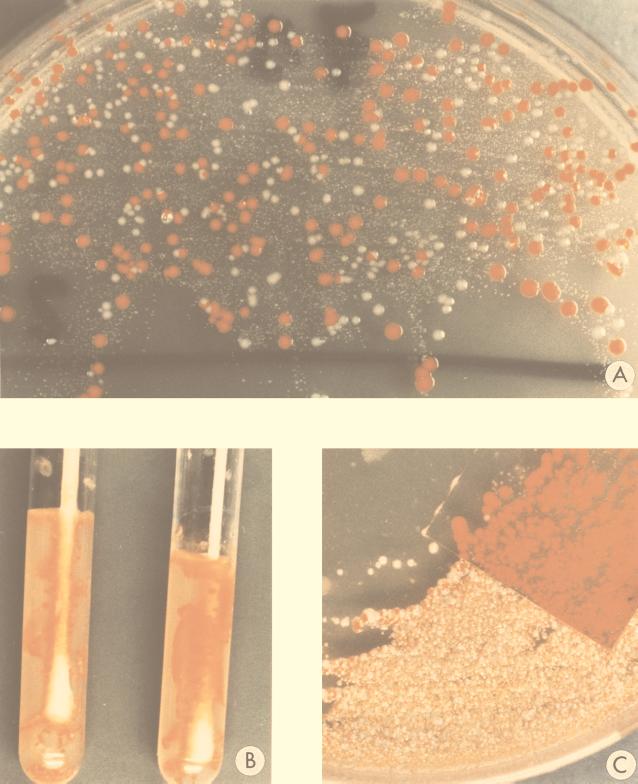
GBS were identified in blood agar by typical beta-hemolysis, Gram staining, catalase reaction, and antigen detection (Streptococcal Grouping Kit; Oxoid, Basingtoke, United Kingdom) and in plates and tubes of GM by its red or orange pigment (Fig. 1).
FIG. 1.
Appearances of colonies of GBS from vaginoanorectal specimens on GM after 18 h of incubation. (A) GM plates incubated under anaerobic conditions. (B) GM tubes. (C) GM plates incubated under aerobic conditions with a cover slide upon the inoculum.
RESULTS
GBS were recovered from 108 of the 800 vaginal specimens (13.5%) and from 89 of the 450 vaginoanorectal specimens (19.8%) by one or several of the culture techniques assayed. Although only 54% of GBS-positive samples in GM tubes were detected after 10 h, all positives samples in GM (plates and tubes) were detected after 18 h of incubation, and all GBS were detected in blood agar. Among the 450 vaginoanorectal samples, the 85 samples that were GBS positive by direct detection in GM plates were always positive in both GM plates: the one incubated anerobically and the other incubated aerobically with a cover slide upon the inoculum. Rates of recovery of GBS and the sensitivities of the different methods are shown in Table 1. Nonsignificant differences (P > 0.05; McNemar test) in GBS isolation frequency between direct sampling in GM (plates or tubes) and enrichment in selective broth for vaginal specimens were noted. However, for vaginoanorectal specimens significant differences (P < 0.01; Cochran test) were found. The method with maximum sensitivity for detection of GBS from vaginoanorectal swabs (96%) was the direct use of GM in either plates or tubes.
TABLE 1.
Comparison of GBS culture detection methods with vaginal and vaginoanorectal samples from 1,250 pregnant women
| Specimen type (total no. of specimens) | No. of positive cultures detected (relative sensitivity [%] vs all methods)
|
||||||
|---|---|---|---|---|---|---|---|
| Direct inoculation in GM
|
Subculture from selective broth onto:
|
All methods | |||||
| Plates anaerobiosis | Plates with cover slide, aerobiosis | Tubes | Blood agar plates | GM plates, anaerobiosis | GM tubes | ||
| vaginal | |||||||
| First phase (400) | 52 (93) | NDa | ND | 54 (96) | 54 (96) | ND | 56 (100) |
| Second phase (400) | ND | ND | 50 (96) | 52 (100) | 52 (100) | ND | 52 (100) |
| Vaginoanorectal (450) | 85 (96) | 85 (96) | 85 (96) | 73 (82) | 79 (89) | 79 (89) | 89 (100) |
ND, not done.
There were discrepancies for 16 of the 89 GBS-positive vaginoanorectal samples: (i) for 4 of these 16 samples GBS were detected only after selective enrichment and only in GM (plates and tubes), (ii) for 2 of the 16 samples GBS were detected after selective enrichment in GM (plates and tubes) only (not in the blood agar plate) and also by direct sampling of GM (plates and tubes), and (iii) for 10 of the 16 samples GBS were not detected after selective enrichment either in blood agar or in GM (plates or tubes). The following results were obtained for the GM plates directly inoculated with these 10 vaginoanorectal samples (for which GBS were not recovered from the selective broth): (i) for 5 of these samples there was a heavy growth of enterococci, (ii) for 4 samples there was a heavy growth of enterococci plus members of the family Enterobacteriaceae, and (iii) for 1 sample there was scanty growth of GBS, enterococci, and Proteus. The MIC of gentamicin for the last GBS strain was determined on Todd-Hewitt broth with an inoculum of 100 CFU/ml and was found to be 2 μg/ml (lower than the gentamicin concentration in the selective enrichment broth used). Furthermore, the antigen detection test could not be carried out directly with samples from the blood agar plate for 13 GBS-positive vaginoanorectal samples because of a heavy growth of fecal bacteria, and it was necessary to subculture GBS onto another blood agar plate.
DISCUSSION
This study has confirmed previous reports (12, 19) that showed that the ability of GM (either plates or tubes) to detect GBS in vaginal samples is similar to those of selective broths. However, with regard to vaginoanorectal specimens, the results of the present study are similar to those of Dunne and Holland-Staley (5), who reported the lower sensitivity of gentamicin-nalidixic selective broth for the detection of GBS compared to that of direct plating in neomicin-nalidixic blood agar. The failure of selective broth to recover GBS occurs mainly with specimens with which there is a heavy growth of enterococci on culture. In these cases GBS are probably overgrown by enterococci and cannot be observed in blood agar plate subcultures. In this study the only case in which the selective broth failed to recover a GBS strain and in which there was no heavy growth of fecal bacteria by direct plating on a GM plate might be explained by the low inoculum of GBS and because the low gentamicin MIC for this strain did not allow it to thrive in the selective broth. Similar problems for GBS detection have previously been reported with gentamicin-containing selective broths (5, 7, 8, 16, 17). The time to detection of GBS is usually 2 days when selective broth and subculture onto a blood agar plate are used: 18 h for incubation in the broth and a further 24 h for incubation on the blood agar plate. It is then necessary to carry out an antigen detection test. Moreover, when there is a heavy growth of other bacteria in the blood agar plate, a further 24 h is required to retrieve GBS colonies that can be isolated. Although in this study only 54% of GBS-positive samples detected directly in GM tubes were detected in 10 h, all GBS detected directly in GM (tubes and plates) were identified within 18 h of receipt of the specimen. For bloody specimens the initial reading of GM tubes may be misleading, and these tubes should always be assessed after at least 18 h. In contrast, with selective broth cultures a minimum of 2 days and sometimes 3 days was required for the identification of positive samples. A similar time for detection of GBS by the selective broth technique has also been reported recently (5).
Cultures of GBS incubated aerobically under a cover slide in GM plates exhibited the same pigment intensity as cultures incubated anaerobically (Fig. 1C). This trick allows aerobic incubation of GM plates and eliminates the inconvenience of using anaerobiosic incubation. This phenomenon may perhaps be explained because growth under a cover slide keeps the developing GBS cells coplanar (3), and this restriction could produce cell stress and trigger pigment production (20, 23).
This work has confirmed that human hemolytic GBS can be detected directly by using GM plates or tubes after overnight incubation at least as reliably as they can be detected after 2 to 3 days of incubation in a selective enrichment broth and subculture onto blood agar plates. GM can be used as (i) GM plates incubated anaerobically, (ii) GM plates incubated aerobically under a cover slide, or (iii) GM tubes.
In addition, identification of GBS in GM is straightforward (because of its characteristic red-orange color), resulting in an important savings of labor and the cost of reagents otherwise necessary for the accurate identification of GBS (12).
Although it occurs very infrequently, nonhemolytic, nonpigmented GBS have been implicated in cases of neonatal disease (18). Although these strains grow perfectly in GM (4, 15), methods that do not rely on either hemolysis or pigment production must be used to detect them.
REFERENCES
- 1.Baker C J, Clark D J, Barret F F. Selective broth medium for isolation of group B streptococci. Appl Microbiol. 1973;26:884–885. doi: 10.1128/am.26.6.884-885.1973. [DOI] [PMC free article] [PubMed] [Google Scholar]
- 2.Centers for Disease Control and Prevention. Prevention of perinatal group B streptococcal disease: a public health perspective. Morbid Mortal Weekly Rep. 1996;45(No. RR-7):1–24. [PubMed] [Google Scholar]
- 3.Codner R C. Solid and solidified growth media in microbiology. In: Norris J R, Ribbons D W, editors. Methods in microbiology. Vol. 1. London, United Kingdom: Academic Press; 1969. pp. 427–454. [Google Scholar]
- 4.Cueto M, Sánchez M J, Serrano J, Aguilar J M, Martinez R, Rosa M. Bacteremia caused by nonhemolytic group B streptococci. Clin Microbiol News. 1996;18(7):55–56. [Google Scholar]
- 5.Dunne W M, Holland-Staley C A. Comparison of NNA agar culture and selective broth culture for detection of group B streptococcal colonization in women. J Clin Microbiol. 1998;36:2298–2300. doi: 10.1128/jcm.36.8.2298-2300.1998. [DOI] [PMC free article] [PubMed] [Google Scholar]
- 6.Fallon R J. The rapid identification of group B streptococci. J Clin Pathol. 1975;27:902–905. doi: 10.1136/jcp.27.11.902. [DOI] [PMC free article] [PubMed] [Google Scholar]
- 7.Fenton L J, Arper M H. Evaluation of colistin and nalidixic acid in Todd-Hewitt broth for selective isolation of group B streptococci. J Clin Microbiol. 1979;9:167–169. doi: 10.1128/jcm.9.2.167-169.1979. [DOI] [PMC free article] [PubMed] [Google Scholar]
- 8.Gray B M, Pass M A, Dillon H C. Laboratory and field evaluation of selective media for isolation of group B streptococci. J Clin Microbiol. 1979;9:466–470. doi: 10.1128/jcm.9.4.466-470.1979. [DOI] [PMC free article] [PubMed] [Google Scholar]
- 9.Haugh R H, Soderlund E. Pigment production in group B streptococci. Acta Pathol Microbiol Scand. 1977;85:286–288. doi: 10.1111/j.1699-0463.1977.tb01976.x. [DOI] [PubMed] [Google Scholar]
- 10.Islam A K M S. Rapid recognition of group B streptococci. Lancet. 1977;i:256–257. doi: 10.1016/s0140-6736(77)91055-8. [DOI] [PubMed] [Google Scholar]
- 11.Jones D E, Friedl E M, Kanarek K S, Williams J K, Lim D V. Rapid identification of pregnant women heavily colonized with group B streptococci. J Clin Microbiol. 1983;18:558–560. doi: 10.1128/jcm.18.3.558-560.1983. [DOI] [PMC free article] [PubMed] [Google Scholar]
- 12.Kelly V N, Garland S M. Evaluation of New Granada Medium (modified) for the antenatal detection of group B streptococcus. Pathology. 1994;26:487–489. doi: 10.1080/00313029400169242. [DOI] [PubMed] [Google Scholar]
- 13.Koneman E W, Allen D D, Janda W M, Schreckenberger P C, Winn W C. Color atlas and textbook of diagnostic microbiology. 5th ed. Philadelphia, Pa: Lippincott; 1997. Gram positive cocci; p. 608. [Google Scholar]
- 14.Merrit K, Jacobs N J. Improved medium for detecting pigment production by group B streptococci. J Clin Microbiol. 1976;4:379–380. doi: 10.1128/jcm.4.4.379-380.1976. [DOI] [PMC free article] [PubMed] [Google Scholar]
- 15.Miranda C, Gámez M I, Navarro J M, Rosa-Fraile M. Endocarditis caused by nonhemolytic group B streptococcus. J Clin Microbiol. 1997;35:1016–1017. doi: 10.1128/jcm.35.6.1616-1617.1997. [DOI] [PMC free article] [PubMed] [Google Scholar]
- 16.Persson K M S. Antimicrobial susceptibility of group B streptococci. Eur J Clin Microbiol. 1986;5:165–166. doi: 10.1007/BF02013976. [DOI] [PubMed] [Google Scholar]
- 17.Persson M S, Forsgren A. Evaluation of culture methods for isolation of group B streptococci. Diagn Microbiol Infect Dis. 1987;6:175–177. doi: 10.1016/0732-8893(87)90104-0. [DOI] [PubMed] [Google Scholar]
- 18.Roe M H, Todd J K, Favara B E. Nonhemolytic group B streptococcal infections. J Pediatr. 1976;89:75–77. doi: 10.1016/s0022-3476(76)80931-6. [DOI] [PubMed] [Google Scholar]
- 19.Rosa M, Pérez M, Carazo C, Pareja L, Peis J I, Hernández F. New Granada medium for detection and identification of group B streptococci. J Clin Microbiol. 1992;30:1019–1021. doi: 10.1128/jcm.30.4.1019-1021.1992. [DOI] [PMC free article] [PubMed] [Google Scholar]
- 20.Russell N J. Function of lipids: structural roles and membrane functions. In: Ratledge C, Wilkinson S G, editors. Microbial lipids. Vol. 2. London, United Kingdom: Academic Press; 1989. pp. 279–365. [Google Scholar]
- 21.Schuchat A. Epidemiology of group B streptococcal disease in the United States: shifting paradigms. Clin Microbiol Rev. 1998;11:497–513. doi: 10.1128/cmr.11.3.497. [DOI] [PMC free article] [PubMed] [Google Scholar]
- 22.Tapsall J W. Relationship between pigment production and haemolysin formation by Lancefield group B streptococci. J Med Microbiol. 1987;24:83–87. doi: 10.1099/00222615-24-1-83. [DOI] [PubMed] [Google Scholar]
- 23.Taylor R F. Bacterial triterpenoids. Microbiol Rev. 1984;48:181–198. doi: 10.1128/mr.48.3.181-198.1984. [DOI] [PMC free article] [PubMed] [Google Scholar]
- 24.Yancey M K, Schuchat A, Brown L K, Ventura V I, Markenson G R. The accuracy of late antenatal screening cultures in predicting genital group B streptococcal colonization at delivery. Obstet Gynecol. 1996;88:811–815. doi: 10.1016/0029-7844(96)00320-1. [DOI] [PubMed] [Google Scholar]


