Abstract
BACKGROUND AND AIM:
Oxidative stress leads to a compensatory increase in levels of serum ceruloplasmin in patients with such imbalances. Greater than normal serum ceruloplasmin levels are noticed in numerous cancers including the leukemias and Hodgkin's lymphoma. The purpose of the present study was to estimate and evaluate the efficacy of serum ceruloplasmin levels as a potential biomarker in the early detection of oral potentially malignant epithelial lesions (PMELs) including leukoplakia, oral submucous fibrosis (OSMF), and oral squamous cell carcinoma (OSCC) patients.
MATERIALS AND METHODS:
The present observational study was conducted over a period of 2 years wherein 100 subjects aged between 18 to 60 years were divided into four groups with Group A consisting of 25 healthy controls, Group B and C with 25 patients each, clinically diagnosed with oral leukoplakia and OSMF and Group D with 25 patients clinically diagnosed and histopathologically proven OSCC. The patients were subjected to incisional biopsy after routine hematological investigation while the same sera samples were used for analysis of serum ceruloplasmin levels.
STATISTICAL ANALYSIS USED:
Comparison of serum ceruloplasmin levels between the groups was performed using one way analysis of variance (one way ANOVA) test while P < 0.05 was considered statistically significant.
RESULTS:
The mean serum ceruloplasmin levels were found to be 43.19 ± 1.90mg/dl in subjects of group A, 47.68 ± 1.51mg/dl in group B, 47.74 ± 1.45mg/dl in group C and 47.73 ± 0.74mg/dl in group D. Using one-way ANOVA, statistically significant variations were found in the values of mean serum ceruloplasmin levels in subjects of the four groups (F-value = 59.58, P = 0.0001).
CONCLUSIONS:
The observations of the present study revealed that serum ceruloplasmin levels were found to be raised in all 3 study groups including oral leukoplakia, OSMF and OSCC as compared to the controls while the results were found to be statistically significant.
Keywords: Biomarker, early detection, oral potentially malignant epithelial lesions, oral squamous cell carcinoma, Serum ceruloplasmin
Introduction
Oral potentially malignant epithelial lesions (PMELs) are defined as those lesions and/or, conditions of oral mucosa that show features of epithelial dysplasia but are not frank malignant lesions. [1] The term pre-cancerous lesion/condition has been discarded since not all these lesions/conditions turn into malignancies. Thus, these changes of the mucosa are referred to as PMELs. [2,3] The most common lesion among these PMELs is oral leukoplakia while the frequent sites affected are the buccal mucosa, gingiva (alveolar mucosa) and vermilion border of lip. Leukoplakia is a white patch or, plaque that cannot be characterized clinically or, histopathologically as any other disease. [4] The rate of malignant transformation of leukoplakia has been rated as around 0.13-34%. [5] Erythroplakia is nothing apart from an advanced stage or, variant of leukoplakia which has predominant red elements in it indicating even higher chances of dysplasia and being far more prone for malignant transformation. [4,5]
Likewise, oral submucous fibrosis (OSMF) is an insidious chronic disease process affecting any part of the oral mucosa and associated with juxta-epithelial inflammatory reaction followed by fibro-elastic changes in lamina propria with epithelial atrophy. [6] The condition is characterized by burning sensation of oral mucosa assisted with ulceration and pain, blanching of mucosa along with depapillation of tongue, depigmentation of mucosa and progressive reduction in mouth opening. [2] The overall prevalence of OSMF in India is estimated to be about 0.2%–0.5% and the prevalence is seen to vary with gender being 0.2%–2.3% in males while 1.2%–4.57% in females. It shows a high degree of malignant potential which ranges between 2.3% and 7.6%. [7]
Oral cancer is the most common malignancy known in the head and neck region and is one of the major causes of deaths worldwide. Approximately 80,000 new cases of oral cancers are diagnosed each year, mainly, due to consumption of different forms of tobacco products such as gutkha, quid, snuff or, misri. [8] Annually, 1,30,000 people succumb to oral cancers which translates into approximately 14 deaths per hour in India. [9] The most commonly encountered oral neoplasm is oral squamous cell carcinoma (OSCC) and it accounts for 95% of all oral cancers reported. [5] Squamous cell carcinoma has been defined by Pindborg and Sirsat [6] as a malignant epithelial neoplasm exhibiting squamous differentiation characterized by formation of keratin and/or, presence of intercellular bridges. All PMELs, eventually, progress to develop invasive OSCCs. To predict this aggressiveness, grading of the neoplasm is done based on assessment of the degree of keratinization, cellular, and nuclear pleomorphism and mitotic activity which help in the assessment of prognosis as well as deciding treatment guidelines for the disease. [5]
Despite recent advances in cancer treatments, the outcome and prognosis of OSCC is still poor. The lacuna for this lies in delayed and late diagnosis of neoplasm when the tumor is already in the advanced stages of disease. [10] An early enough diagnosis is, thus, highly warranted to initiate treatment in the initial stages itself to arrest progression of the malignant process. The stability, progress of PMELs into frank malignancies and/or, their regression, though, are not predictable by clinical and histological features and here, comes the role of tumor markers which are certain specific substances released either by tumor or, host, while combating the tumor, into the serum. The identification of such substances which can predict disease progression is, thus, of utmost importance in the management of these lesions. [11]
Classically, a marker is synthesized by tumor cells and released into circulation in large quantities during the process. Altered concentration of these biomarkers in the serum or, saliva of an individual, then, gives signal of the future alarming condition pertaining to the process of frank malignant transformations. [12] Ceruloplasmin exhibits a copper-dependent oxidase activity associated with the possible oxidation of ferrous (Fe2+) ions into ferric (Fe3+) ions, therefore, assisting in its transport in plasma in association with transferrin, capable of carrying iron only in ferric state. [13] Oxidative stress is an imbalance resulting out of free radical damage and disruption of oxidant-antioxidant balance in the body. [14] It has been observed that oxidative stress might lead to compensatory increased levels of serum ceruloplasmin in patients with such imbalances. Greater than normal serum ceruloplasmin levels are noticed in numerous cancers including the leukemias, Hodgkin's lymphoma as well as during copper toxicity or, zinc deficiency, acute and chronic inflammations, rheumatoid arthritis and secondary to drugs such as carbamazepine, phenobarbital, and valproic acid. [15,16] Serum ceruloplasmin levels are reduced in patients with hepatic disorders due to reduced synthesis, in Wilson's disease (copper storage disease), overdose of Vitamin C or, in aceruloplasminemia. [17] The purpose of the present study was to estimate and evaluate the efficacy of serum ceruloplasmin levels as a potential biomarker in the early detection of oral PMELs including oral leukoplakia, OSMF, and OSCC patients.
Materials and Methods
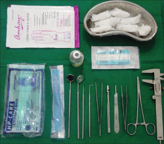
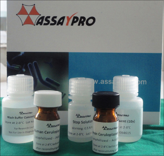
The present observational study was conducted over a period of 2 years wherein 100 subjects aged between 18 to 60 years clinically diagnosed and histopathologically confirmed with oral leukoplakia, OSMF and OSCC were divided into 4 groups with each group consisting of 25 patients as Group A consisting of 25 healthy controls, Group B with 25 patients clinically diagnosed with oral leukoplakia, Group C with 25 patients clinically diagnosed with OSMF and Group D with 25 patients clinically diagnosed and histopathologically proven OSCC. Subjects with present or, past history of any major illness such as liver disease, diabetes, hypertension, and tuberculosis, subjects undergoing radiotherapy or, chemotherapy for cancer, patients with a history of malignancy other than oral cancers and those who were >60 years of age were excluded because of possible immunocompromise. Biopsy was considered as the gold standard for confirmation of diagnosis. Ethical clearance was obtained from the Institutional Ethics Committee while the subjects were informed in detail regarding the need and protocol of study and a written, informed consent was obtained from them. All the subjects, the patients and controls, were examined thoroughly [Figure 1] and a detailed history was recorded. The patients diagnosed with oral leukoplakia, OSMF and OSCC were, then, subjected to incisional biopsy taking tissue from the periphery of lesions along with adjacent normal tissue after routine hematological investigations. The same sera samples were, then, used for the analysis of serum ceruloplasmin levels in patients while sera was drawn from the controls and similar procedure was followed for analysis of serum ceruloplasmin levels in both patients and controls using human ceruloplasmin elisa kit manufactured by cusabio assay max [Figure 2].
Figure 1.

Armamentarium for clinical examination
Figure 2.

Human ceruloplasmin enzyme-linked immuno-sorbent assay kit (assay max) for estimation of serum ceruloplasmin levels
Statistical Analysis Used: The statistical analysis was done using the Statistical Package for Social Sciences [SPSS version 17.0 (SPSS Inc., Chicago, IL, USA), Epi-Info 6.0 version] and Graph Pad Prism version 5.0. Comparison of serum ceruloplasmin levels between groups was performed using one way analysis of variance (one-way ANOVA) test (F-Test) while frequencies were compared with the help of Chi-square test. Inter-group comparisons and multiple comparisons were done by Tukey's test. P <0.05 was considered statistically significant.
Results
Table 1 reveals age-wise distribution of patients wherein statistically significant differences were observed in the mean age of patients in 4 groups on using Chi-square test (ℵ 2 -value = 69.50, P = 0.0001). Likewise, Table 2 reveals gender-wise distribution of patients wherein using Chi-square analysis, statistically significant differences were found as far as gender of patients was concerned in all 4 groups (ℵ 2 -value = 8.94, P = 0.030).
Table 1.
Age-wise distribution of patients
| Age group (years) | Group A, n (%) | Group B, n (%) | Group C, n (%) | Group D, n (%) | χ2, P |
|---|---|---|---|---|---|
| 20-29 | 16 (64) | 2 (8) | 9 (36) | 0 | 69.50, 0.0001* |
| 30-39 | 8 (32) | 6 (24) | 5 (20) | 1 (4) | |
| 40-49 | 1 (4) | 13 (52) | 7 (28) | 6 (24) | |
| 50-59 | 0 | 4 (16) | 4 (16) | 16 (64) | |
| ≥60 | 0 | 0 | 0 | 2 (8) | |
| Total | 25 (100) | 25 (100) | 25 (100) | 25 (100) | |
| Mean±SD | 28.36±5.79 | 41.32±8.02 | 36.52±10.27 | 53.0±5.46 | |
| Range | 20-40 | 28-55 | 22-55 | 39-60 |
*P<0.05 statistically significant. SD: Standard deviation
Table 2.
Gender-wise distribution of patients
| Gender | Group A, n (%) | Group B, n (%) | Group C, n (%) | Group D, n (%) | χ2, P |
|---|---|---|---|---|---|
| Male | 16 (64) | 24 (96) | 21 (84) | 21 (84) | 8.94, 0.030* |
| Female | 9 (36) | 1 (4) | 4 (16) | 4 (16) | |
| Total | 25 (100) | 25 (100) | 25 (100) | 25 (100) |
*P<0.05 statistically significant
Table 3 reveals habit-wise distribution of patients showing 16% of subjects in group A, 68% in group B, 96% in group C and 36% in group D were having a history of gutkha chewing while 4% of subjects in Group A and C each and 8% in Group D had history of betel nut chewing. Furthermore, 4% of subjects in group A and 32% of subjects each in Group B and Group D had history of smoking while 24% of subjects in group D had history of alcohol consumption. Furthermore, 76% of subjects in group A had no such deleterious habit. Analyzing the findings using Chi-square test revealed statistically significant differences in relation to habit-wise distribution of subjects as well in all 4 groups (ℵ 2 -value = 107.60, P = 0.0001).
Table 3.
Habit-wise distribution of patients
| Habit | Group A, n (%) | Group B, n (%) | Group C, n (%) | Group D, n (%) | χ2, P |
|---|---|---|---|---|---|
| Gutkha | 4 (16) | 17 (68) | 24 (96) | 9 (36) | 107.60, 0.0001* |
| Betel nut | 1 (4) | 0 | 1 (4) | 2 (8) | |
| Smoking | 1 (4) | 8 (32) | 0 (0) | 8 (32) | |
| Alcohol | 0 | 0 | 0 | 6 (24) | |
| No habit | 19 (76) | 0 | 0 | 0 | |
| Total | 25 (100) | 25 (100) | 25 (100) | 25 (100) |
*P<0.05 statistically significant
Table 4 reveals the descriptive statistics comparing mean serum ceruloplasmin levels in the 4 groups with the mean serum ceruloplasmin levels to be 43.19 ± 1.90mg/dl in subjects of Group A, 47.68 ± 1.51mg/dl in group B, 47.74 ± 1.45mg/dl in Group C and 47.73 ± 0.74mg/dl in group D. Using one-way ANOVA, statistically significant variations were found in the values of mean serum ceruloplasmin levels in subjects of the 4 groups (F-value = 59.58, P = 0.0001).
Table 4.
Descriptive statistics revealing serum ceruloplasmin levels in various groups
| Group | n | Mean | SD | SE | Minimum–maximum |
|---|---|---|---|---|---|
| Group A | 25 | 43.19 | 1.90 | 0.38 | 40.00-45.90 |
| Group B | 25 | 47.68 | 1.51 | 0.30 | 42.00-49.50 |
| Group C | 25 | 47.74 | 1.45 | 0.29 | 44.70-49.70 |
| Group D | 25 | 47.73 | 0.74 | 0.14 | 46.10-48.90 |
|
| |||||
| One-way ANOVA | |||||
|
| |||||
| Source of variation | Sum of squares | df | Mean square | F | P |
|
| |||||
| Between groups | 383.58 | 3 | 127.86 | 59.58 | 0.0001* |
| Within groups | 206.006 | 96 | 2.14 | ||
| Total | 589.58 | 99 | |||
|
| |||||
| Multiple comparisons: Tukey’s test | |||||
|
| |||||
| Group | Mean difference (I−J) | SE | P | 95% CI (lower bound–upper bound) | |
|
| |||||
| Group A | |||||
| Group B | 4.48 | 0.41 | 0.0001* | 3.40-5.56 | |
| Group C | 4.54 | 0.41 | 0.0001* | 3.46-5.63 | |
| Group D | 4.53 | 0.41 | 0.0001* | 3.45-5.61 | |
| Group B | |||||
| Group C | 0.06 | 0.41 | 0.999 | 1.01-1.14 | |
| Group D | 0.05 | 0.41 | 0.999 | 1.03-1.13 | |
| Group C | |||||
| Group D | 0.01 | 0.41 | 1.000 | −1.09-1.07 | |
*P<0.05 statistically significant. SD: Standard deviation, SE: Standard error, CI: Confidence interval, ANOVA: Analysis of variance
Discussion
Cancer, a disorder of cellular behavior, is characterized by alteration of serum glycoproteins and cell surface glycosylation and is associated with various types of transformation processes. Early detection and early treatment of oral PMELs and oral cancers not only reduces mortality, but, also, renders quality life to the survivors. A majority of neoplasms are preventable as well as curable if they are detected in the early enough stages, especially, oral PMELs like oral leukoplakia and OSMF which usually precede frank oral cancers. [18] Biomarkers provide a noninvasive means of diagnosis, facilitate early detection of PMELs or, malignant conditions and their early treatment eventually affects the prognosis these lesions have after treatment. Cell membrane mainly consists of glycoproteins and glycolipids. Glycoproteins are protein-carbohydrate complexes in which oligosaccharides and or, polysaccharides are joined to specific amino acids of proteins by covalent linkages. [18] Till now, many researchers have developed and successfully demonstrated the use of protein markers as biomarkers in the diagnosis and management of oral cancers. The present study evaluated the diagnostic utility of serum ceruloplasmin as a potential cancer marker.
The age of subjects in the control group (A), patients with oral leukoplakia (B), OSMF (C) and OSCC (D) ranged from 20-40 years, 28-55 years, 22-55 years, and 39-60 years respectively in the present study, thus, showcasing a broad range of probability of the occurrence of oral PMELs and OSCC in the affected patients. From the analysis of data, it was evident that OSCC showed progression with ageing and became evident in the 5 th decade of life. This observation was in accordance with the studies conducted by Chittemsetti et al. [19] and Shetty et al. [20]. There are varying reports on sex ratio in different published studies. In the present study, out of 25 oral leukoplakia patients, 24 (96%), 25 OSMF patients, 21 (84%), 25 OSCC patients, 21 (84%) of the patients were male while 1 (4%), 4 (16%), and 4 (16%) of the patients were females respectively indicating a male predominance. The male to female ratio in the OSCC group in the present study was found to be 5.25:1 which was similar to the findings of the study conducted by Shetty et al. [20] who found a higher male prevalence reported for the lesions with a male to female ratio of 5:1. In other studies by Elango et al. [21] and Mehrotra and Yadav, [22] the male to female ratio was found to be 4:1 while the mean age of patients being 55.92 ± 10.17 years.
The occurrence of oral PMELs and oral cancers is seen to be higher in the males and this might be due to the much prevalent habit of chewing gutkha, betel nut, smoking, and drinking alcohol in males as compared to females. Literature is abuzz with correlation of oral malignancies and habits such as smoking and tobacco chewing. Gutkha chewing (36%) and smoking (32%) were the most common habits found in the present study followed by alcohol (24%). Thus, gutkha chewing, smoking and alcohol were found to be the major risk factors in the present study. Similar risk factors in head and neck malignancies have been reported in various other studies including the ones conducted by Shashikanth and Rao [23] and Day and Blot. [24]
Ceruloplasmin is a glycoprotein encoded by CP gene on chromosome no. 3q24 involved in the transport of copper ions in body and also, involved in iron metabolism by virtue of ferroxidase activity. It is synthesized primarily in the liver and contains 6-7 copper ions. Ceruloplasmin is an acute phase reactant and a transport protein. [16] A balance between oxidant carcinogens and endogenous antioxidant defenses is of particular relevance to the process leading to the evolution of carcinogenesis. Oxidative stress is an imbalance between free radical damage and the antioxidant protection in body. Copper and ceruloplasmin have been observed to be significantly increased in numerous cancers. Cupric ions are reported to inhibit the production of singlet oxygen and this is of particular significance because of the latter's ability to cross the cell membrane and its high reactivity toward various biomolecules. [25,26,27,28,29]
Ceruloplasmin levels, as assessed in the present study, showed significantly higher levels in all 3 study groups including oral leukoplakia (B), OSMF (C) and OSCC (D) with the corresponding values being 47.68 ± 1.51mg/dl, 47.74 ± 1.45mg/dl and 47.73 ± 0.74mg/dl respectively as compared to the controls (A), 43.19 ± 1.90mg/dl. The results of the present study suggested that as compared to the controls (A), ceruloplasmin levels remained significantly high (P < 0.05) in all 3 study groups including oral leukoplakia (B), OSMF (C) and OSCC (D), though, were found to be nearly constant. A better correlation could be observed between elevated serum copper levels and some malignant tumors as in the studies conducted by Mailer et al. [30] and Linder et al. [31] This was probably the first evidence which created ceruloplasmin as a potential candidate to be used as a biomarker for oral cancers. Similar correlation was found between increased copper levels and areca nut chewing habit in the study conducted by Arakeri et al. [32]
In the present study, it was observed that controls (A) revealed serum ceruloplasmin levels to be 43.19 ± 1.90mg/dl which was found to be slightly on the higher side when compared to an earlier reported study conducted by Senra Varela et al. [33] who found the mean serum ceruloplasmin levels in the controls to be 32.4mg/dl. In the present study, the mean serum ceruloplasmin levels were significantly higher (P < 0.05) in the OSCC (D) group as compared to the controls, the level of change, though, with respect to the other PMELs, oral leukoplakia (B) and OSMF (C) and OSCC (D), did not represent any significant variation.
Jayadeep et al., [34] also, reported similar findings in relation to serum ceruloplasmin levels in their study on oral pre-cancerous lesions and frank oral cancers highlighting not only the diagnostic significance serum ceruloplasmin levels have in the early detection of malignancies but prognostic significance marking disease progression. The increased serum ceruloplasmin levels indicate elevated antioxidant activity of the serum. Serum ceruloplasmin, apart from being an effective antioxidant protein, is one of the acute phase reactants, the concentration of which increases in the plasma after tissue injury. The acute phase reactants protect the tissue as a whole from the deleterious effects caused due to the release of free radicals and oxidation products, thus, suggesting that the body responds to free radical damage by raising the antioxidant activity of plasma by elevated ceruloplasmin levels. [35]
Decreased levels of copper are of great significance in the etiology of OSMF as has been reported in numerous other studies including the ones conducted by Jani et al. [36] and Patil and Joshi [37] to name a few. The decrease in copper levels obviously brings about a decrease in serum ceruloplasmin levels, thus, influencing iron absorption and mobilization of iron from the liver and other tissue stores. [38] Further deficiency in copper levels, also, affects iron absorption and thus, leads to iron deficiency.
In accordance with the findings of the present study, Akinmoladun et al., [39] also, found increased mean serum ceruloplasmin levels in patients with oral PMELs and frank oral cancers in their study. Increased serum ceruloplasmin levels have, also, been previously reported in various malignancies. [40,41] The reasons for the same, though, not clearly understood, could be due to serum ceruloplasmin being an acute phase reactant apart from being a direct response to increase in copper levels, which, also, had, been similarly reported in various malignancies. [25,26,27,28,29]
Even though not being sensitive for phase base reactions, usefulness of serum ceruloplasmin levels never becomes less as could be seen from the findings of the present study where its sensitivity and specificity had been benchmarked in various oral PMELs and frank OSCC and it is noteworthy that it is expressed by the tumoral cells just as the other established biomarkers and hence, makes it a featured marker of future use in such situations when investigated in detail as, also, concluded by Kunapuli et al. [42] and Abd-el-Fattah et al. [43]. Andrzejewska et al., [44] also reported the said correlation between serum ceruloplasmin levels and various clinical stages of cancer of larynx as, also, in assessing prognosis in such cases as was observed by Krecicki and Leluk [45] who concluded that the determination of serum ceruloplasmin levels could be of use in monitoring of cancer patients.
There are few possible limitations that need to be addressed as far as the present study is concerned. First, the serum ceruloplasmin levels were not correlated with the histopathological grading and staging of the included oral PMELs including oral leukoplakia, OSMF and OSCC. Second, serum ceruloplasmin levels were significantly higher in the oral PMELs as compared to the controls, though, inter-group variations were reported to be nearly constant in all 3 study groups. Third, increase in serum ceruloplasmin levels in humans is seen in chronic inflammatory processes, active hepatitis, biliary liver cirrhosis, and malignant tumors. It becomes imperative, therefore, to detect the correct cause of increase in serum ceruloplasmin levels and thus, render a need to pre-evaluate the hepatic and renal diseases in future research projects. Furthermore, post-treatment serum ceruloplasmin levels were not evaluated, so, their impact on the prognosis of the lesions cannot be commented upon. Serum ceruloplasmin, being in the very primitive stages, thus, needs more researches to contribute as a specific biomarker for oral cancers.
Conclusions
The observations of the present study revealed that serum ceruloplasmin levels were found to be raised in all 3 study groups including oral leukoplakia, OSMF and OSCC as compared to the controls while the results were found to be statistically significant. In addition, a definitive association was found between harmful habits in decreasing order of gutkha chewing, smoking, alcohol consumption, and betel nut to the incidence of oral PMELs and frank OSCC. Thus, it could be concluded from the observations of the present study that serum ceruloplasmin in conjunction with clinical diagnostic procedures can be used as a potentially reliable, adjunctive serological marker for monitoring and assessing oral PMELs and frank OSCC.
Financial support and sponsorship
Nil.
Conflicts of interest
There are no conflicts of interest.
References
- 1.Siar CH, Mah MC, Gill PP. Prevalence of bilateral 'mirror-image' lesions in patients with oral potentially malignant epithelial lesions. Eur Arch Otorhinolaryngol. 2012;269:999–1004. doi: 10.1007/s00405-011-1712-x. [DOI] [PubMed] [Google Scholar]
- 2.Onofre MA, Sposto MR, Navarro CM, Motta ME, Turatti E, Almeida RT. Potentially malignant epithelial oral lesions: Discrepancies between clinical and histological diagnosis. Oral Dis. 1997;3:148–52. doi: 10.1111/j.1601-0825.1997.tb00026.x. [DOI] [PubMed] [Google Scholar]
- 3.Pravda C, Srinivasan H, Koteeswaran D, Manohar LA. Verrucous carcinoma in association with oral submucous fibrosis. Indian J Dent Res. 2011;22:615. doi: 10.4103/0970-9290.90329. [DOI] [PubMed] [Google Scholar]
- 4.Anderson A, Ishak N. Marked variation in malignant transformation rates of oral leukoplakia. Evid Based Dent. 2015;16:102–3. doi: 10.1038/sj.ebd.6401128. [DOI] [PubMed] [Google Scholar]
- 5.Kramer IR, Lucas RB, Pindborg JJ, Sobin LH. Definition of leukoplakia and related lesions: An aid to studies on oral precancer. Oral Surg Oral Med Oral Pathol. 1978;46:518–39. [PubMed] [Google Scholar]
- 6.Pindborg JJ, Sirsat SM. Oral submucous fibrosis. Oral Surg Oral Med Oral Pathol. 1966;22:764–79. doi: 10.1016/0030-4220(66)90367-7. [DOI] [PubMed] [Google Scholar]
- 7.Patil S, Maheshwari S. Proposed new grading of oral submucous fibrosis based on cheek flexibility. J Clin Exp Dent. 2014;6:e255–8. doi: 10.4317/jced.51378. [DOI] [PMC free article] [PubMed] [Google Scholar]
- 8.Krishna A, Singh S, Kumar V, Pal US. Molecular concept in human oral cancer. Natl J Maxillofac Surg. 2015;6:9–15. doi: 10.4103/0975-5950.168235. [DOI] [PMC free article] [PubMed] [Google Scholar]
- 9.Singh S, Singh J, Chandra S, Samadi FM. Prevalence of oral cancer and oral epithelial dysplasia among North Indian population: A retrospective institutional study. J Oral Maxillofac Pathol. 2020;24:87–92. doi: 10.4103/jomfp.JOMFP_347_19. [DOI] [PMC free article] [PubMed] [Google Scholar]
- 10.Rai NP, Anekar J, Shivaraja Shankara YM, Divakar DD, Al Kheraif AA, Ramakrishnaiah R, et al. Comparison of Serum Fucose Levels in Leukoplakia and Oral Cancer Patients. Asian Pac J Cancer Prev. 2015;16:7497–500. doi: 10.7314/apjcp.2015.16.17.7497. [DOI] [PubMed] [Google Scholar]
- 11.Fernández-Olavarría A, Mosquera-Pérez R, Díaz-Sánchez RM, Serrera-Figallo MA, Gutiérrez-Pérez JL, Torres-Lagares D. The role of serum biomarkers in the diagnosis and prognosis of oral cancer: A systematic review. J Clin Exp Dent. 2016;8:e184–93. doi: 10.4317/jced.52736. [DOI] [PMC free article] [PubMed] [Google Scholar]
- 12.Vajaria BN, Patel PS. Glycosylation: A hallmark of cancer? Glycoconj J. 2017;34:147–56. doi: 10.1007/s10719-016-9755-2. [DOI] [PubMed] [Google Scholar]
- 13.Song D, Dunaief JL. Retinal iron homeostasis in health and disease. Front Aging Neurosci. 2013;5:24. doi: 10.3389/fnagi.2013.00024. [DOI] [PMC free article] [PubMed] [Google Scholar]
- 14.Vassiliev V, Harris ZL, Zatta P. Ceruloplasmin in neurodegenerative diseases. Brain Res Brain Res Rev. 2005;49:633–40. doi: 10.1016/j.brainresrev.2005.03.003. [DOI] [PubMed] [Google Scholar]
- 15.O'Brien PJ, Bruce WR. 2nd ed. New York: Springer-Verlag; 2009. Endogenous Toxins: Targets for Disease Treatment and Prevention; pp. 405–6. [Google Scholar]
- 16.Hellman NE, Gitlin JD. Ceruloplasmin metabolism and function. Annu Rev Nutr. 2002;22:439–58. doi: 10.1146/annurev.nutr.22.012502.114457. [DOI] [PubMed] [Google Scholar]
- 17.Gitlin JD. Aceruloplasminemia. Pediatr Res. 1998;44:271–6. doi: 10.1203/00006450-199809000-00001. [DOI] [PubMed] [Google Scholar]
- 18.Bose KS, Gokhale PV, Dwivedi S, Singh M. Quantitative evaluation and correlation of serum glycoconjugates: Protein bound hexoses, sialic acid and fucose in leukoplakia, oral sub mucous fibrosis and oral cancer. J Nat Sci Biol Med. 2013;4:122–5. doi: 10.4103/0976-9668.107275. [DOI] [PMC free article] [PubMed] [Google Scholar]
- 19.Chittemsetti S, Manchikatla PK, Guttikonda V. Estimation of serum sialic acid in oral submucous fibrosis and oral squamous cell carcinoma? J Oral Maxillofac Pathol. 2019;23:156. doi: 10.4103/jomfp.JOMFP_239_18. doi: 10.4103/jomfp.JOMFP_239_18. [DOI] [PMC free article] [PubMed] [Google Scholar]
- 20.Shetty RK, Bhandary SK, Kali A. Significance of Serum L-fucose glycoprotein as Cancer biomarker in head and neck malignancies without distant metastasis. J Clin Diagn Res. 2013;7:2818–20. doi: 10.7860/JCDR/2013/6681.3765. [DOI] [PMC free article] [PubMed] [Google Scholar]
- 21.Elango JK, Gangadharan P, Sumithra S, Kuriakose MA. Trends of head and neck cancers in urban and rural India. Asian Pac J Cancer Prev. 2006;7:108–12. [PubMed] [Google Scholar]
- 22.Mehrotra R, Yadav S. Oral squamous cell carcinoma: Etiology, pathogenesis and prognostic value of genomic alterations. Indian J Cancer. 2006;43:60–6. doi: 10.4103/0019-509x.25886. [DOI] [PubMed] [Google Scholar]
- 23.Shashikanth MC, Rao BB. Study of serum fucose and serum sialic acid levels in oral squamous cell carcinomia. Indian J Dent Res. 1994;5:119–24. [PubMed] [Google Scholar]
- 24.Day GL, Blot WJ. Second primary tumors in patients with oral cancer. Cancer. 1992;70:14–9. doi: 10.1002/1097-0142(19920701)70:1<14::aid-cncr2820700103>3.0.co;2-s. [DOI] [PubMed] [Google Scholar]
- 25.Hrgovcic M, Tessmer CF, Thomas FB, Ong PS, Gamble JF, Shullenberger CC. Serum copper observations in patients with malignant lymphoma. Cancer. 1973;32:1512–24. doi: 10.1002/1097-0142(197312)32:6<1512::aid-cncr2820320631>3.0.co;2-p. [DOI] [PubMed] [Google Scholar]
- 26.Mortazavi SH, Bani-Hashemi A, Mozafari M, Raffi A. Value of serum copper measurement in lymphomas and several other malignancies. Cancer. 1972;29:1193–8. doi: 10.1002/1097-0142(197205)29:5<1193::aid-cncr2820290510>3.0.co;2-6. [DOI] [PubMed] [Google Scholar]
- 27.Sanada S, Ogura K, Kiriyama T, Yoshida O. Serum copper and zinc levels in patients with malignant neoplasm of the urogenital tract. Hinyokika Kiyo. 1985;31:1299–316. [PubMed] [Google Scholar]
- 28.Jacobs AJ, Sommers GM, Axelrod JH, Galakatos AE, Kao MS, Camel HM. Serum copper in ovarian carcinoma. Cancer. 1988;61:1015–7. doi: 10.1002/1097-0142(19880301)61:5<1015::aid-cncr2820610526>3.0.co;2-t. [DOI] [PubMed] [Google Scholar]
- 29.Margalioth EJ, Udassin R, Cohen C, Maor J, Anteby SO, Schenker JG. Serum copper level in gynecologic malignancies. Am J Obstet Gynecol. 1987;157:93–6. doi: 10.1016/s0002-9378(87)80353-8. [DOI] [PubMed] [Google Scholar]
- 30.Mailer C, Swartz HM, Konieczny M, Ambegaonkar S, Moore VL. Identity of the paramagnetic element found in increased concentrations in plasma of cancer patients and its relationship to other pathological processes. Cancer Res. 1974;34:637–42. [PubMed] [Google Scholar]
- 31.Linder MC, Moor JR, Wright K. Ceruloplasmin assays in diagnosis and treatment of human lung, breast, and gastrointestinal cancers. J Natl Cancer Inst. 1981;67:263–75. [PubMed] [Google Scholar]
- 32.Arakeri G, Patil SG, Ramesh DN, Hunasgi S, Brennan PA. Evaluation of the possible role of copper ions in drinking water in the pathogenesis of oral submucous fibrosis: A pilot study. Br J Oral Maxillofac Surg. 2014;52:24–8. doi: 10.1016/j.bjoms.2013.01.010. [DOI] [PubMed] [Google Scholar]
- 33.Senra Varela A, Lopez Saez JJ, Quintela Senra D. Serum ceruloplasmin as a diagnostic marker of cancer. Cancer Lett. 1997;121:139–45. doi: 10.1016/s0304-3835(97)00340-6. [DOI] [PubMed] [Google Scholar]
- 34.Jayadeep A, Raveendran Pillai K, Kannan S, Nalinakumari KR, Mathew B, Krishnan Nair M, et al. Serum levels of copper, zinc, iron and ceruplasmin in oral leukoplakia and squamous cell carcinoma. J Exp Clin Cancer Res. 1997;16:295–300. [PubMed] [Google Scholar]
- 35.Rajguru JP, Mouneshkumar CD, Radhakrishnan IC, Negi BS, Maya D, Hajibabaei S, et al. Tumor markers in oral cancer: A review. J Family Med Prim Care. 2020;9:492–6. doi: 10.4103/jfmpc.jfmpc_1036_19. [DOI] [PMC free article] [PubMed] [Google Scholar]
- 36.Jani YV, Chaudhary AR, Dudhia BB, Bhatia PV, Soni NC, Patel PS. Evaluation of role of trace elements in oral submucous fibrosis patients: A study on Gujarati population. J Oral Maxillofac Pathol. 2017;21:455. doi: 10.4103/jomfp.JOMFP_106_14. [DOI] [PMC free article] [PubMed] [Google Scholar]
- 37.Patil DJ, Joshi M. Evaluation of hematological profile in oral submucous fibrosis: A cross-sectional study? J Oral Maxillofac Pathol. 2020;24:575. doi: 10.4103/jomfp.JOMFP_65_20. doi: 10.4103/jomfp.JOMFP_65_20. [DOI] [PMC free article] [PubMed] [Google Scholar]
- 38.Swaminathan M. India: Geetha Book House; 1981. Minerals in Biochemistry for Medical Students; pp. 358–410. [Google Scholar]
- 39.Akinmoladun VI, Arinola OG, Elumelu-Kupoluyi T, Eriba LO. Evaluation of humoral immunity in oral cancer patients from a nigerian referral centre. J Maxillofac Oral Surg. 2013;12:410–3. doi: 10.1007/s12663-012-0440-0. [DOI] [PMC free article] [PubMed] [Google Scholar]
- 40.McIntire KR. Use of multiple immunoassays for circulating tumor markers. In: Herberman RB, McIntire KR, editors. Immuno-Diagnosis of Cancer. New York: Marcel Dekker; 1979. pp. 521–39. [Google Scholar]
- 41.Lamoureux G, Mandeville R, Poisson R, Legault-Poisson S, Jolicoeur R. Biologic markers and breast cancer: A multi-parametric study. 1. Increased serum protein levels. Cancer. 1982;49:502–12. doi: 10.1002/1097-0142(19820201)49:3<502::aid-cncr2820490318>3.0.co;2-d. [DOI] [PubMed] [Google Scholar]
- 42.Kunapuli SP, Singh H, Singh P, Kumar A. Ceruloplasmin gene expression in human cancer cells. Life Sci. 1987;40:2225–8. doi: 10.1016/0024-3205(87)90057-9. [DOI] [PubMed] [Google Scholar]
- 43.Abd-el-Fattah M, Scherer R, Fouad FM, Ruhenstroth-Bauer G. Kinetics of the acute-phase reaction in rats after tumor transplantation. Cancer Res. 1981;41:2548–55. [PubMed] [Google Scholar]
- 44.Andrzejewska H, Klonowski S, Tomaszewski J. Zachowanie sie aktywności enzymatycznej ceruloplazminy w surowicy krwi chorych na raka krtani [Serum ceruloplasmin activity in patients with cancer of the larynx] Otolaryngol Pol. 1992;46:138–44. [PubMed] [Google Scholar]
- 45.Krecicki T, Leluk M. Acute phase reactant proteins: An aid to monitoring surgical treatment of laryngeal carcinoma. J Laryngol Otol. 1992;106:613–5. doi: 10.1017/s002221510012033x. [DOI] [PubMed] [Google Scholar]


