Figure 1.
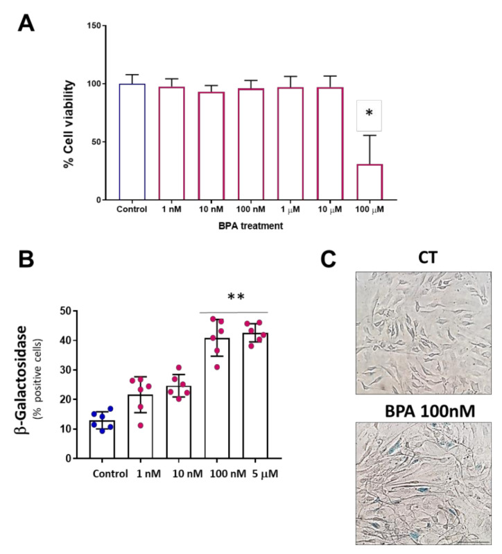
BPA induces cellular senescence at low concentrations. (A) MTT viability assay in MAEC treated with BPA 5 days at concentrations ranging from 1nM to 100 µM. A significant reduction in cell viability is observed at 100 µM. Data are the means ± SD (n = 3 with a triplicate per experimental condition). p was determined by a Kruskal–Wallis test with Dunn’s multiple comparisons test. * p < 0.0001. (B) Senescence-associated β-Gal assay in MAEC treated with BPA 5 days at the indicated concentrations. Note that the maximum effect on cell senescence was observed at concentrations of 100 nM and 5 µM. Data points represent the mean ± SD (n = 6 in triplicate). p was determined by a Kruskal–Wallis test with Dunn’s multiple comparisons test. ** p < 0.001. (C) Representative microphotographs of the senescence assay (scale bar = 50 µm). Senescent cells show the characteristic staining of β-Gal in blue color, CT means control group.

