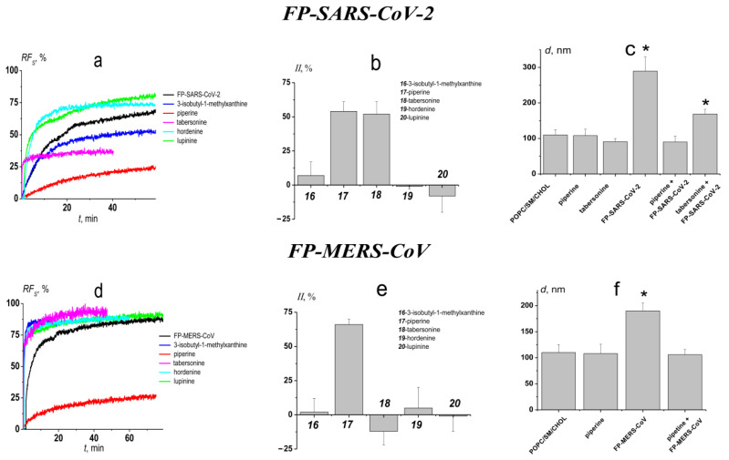
Figure 5.
The effects of the alkaloids on FP-SARS-CoV-2 (upper panel) and FP-MERS-CoV (lower panel) mediated fusion of POPC/SM/CHOL (60/20/20 mol%) vesicles. (a,d) The time dependence of relative fluorescence of calcein (RFS, %) leaked due to the fusion induced by 50 μM of FP-SARS-CoV-2; (a) and 200 μM of FP-MERS-CoV; (d) in the absence and presence of alkaloids. Liposomes were incubated with 400 μM of alkaloids for 30 min before addition of the peptides. The relationship between the color line and the alkaloid is given in the figure. (b,e) The inhibition index (II) characterizing the ability of tested alkaloids to suppress the fusion induced by 50 μM of FP-SARS-CoV-2 (b); and 200 μM of FP-MERS-CoV (e). (c,f) The diameter (d, nm) of the POPC/SM/CHOL liposomes before and after addition of 50 μM of FP-SARS-CoV-2 (c) and 200 μM of FP-MERS-CoV (f) to vesicles pretreated with 400 μM of piperine or tabersonine. *—p ≤ 0.01 (Mann–Whitney–Wilcoxon’s test, untreated liposomes vs. vesicles in the presence of fusion peptides or/and alkaloids).

