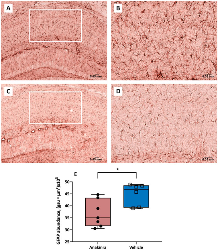
Figure 4.
Abundance of GFAP-positive astrocytes in the CA1 region of hippocampus of mice with autoimmune seizures. (A-D) Representative GFAP immunostaining of the CA1 region in vehicle-treated (upper panel) and anakinra-treated (lower panel) mice with seizures induced by continuous infusion of anti-NMDAR antibodies at 10 X (A, C) and 20 X (B, D). (E) Anakinra reduced the expression of GFAP in the CA1 region of hippocampus in mice with seizures. The abundance of GFAP labeling in the CA1 region was determined as the sum of the products of mean pixel intensity (grey scale units, gsu) and area of each event (μ2) in a fixed scan area. N = 7 (anakinra-treated), n = 6 (vehicle-treated). * p<0.05, Student’s t-test. Error bars represent mean ± SEM.

