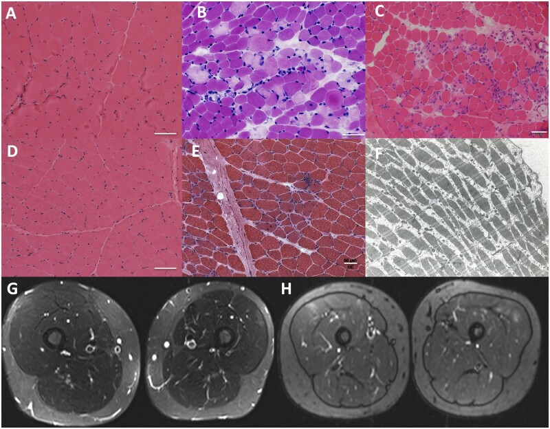
Figure 1.
Histological and imaging findings in patients with MLIP pathogenic variants. Muscle biopsies varied from showing normal to minimal findings in Patients P1 and P3 (A and D, respectively; haematoxylin and eosin, ×200) to revealing focal areas of necrosis and regeneration of muscle fibres in Patients P2, P5 and P4 (B, C and E, respectively; haematoxylin and eosin, ×200). Electron microscopy imaging performed on Patient P6 (F) did not reveal ultrastructural abnormalities. MRI of thighs in Patients P1 and P3 show mild signal hyperintensity in the vastus lateralis (G and H, respectively; G: STIR sequence; H: TIRM sequence).

