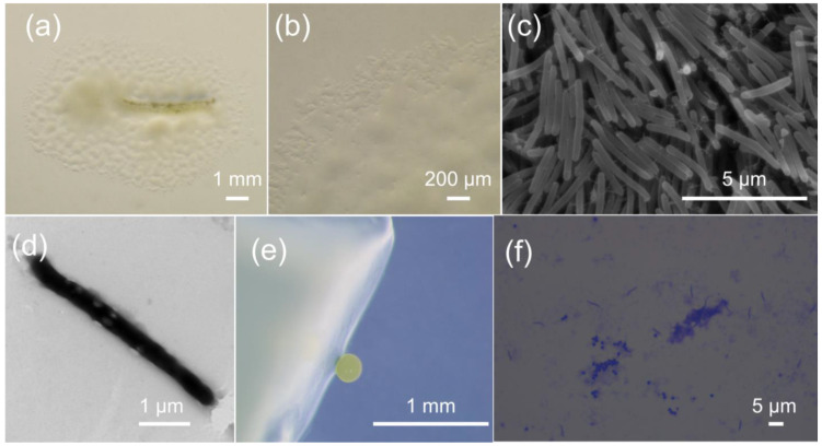
Figure 1.
Growth and morphology of strain M34. (a) The spreading swarm colony of M34 on VY/2 agar after 1 week of growth. (b) Swarm colony edge on VY/2 agar with defined veins and flared edges. The scanning electron micrograph (c) and transmission electron micrograph (d) of M34 vegetative cells showing slender and flexuous-shaped rods. (e) The pale lemon-yellow and roundish fruiting bodies formed on VY/2 agar. (f) The crystal violet stained peripheral rods and roundish myxospores from cracked fruiting bodies.

