Abstract
The new-generation ACURATE neo2 system was commercially released in September 2020. In this study, we sought to compare the aortic regurgitation (AR) severity of the ACURATE neo2 versus the ACURATE neo transcatheter heart valve, using quantitative videodensitometric angiography (qAR). This is a retrospective, Corelab analysis of final post-transcatheter aortic valve implantation (TAVI) aortograms of patients treated with the ACURATE neo2 and ACURATE neo systems. The ACURATE neo2 cohort comprised consecutive patients treated between September 2020 and January 2021 at two centers. The ACURATE neo cohort included consecutive patients treated before September 2020. Our primary objective was to compare AR severity on qAR following TAVI with ACURATE neo2 and ACURATE neo. Out of 401 aortograms, 228 (56.9%) were analyzable, with 120 in the ACURATE neo2 cohort, and 108 in the ACURATE neo cohort. The mean AR fraction was 4.4 ± 4.8% in the neo2 cohort, and 9.9 ± 8.2% in the neo cohort (p < 0.001). Furthermore, moderate or severe AR (qAR > 17%) was detected in 2 aortograms (1.7%) in the neo2 cohort and 15 aortograms (13.9%) in the neo cohort (p < 0.001). Quantitative aortography shows a lower rate of moderate or severe paravalvular AR in what is the first European experience of the new-generation, self-expanding ACURATE neo2 when compared to the first-generation ACURATE neo. Moreover, aortographic data need to be correlated and compared to Core Laboratory-adjudicated 30-day echocardiographic data.
Keywords: transcatheter aortic valve implantation, transcatheter heart valve, aortic regurgitation, ACURATE neo, videodensitometry
1. Introduction
Moderate or severe aortic regurgitation (AR) following transcatheter aortic valve implantation (TAVI) has been associated with increased short- and long-term mortality [1]. In the randomized SCOPE-2 trial [2] comparing the ACURATE neo transcatheter heart valve (THV) (Boston Scientific Corporation, Natick, Massachusetts, USA) with the Evolut THV series (Medtronic, Minneapolis, MN, USA), the rates of cardiac death were 2.8% vs. 0.8% (p = 0.03) at 30 days and 8.4% vs. 3.9% (p = 0.01) at one year, respectively. Excess mortality was partially attributed to the higher, 30-day rate of moderate or severe paravalvular AR in the ACURATE neo arm (10% vs. 3%; p = 0.002). The newly designed, self-expanding ACURATE neo2 THV (Boston Scientific Corporation, Natick, MA, USA) is equipped with inner and outer pericardial skirts extended to cover the waist of the stent in order to improve conformability to calcified and irregular aortic valve anatomy, thereby preventing or mitigating paravalvular AR.
Quantitative videodensitometric angiographic assessment of aortic regurgitation (qAR) relies on time–density curves recorded in the region of reference (aortic root) and in the region of interest (left ventricular outflow tract (LVOT)) [3,4,5]. The qAR has been extensively vetted and validated in vitro [6,7], in animal models [8], and in a clinical setting in comparison to transthoracic and transoesophageal echocardiography [9,10], as well as cardiac magnetic resonance imaging [11]. Furthermore, the long-term vital prognostic value of a threshold of 17% in AR has been reported [12]. The improvement in AR following post-balloon dilatation has also been assessed with this technique, and its impact on long-term prognosis has been demonstrated [13].
In the present study, we aim to compare the severity of paravalvular AR, as assessed by qAR, in two cohorts of patients treated either with the new-generation ACURATE neo2 THV or the first-generation ACURATE neo THV.
2. Materials and Methods
This is a retrospective analysis of the final post-TAVI aortogram from patients treated with TAVI using the ACURATE neo2 and ACURATE neo THVs in a Core Laboratory, independent of industry. The ACURATE neo2 cohort comprised consecutive patients treated between September 2020 and January 2021 at two centers (Karolinska University hospital, Stockholm, Sweden and Kerckhoff Heart Center, Bad Nauheim, Germany), and participating in the multicenter Early Neo2 Registry (NCT04810195). Likewise, the ACURATE neo cohort included consecutive patients treated before September 2020. The consecutive recruitment of patients was a prerequisite for this analysis. Patients with severe aortic stenosis (AS) were treated with TAVI, and this was based on the decision of the local heart team. The study protocol was developed in accordance with the Declaration of Helsinki, and was approved by the ethics committee of each participating hospital. Data acquisition and analysis were performed in compliance with protocols approved by the Ethical Committee of the Karolinska University (NCT04810195). Written informed consent was obtained from all participants prior to the study.
A quantitative angiographic videodensitometric assessment of paravalvular AR was performed using the CAAS A-Valve 2.0.2 (Pie Medical Imaging BV, Maastricht, The Netherlands). Details of the Core Laboratory methodology are described elsewhere [9,10,11,12,13,14,15,16,17,18]. Aortographic data were analyzed in an independent Core Laboratory (CORRIB Research Center for Advanced Imaging and Core Lab, Galway, Ireland) by experienced analysts who were blinded to the investigators and to other clinical data. When analyzing the angiographies, the difference between ACURATE neo2 and ACURATE neo THVs was not detectable.
ACURATE neo2 and ACURATE neo THV sizing was conducted according to manufacturer instructions, and based on the preprocedural multidetector computed tomographic and echocardiographic measurements. A perimeter-derived mean annulus diameter was used for size selection. Computed tomography (CT) acquisition and analysis were performed according to the local practice of each participating site. TAVI procedures were performed via the transfemoral approach in all patients. Used THVs included the ACURATE neo2 (23, 25, and 27 mm) and the ACURATE neo (23, 25, and 27 mm).
The main outcome of the study was understanding the severity of paravalvular AR, assessed by qAR following TAVI. Both the absolute value of AR fraction (between 0 and 100%) as well as grade of severity (none or trace; mild; moderate or severe) were used to compare THV performance between the ACURATE neo2 and the ACURATE neo THVs. The stratification of continuous variable regurgitation fractioninto categorical variables was performed according to the following predetermined threshold criteria: (1) none or trace regurgitation (qAR < 6%); (2) mild (6% ≤ qAR ≤ 17%); and (3) moderate or severe (qAR > 17%) [9,10,11,12,13,14,15,16,17,18]. No other outcome variables were assessed in this study.
Categorical variables were reported as numeric values and percentages, and compared with the chi-square test or Fisher’s exact test as appropriate. The mean ± standard deviation for continuous variables was compared using the Student t-test or the Mann–Whitney U-test, depending on the variable distribution. We compared baseline and procedural characteristics for potential selection bias between the ACURATE neo2 and ACURATE neo cohorts. The proportion of patients with moderate or severe AR (qAR > 17%) following TAVI was compared using the chi-square test. A two-sided p value of 0.05 was considered indicative of statistical significance. Statistical analyses were performed with SPSS version 26.0 (IBM, Armonk, New York, NY, USA).
3. Results
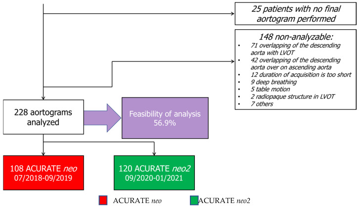
Among the 401 patients included in this study, no final aortogram injection was performed in 25 (6.2%) patients, while qAR was analyzable in 228 (60.6%) patients, including 120 and 108 patients treated with the ACURATE neo2 and ACURATE neo THVs, respectively. The common causes of the non-analyzability of post-TAVI aortograms are listed in Figure 1. Out of 148 non-analyzable cases, the main reasons provided were the overlapping of the descending aorta with LVOT (48.0%) and the overlapping of the descending aorta on ascending aorta (28.4%) (Figure 1). The mean age and the Euro score II were not significantly different between the ACURATE neo2 and ACURATE neo cohorts (80.9 ± 6.1 vs. 80.4 ± 6.2, p = 0.485, and 4.6 ± 3.7 vs. 5.5 ± 6.7, p = 0.237). Baseline characteristics, including cardiovascular risk factors, comorbidities, and hemodynamic parameters on echocardiography, were similar between patients treated with the ACURATE neo2 and ACURATE neo THVs (Table 1). Likewise, procedural characteristics were similar between the two cohorts, with the exception that predilatation was used less frequently (70.0% vs. 100%, p < 0.001) in the ACURATE neo2 cohort (Table 1). Post-procedure, there were no significant differences in complications that followed TAVI between the two cohorts (Table 1).
Figure 1.
A flowchart of Core Laboratory quantitative assessment of AR.
Table 1.
The baseline and procedural characteristics between qAR-analyzable patients after ACURATE neo2 and ACURATE neo implantation.
| ACURATE neo2 N = 120 |
ACURATE neo N = 108 |
p-Value | |
|---|---|---|---|
| Baseline characteristics | |||
| Age | 80.9 ± 6.1 | 80.4 ± 6.2 | 0.485 |
| Man | 43 (35.8) | 51 (47.2) | 0.081 |
| Body weight, kg | 72.3 ± 14.1 | 70.9 ± 13.2 | 0.440 |
| Body height, cm | 167.3 ± 9.1 | 167.0 ± 9.4 | 0.791 |
| Hypertension | 96 (80.0) | 82 (75.9) | 0.458 |
| Diabetes mellitus | 41 (34.2) | 30 (27.8) | 0.298 |
| Atrial fibrillation | 48 (40.0) | 39 (36.1) | 0.546 |
| Prior stroke | 14 (11.7) | 10 (9.3) | 0.554 |
| Prior pacemaker implantation | 13 (10.8) | 16 (14.8) | 0.368 |
| Prior cardiac surgery | 19 (15.8) | 14 (13.0) | 0.539 |
| Previous percutaneous coronary intervention | 28 (23.3) | 33 (30.6) | 0.219 |
| Chronic obstructive pulmonary disease | 21 (17.5) | 18 (16.7) | 0.867 |
| NYHA 3 or 4 | 88 (73.3) | 68 (63.0) | 0.093 |
| Creatinine clearance, mL/min | 92.0 ± 33.2 | 85.1 ± 23.7 | 0.078 |
| Euro score II, % | 4.8 ± 3.7 | 5.5 ± 6.7 | 0.374 |
| Baseline Echocardiographic Parameters | |||
| Left ventricular ejection fraction <50% | 16 (13.3) | 24 (22.2) | 0.078 |
| LV Aorta mean gradient, mmHg | 44.3 ± 15.3 | 47.1 ± 12.6 | 0.140 |
| Systolic pulmonary artery pressure, mmHg | 29.4 ± 23.4 | 30.5 ± 20.7 | 0.717 |
| Aortic regurgitation before TAVI | 0.511 | ||
| None or trace | 33 (47.8) | 36 (52.2) | |
| Mild | 72 (60.0) | 63 (58.9) | |
| Moderate | 12 (10.0) | 7 (6.5) | |
| Severe | 3 (2.5) | 1 (0.9) | |
| Mitral regurgitation before TAVI | 0.508 | ||
| None or trace | 16 (13.3) | 21 (19.8) | |
| Mild | 86 (71.7) | 70 (66.0) | |
| Moderate | 17 (14.2) | 13 (12.3) | |
| Severe | 1 (0.8) | 2 (1.9) | |
| Baseline Computed Tomography Findings | |||
| Perimeter derived mean annulus diameter, mm | 23.6 ± 1.8 | 23.9 ± 1.7 | 0.124 |
| Bicuspid aortic valve | 12 (10.0) | 11 (10.2) | 0.963 |
| Procedural Characteristics | |||
| Predilatation | 84 (70.0) | 108 (100) | <0.001 |
| Predilatation balloon size, mm | 22.6 ± 1.7 | 22.2 ± 1.6 | 0.114 |
| Postdilatation | 52 (43.3) | 60 (55.6) | 0.065 |
| Postdilatation balloon size, mm | 23.0 ± 1.7 | 23.0 ± 1.7 | 0.952 |
| Implanted THV size, mm | 25.2 ± 1.6 | 25.6 ± 1.5 | 0.054 |
| Complications Following TAVI | |||
| Valve embolization | 3 (2.5) | 1 (0.9) | 0.366 |
| Need for second TAVI valve | 2 (1.7) | 0 | 0.276 |
| Cardiac tamponade | 1 (0.8) | 0 | 0.526 |
| New permanent pacemaker implantation | 5 (4.2) | 8 (7.4) | 0.292 |
| Major vascular complications | 2 (1.7) | 0 | 0.276 |
| Major bleeding | 2 (1.7) | 1 (0.9) | 0.624 |
| Stroke | 4 (3.3) | 0 | 0.075 |
| Mortality up to 30 days | 0 | 0 | - |
NYHA: New York Heart Association; TAVI: transcatheter aortic valve replacement; LV: left ventricular; THV: transcatheter heart valve.
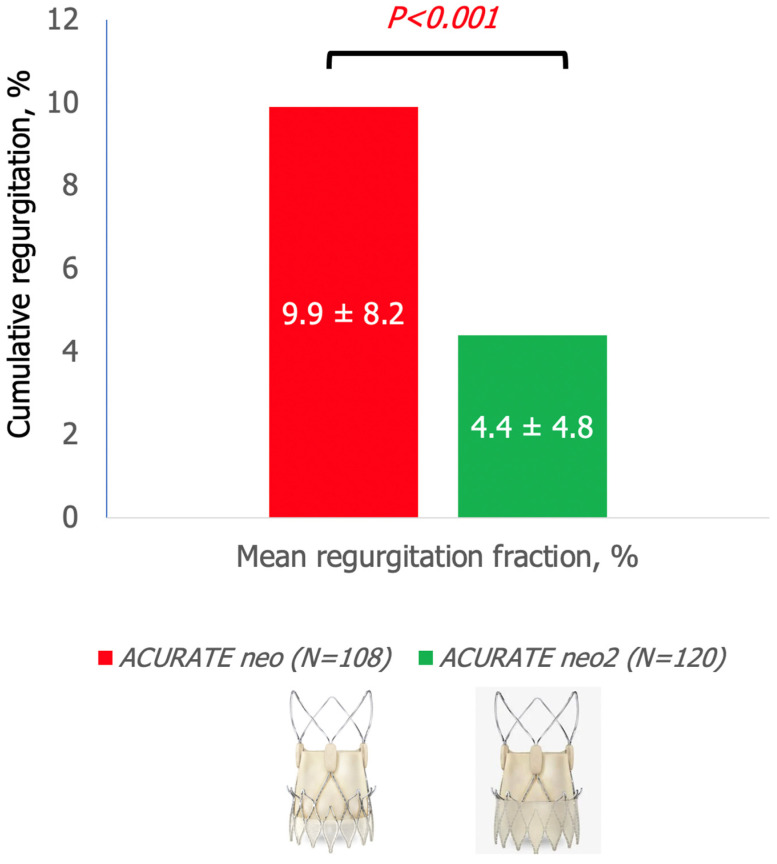
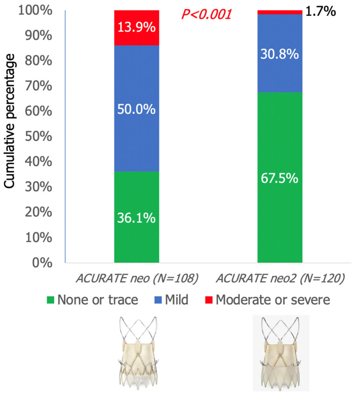
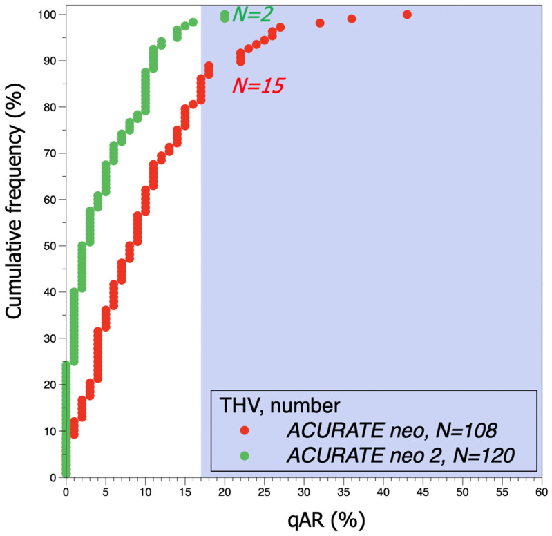
The mean post-TAVI aortic regurgitation fraction was lower in the ACURATE neo2 when compared with the ACURATE neo (4.4 ± 4.8% vs. 9.9 ± 8.2%; p < 0.001) (Figure 2). In addition, the rate of moderate or severe AR was lower for the ACURATE neo2 than for the ACURATE neo (1.7% vs. 13.9%, p < 0.001) (Figure 3 and Figure 4).
Figure 2.
Comparison of mean regurgitation fraction on qAR following TAVI between ACURATE neo and ACURATE neo2 THVs. Mean aortic regurgitation fraction following TAVI obtained by qAR (4.4 ± 4.8% vs. 9.9% ± 8.2%; p < 0.001) was lower in the ACURATE neo2 THV cohort when compared with the ACURATE neo THV cohort.
Figure 3.
Cumulative percentage of AR severity grade on qAR following TAVI for ACURATE neo and ACURATE neo2 THVs. Moderate or severe qAR was seen in 1.7% vs. 13.9% (p < 0.001) in the ACURATE neo2 THV cohort when compared to the ACURATE neo THV cohort, respectively.
Figure 4.
Cumulative frequency curves of qAR after TAVI for ACURATE neo and ACURATE neo2 THVs. The shaded background shows the area above 17% of qAR, indicating moderate or severe AR. Moderate or severe qAR was seen in 2 vs. 15 patients (p < 0.001) in the ACURATE neo2 THV cohort when compared to the ACURATE neo THV cohort, respectively. qAR: quantitative angiographic aortic regurgitation; AR: aortic regurgitation; THV: transcatheter heart valve; TAVI: transcatheter aortic valve replacement; LVOT: left ventricular outflow tract.
4. Discussion
To the best of our knowledge, this is the first study to compare the post-TAVI paravalvular AR of the self-expanding ACURATE neo2 THV with the first-generation ACURATE neo THV. The quantitative aortographic analysis reveals a 12.2% absolute risk reduction in the rate of moderate or severe AR with the ACURATE neo2 when compared with the ACURATE neo THV.
The new-generation ACURATE neo2 THV system was commercially released in September 2020 to replace the first-generation ACURATE neo THV. The newly designed valve system features the same self-expanding nitinol frame, porcine pericardial leaflets, and delivery system as the earlier generation ACURATE neo THV, with the exception of a modified skirt material and coverage [19]. The newly designed ACURATE neo2 is equipped with a 60% larger inner and outer skirt that covers the inflow and the waist of the stent. Furthermore, the redesigned skirt is made of a specific material to comply with the calcified and irregular annulus anatomy in the device landing zone. The ACURATE neo2 is also equipped with radiopaque positioning markers for accurate placement, which might have aided in mitigating the severity of paravalvular AR of the valve. Our analysis demonstrated that the ACURATE neo2 THV is associated with a significant reduction in the aortic regurgitation fraction and a lower rate of moderate or severe paravalvular AR, in comparison with the ACURATE neo THV. This can be explained by how the internal skirt of the ACURATE neo2 THV prevents the bioprosthetic valve from inadvertent damage caused by native calcium spicules, and thus minimizes propensity for AR. Additionally, as mentioned previously, the extended frame coverage of the ACURATE neo2 by the external skirt mitigates paravalvular AR by facilitating the plugging of micro-channels at the THV anchor site.
We used qAR, a quantitative videodensitometric aortography software, in this comparison study. In the prospective RESPOND study, the qAR displayed a good relationship with the Core Laboratory-adjudicated echocardiographic, providing a more granular discrimination of regurgitation within the same strata of regurgitation as assessed by echocardiography [15]. Furthermore, this qAR is used as part of the primary composite end-point in the study protocol of the randomized LANDMARK trial (NCT04275726), comparing the Myval THV with the Evolut and Sapien 3 THV series [20].
This study included consecutive patients treated with TAVI at two European centers using the ACURATE neo THV systems. Essentially, the first series of patients treated with the ACURATE neo2 THV, representing the index cohort, were compared to the latest series of patients treated with the ACURATE neo THV. The change from the earlier-generation ACURATE neo to the newly designed THV occurred in September 2020. Baseline characteristics, including the aortic annulus perimeter on CT scan, were similar between the two cohorts. In addition, all procedures were performed with the same highly experienced operators in performing TAVI using the ACURATE neo THV systems. The only significant difference between the two cohorts was the reduced use of predilatation in the ACURATE neo2 when compared with the ACURATE neo. It is unlikely that the infrequent use of predilatation in the ACURATE neo2 cohort played a role in the reduction in AR severity. However, the association between predilatation and the post-TAVI AR severity has yet to be investigated.
Our findings, although meticulously analyzed by highly experienced observers in a Core Laboratory setting, should be considered as hypothesis-generating, and thereby should be interpreted in line with the following study limitations. Firstly, these data are derived from two large-volume European centers, and by TAVI operators highly experienced in using the ACURATE THV systems. Therefore, the generalizability of our findings of improved performance of the ACURATE neo 2 THV system needs further confirmation in a larger population, including more operators and more centers. In addition, this study was focused on comparing the acute AR severity between the two cohorts, and no post-TAVI echocardiographic data were reported. Therefore, the next logical step is to correlate and compare aortographic data to Core Laboratory-adjudicated 30-day echocardiographic data. Finally, the durability of the ACURATE neo2 THV was not investigated. Further studies comprising at least one year of clinical and echocardiographic follow-up, including an independent clinical event committee and Core Laboratory adjudications, are needed to ascertain our preliminary findings on the improved performance of ACURATE neo2 THV. However, the durability of the device of up to 10 years will be investigated in the ongoing, randomized ACURATE–IDE trial (NCT03735667).
5. Conclusions
In conclusion, quantitative aortography shows a lower rate of moderate or severe paravalvular AR in what is the first European experience of the new-generation, self-expanding ACURATE neo2 THV when compared to the first-generation ACURATE neo THV. Further investigation is needed to confirm this finding. In addition, aortographic data need to be correlated and compared to Core Laboratory-adjudicated 30-day echocardiographic data.
Acknowledgments
We thank Emeline Zeller, clinical trial coordinator at CORRIB Core Lab for her contribution to this study.
Author Contributions
Conceptualization: O.S. and A.R.; Methodology: O.S.; Formal analysis: H.K., M.A., A.E., H.E., R.W.; Investigation: O.S., A.R. and W.-K.K.; Writing—original draft preparation: O.S. and H.K.; Writing—review and editing: All authors; Statistical analysis: O.S. and H.K.; Supervision: O.S., A.R., W.-K.K. and P.W.S.; Project administration: O.S. All authors have read and agreed to the published version of the manuscript.
Funding
This research received funding from Science Foundation of Ireland (SFI) via CÚRAM.
Institutional Review Board Statement
The study protocol was developed in accordance with the Declaration of Helsinki, and was approved by the ethics committee of each participating hospital. Data acquisition and analysis were performed in compliance with protocols approved by the Ethical Committee of the Karolinska University (NCT04810195). Written informed consent was obtained from all participants prior to the study.
Informed Consent Statement
Written informed consent was obtained from all participants prior to the study.
Data Availability Statement
Data is contained within the article.
Conflicts of Interest
Andreas Rück reports grants from Boston Scientific during the conduct of the study, and reports grants, personal fees and non-financial support from Boston Scientific, non-financial support from Medtronic, and personal fees from Edwards outside of the submitted work. Won-Keun Kim is a proctor/speaker/advisory board member to Abbott, Boston, Edwards, Medtronic, Meril, and Shockwave. Dinos Verouhis reports grants from Boston Scientific during the conduct of the study. Nawzad Saleh reports grants from Boston Scientific during the conduct of the study, and grants, personal fees, and non-financial support from Boston Scientific outside of the submitted work. Darren Mylotte is a consultant for Medtronic, Boston Scientific, and Microport. Patrick Serruys reports personal fees from SMT, Philips/Volcano, Xeltis, Novartis and Merillife. Osama Soliman is Science Foundation of Ireland Funded investigator, and he reports several institutional research grants outside of the submitted work. All other authors declare no conflict of interest.
Footnotes
Publisher’s Note: MDPI stays neutral with regard to jurisdictional claims in published maps and institutional affiliations.
References
- 1.Adams D.H., Popma J.J., Reardon M.J., Yakubov S.J., Coselli J.S., Deeb G.M., Gleason T.G., Buchbinder M., Hermiller J., Jr., Kleiman N.S., et al. Transcatheter aortic-valve replacement with a self-expanding prosthesis. N. Engl. J. Med. 2014;370:1790–1798. doi: 10.1056/NEJMoa1400590. [DOI] [PubMed] [Google Scholar]
- 2.Tamburino C., Bleiziffer S., Thiele H., Scholtz S., Hildick-Smith D., Cunnington M., Wolf A., Barbanti M., Tchetche D., Garot P., et al. Comparison of Self-Expanding Bioprostheses for Transcatheter Aortic Valve Replacement in Patients with Symptomatic Severe Aortic Stenosis: The SCOPE 2 Randomized Clinical Trial. Circulation. 2020;142:2431–2442. doi: 10.1161/CIRCULATIONAHA.120.051547. [DOI] [PubMed] [Google Scholar]
- 3.von Bernuth G., Tsakiris A.G., Wood E.H. Quantitation of experimental aortic regurgitation by roentgen videodensitometry. Am. J. Cardiol. 1973;31:265–272. doi: 10.1016/0002-9149(73)91040-0. [DOI] [PubMed] [Google Scholar]
- 4.Klein L.W., Agarwal J.B., Stets G., Rubinstein R.I., Weintraub W.S., Helfant R.H. Videodensitometric quantitation of aortic regurgitation by digital subtraction aortography using a computer-based method analyzing time-density curves. Am. J. Cardiol. 1986;58:753–756. doi: 10.1016/0002-9149(86)90350-4. [DOI] [PubMed] [Google Scholar]
- 5.Grayburn P.A., Nissen S.E., Elion J.L., Evans J., DeMaria A.N. Quantitation of aortic regurgitation by computer analysis of digital subtraction angiography. J. Am. Coll. Cardiol. 1987;10:1122–1127. doi: 10.1016/S0735-1097(87)80355-8. [DOI] [PubMed] [Google Scholar]
- 6.Miyazaki Y., Abdelghani M., de Boer E.S., Aben J.P., van Sloun M., Suchecki T., van’t Veer M., Collet C., Asano T., Katagiri Y., et al. A novel synchronised diastolic injection method to reduce contrast volume during aortography for aortic regurgitation assessment: In vitro experiment of a transcatheter heart valve model. EuroIntervention. 2017;13:1288–1295. doi: 10.4244/EIJ-D-17-00355. [DOI] [PubMed] [Google Scholar]
- 7.Abdelghani M., Miyazaki Y., de Boer E.S., Aben J.P., van Sloun M., Suchecki T., van’t Veer M., Soliman O., Onuma Y., de Winter R., et al. Videodensitometric quantification of paravalvular regurgitation of a transcatheter aortic valve: In vitro validation. EuroIntervention. 2018;13:1527–1535. doi: 10.4244/EIJ-D-17-00595. [DOI] [PubMed] [Google Scholar]
- 8.Modolo R., Miyazaki Y., Chang C.C., Te Lintel Hekkert M., van Sloun M., Suchecki T., Aben J.P., Soliman O.I., Onuma Y., Duncker D.J., et al. Feasibility study of a synchronized diastolic injection with low contrast volume for proper quantitative assessment of aortic regurgitation in porcine models. Catheter. Cardiovasc. Interv. 2019;93:963–970. doi: 10.1002/ccd.27972. [DOI] [PubMed] [Google Scholar]
- 9.Abdelghani M., Tateishi H., Miyazaki Y., Cavalcante R., Soliman O.I.I., Tijssen J.G., de Winter R.J., Baan J., Jr., Onuma Y., Campos C.M., et al. Angiographic assessment of aortic regurgitation by video-densitometry in the setting of TAVI: Echocardiographic and clinical correlates. Catheter. Cardiovasc. Interv. 2017;90:650–659. doi: 10.1002/ccd.26926. [DOI] [PubMed] [Google Scholar]
- 10.Tateishi H., Miyazaki Y., Okamura T., Abdelghani M., Modolo R., Wada Y., Okuda S., Omuro A., Ariyoshi T., Fujii A., et al. Inter-Technique Consistency and Prognostic Value of Intra-Procedural Angiographic and Echocardiographic Assessment of Aortic Regurgitation After Transcatheter Aortic Valve Implantation. Circ. J. 2018;82:2317–2325. doi: 10.1253/circj.CJ-17-1376. [DOI] [PubMed] [Google Scholar]
- 11.Abdel-Wahab M., Abdelghani M., Miyazaki Y., Holy E.W., Merten C., Zachow D., Tonino P., Rutten M.C.M., van de Vosse F.N., Morel M.A., et al. A Novel Angiographic Quantification of Aortic Regurgitation After TAVR Provides an Accurate Estimation of Regurgitation Fraction Derived From Cardiac Magnetic Resonance Imaging. JACC Cardiovasc. Interv. 2018;11:287–297. doi: 10.1016/j.jcin.2017.08.045. [DOI] [PubMed] [Google Scholar]
- 12.Tateishi H., Campos C.M., Abdelghani M., Leite R.S., Mangione J.A., Bary L., Soliman O.I., Spitzer E., Perin M.A., Onuma Y., et al. Video densitometric assessment of aortic regurgitation after transcatheter aortic valve implantation: Results from the Brazilian TAVI registry. EuroIntervention. 2016;11:1409–1418. doi: 10.4244/EIJV11I12A271. [DOI] [PubMed] [Google Scholar]
- 13.Miyazaki Y., Modolo R., Abdelghani M., Tateishi H., Cavalcante R., Collet C., Asano T., Katagiri Y., Tenekecioglu E., Sarmento-Leite R., et al. The Role of Quantitative Aortographic Assessment of Aortic Regurgitation by Videodensitometry in the Guidance of Transcatheter Aortic Valve Implantation. Arq. Bras. Cardiol. 2018;111:193–202. doi: 10.5935/abc.20180139. [DOI] [PMC free article] [PubMed] [Google Scholar]
- 14.Modolo R., Chang C.C., Tateishi H., Miyazaki Y., Pighi M., Abdelghani M., Roos M.A., Wolff Q., Wykrzykowska J.J., de Winter R.J., et al. Quantitative aortography for assessing aortic regurgitation after transcatheter aortic valve implantation: Results of the multicentre ASSESS-REGURGE Registry. EuroIntervention. 2019;15:420–426. doi: 10.4244/EIJ-D-19-00362. [DOI] [PubMed] [Google Scholar]
- 15.Modolo R., Serruys P.W., Chang C.C., Wohrle J., Hildick-Smith D., Bleiziffer S., Blackman D.J., Abdel-Wahab M., Onuma Y., Soliman O., et al. Quantitative Assessment of Aortic Regurgitation After Transcatheter Aortic Valve Replacement With Videodensitometry in a Large, Real-World Study Population: Subanalysis of RESPOND and Echocardiogram Association. JACC Cardiovasc. Interv. 2019;12:216–218. doi: 10.1016/j.jcin.2018.11.004. [DOI] [PubMed] [Google Scholar]
- 16.Modolo R., Chang C.C., Abdelghani M., Kawashima H., Ono M., Tateishi H., Miyazaki Y., Pighi M., Wykrzykowska J.J., de Winter R.J., et al. Quantitative Assessment of Acute Regurgitation Following TAVR: A Multicenter Pooled Analysis of 2258 Valves. JACC Cardiovasc. Interv. 2020;13:1303–1311. doi: 10.1016/j.jcin.2020.03.002. [DOI] [PubMed] [Google Scholar]
- 17.Modolo R., Chang C.C., Onuma Y., Schultz C., Tateishi H., Abdelghani M., Miyazaki Y., Aben J.P., Rutten M.C.M., Pighi M., et al. Quantitative aortography assessment of aortic regurgitation. EuroIntervention. 2020;16:e738–e756. doi: 10.4244/EIJ-D-19-00879. [DOI] [PubMed] [Google Scholar]
- 18.Kawashima H., Wang R., Mylotte D., Jagielak D., De Marco F., Ielasi A., Onuma Y., den Heijer P., Terkelsen C.J., Wijns W., et al. Quantitative Angiographic Assessment of Aortic Regurgitation after Transcatheter Aortic Valve Implantation among Three Balloon-Expandable Valves. Glob. Heart. 2021;16:20. doi: 10.5334/gh.959. [DOI] [PMC free article] [PubMed] [Google Scholar]
- 19.Helge M. TAVI for severe aortic valve stenosis with the ACURATE neo2 valve system: 30-day safety and performance outcomes; Proceedings of the PCR London Valves 2018; London, UK. 8–11 September 2018. [Google Scholar]
- 20.Kawashima H., Soliman O., Wang R., Ono M., Hara H., Gao C., Zeller E., Thakkar A., Tamburino C., Bedogni F., et al. Rationale and design of a randomized clinical trial comparing safety and efficacy of myval transcatheter heart valve versus contemporary transcatheter heart valves in patients with severe symptomatic aortic valve stenosis: The LANDMARK trial. Am. Heart J. 2021;232:23–38. doi: 10.1016/j.ahj.2020.11.001. [DOI] [PubMed] [Google Scholar]
Associated Data
This section collects any data citations, data availability statements, or supplementary materials included in this article.
Data Availability Statement
Data is contained within the article.


