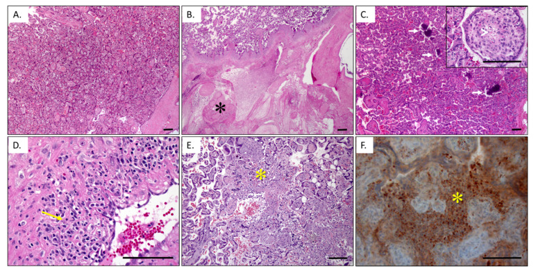
Figure 1.
Zika related uteroplacental pathology. (A) Gestational age-matched negative control placental histology. In contrast, Zika infected placentas (B,C) are more likely to have lobular infarctions (*) and villous stromal microcalcifications (arrows). Early stages of villous stromal cell death are also seen (inset arrowhead). (D) Some cases have maternal decidual leukocytoclastic vasculitis composed of lymphocytes, plasma cells, and eosinophils (arrow). (E) Preliminary data in NHP models suggest a potential relationship with chronic histiocytic intervillositis (*), which is supported by CD68-positive macrophages (*) in the intervillous space around viable villi (F). Photomicrographs of hemotoxylin- and eosin-stained sections (A–E), as well as immunohistochemical stained section with hematoxylin counterstain. Bar is 100 µm.

