Abstract
The antigenic composition of the hepatitis E virus (HEV) protein encoded by open reading frame 2 (ORF2) was determined by using synthetic peptides. Three sets of overlapping 18-, 25-, and 30-mer peptides, with each set spanning the entire ORF2 protein of the HEV Burma strain, were synthesized. All synthetic peptides were tested by enzyme immunoassay against a panel of 32 anti-HEV-positive serum specimens obtained from acutely HEV-infected persons. Six antigenic domains within the ORF2 protein were identified. Domains 1 and 6 located at the N and C termini of the ORF2 protein, respectively, contain strong immunoglobulin G (IgG) and IgM antigenic epitopes that can be efficiently modeled with peptides of different sizes. In contrast, antigenic epitopes identified within the two central domains (3 and 4) were modeled more efficiently with 30-mer peptides than with either 18- or 25-mers. Domain 2 located at amino acids (aa) 143 to 222 was modeled best with 25-mer peptides. A few 30-mer synthetic peptides derived from domain 5 identified at aa 490 to 579 demonstrated strong IgM antigenic reactivity. Several 30-mer synthetic peptides derived from domains 1, 4, and 6 immunoreacted with IgG or IgM with more than 70% of anti-HEV-positive serum specimens. Thus, the results of this study demonstrate the existence of six diagnostically relevant antigenic domains within the HEV ORF2 protein.
Hepatitis E virus (HEV) is an agent of enterically transmitted non-A, non-B hepatitis (6, 7, 35, 37), which is a serious problem in many developing countries (6, 7, 41). The HEV genome is a single-stranded, positive-sense RNA molecule of approximately 7.5 kb (35, 37). Three open reading frames (ORF) were identified within the HEV genome (39): ORF1 encodes nonstructural proteins, ORF2 encodes the putative structural protein(s) (37, 39), and ORF3 encodes a protein of unknown function.
The antigenic composition of HEV proteins has been examined with synthetic peptides of different sizes (9, 18–21) and recombinant proteins (24, 25, 33, 36, 43). One of the most comprehensive studies was performed with overlapping 10-mer synthetic peptides spanning the entire HEV ORF1-, ORF2-, and ORF3-encoded proteins (18). This study found that the ORF1 protein contains at least 12 antigenic regions that can be efficiently modeled with short synthetic peptides. Only three antigenic regions were identified within the HEV ORF2-encoded protein, at amino acids (aa) 25 to 38, 341 to 354, and 517 to 530. Each region was modeled with two overlapping synthetic peptides. Another antigenic region at aa 105 to 122, which was modeled with three overlapping peptides, was revealed within the ORF3-encoded protein (18). Whereas no additional studies have been conducted to further elaborate the antigenic composition of the ORF1-encoded protein, antigenic epitopes from the ORF2- and ORF3-encoded proteins have been extensively explored (9, 19–21, 24, 25, 33, 36, 43). The antigenic region within the ORF3 protein at aa 105 to 122, which was found with 10-mer peptides (18), was also identified in other studies with synthetic peptides of different sizes (9, 19) and recombinant proteins (43). However, even though a thorough scan of the ORF3 protein with 10-mer peptides revealed just this single antigenic region (18), two additional antigenic regions at aa 31 to 40 and 63 to 76 were identified within the ORF3 protein with synthetic peptides of different sizes (19). In contrast to the ORF3 protein, only one of three antigenic epitopes found with 10-mer peptides within the ORF2 protein at aa 517 to 530 (18) has been confirmed with peptides of different sizes (20). The other two antigenic regions identified with 10-mer peptides were located at aa 25 to 38 and 341 to 354 (18). Strong antigenic reactivity was recently associated with an approximately 100-aa N-terminal region of the ORF2 protein (25), thus confirming the existence of a strong antigenic region located at the N terminus. On the other hand, 10-mer peptides (18) could not confirm the existence of strong antigenic epitopes at aa 319 to 340 (19), 394 to 470 (20), and 631 to 660 (9, 19, 43), identified with synthetic peptides and recombinant proteins. Collectively, these findings demonstrate the inconsistent modeling of some antigenic epitopes from the HEV ORF2 and ORF3 proteins with synthetic peptides of different sizes and with recombinant proteins.
The ORF2 protein expressed in the baculovirus expression system (14, 40) or vaccinia expression system (8) or fragments of this protein expressed in Escherichia coli (10, 13, 24, 25, 27, 31, 33) have been used as important diagnostic reagents for the development of diagnostic tests to detect anti-HEV activity in serum specimens. Some of these proteins have also been identified as important vaccine candidates eliciting protective immunity in animal models (34, 41). An intensive analysis of a large set of recombinant polypeptides containing different parts of the HEV ORF2 protein clearly showed that the antigenic properties of some fragments of this protein differ from the antigenic properties of the whole protein. For example, some fragments could bind anti-HEV antibodies from convalescent-phase serum specimens with a much higher efficacy than that of the full-length protein expressed in the same expression system (25), suggesting that ORF2 fragments may be better suited as diagnostic reagents than the full-length protein. In accordance with these findings, it was recently shown by using an in vitro seroneutralization test that various HEV strains were neutralized more efficiently with antibodies derived against ORF2 fragments expressed in E. coli than with antibodies derived against an ORF2 full-length protein expressed in the baculovirus expression system (28). These findings suggest that the antigenic composition of the HEV ORF2 protein is complex and that different antigenic epitopes can be efficiently modeled with different fragments of this protein as represented with either synthetic peptides or recombinant proteins.
In this paper, we report the identification of six antigenic domains within the ORF2-encoded protein as modeled with three sets of overlapping synthetic peptides of different sizes, with each set spanning the entire ORF2 protein. The most diagnostically relevant antigenic regions that were efficiently modeled with synthetic peptides were found within domains 1, 4, and 6. The combination of only a few peptides derived from these domains may be used to develop diagnostic tests to detect anti-HEV activity in serum specimens obtained from acutely infected patients.
MATERIALS AND METHODS
Serum specimens.
All anti-HEV-positive serum specimens were obtained from a collection reposited in the Hepatitis Branch, Division of Viral and Rickettsial Diseases, National Center for Infectious Diseases, Centers for Disease Control and Prevention, Atlanta, Ga. Ten serum specimens were obtained from 10 acutely HEV-infected patients during an outbreak of HEV infection in central Asia (collected by M. O. Favorov). Twenty-two anti-HEV-positive specimens were obtained from an outbreak of HEV infection in India from 22 persons with acute hepatitis E. Serum specimens were selected from these two regions of the world because the HEV strains circulating in these regions are the most homologous to the HEV Burma strain (2, 3, 30), whose sequence was used to design synthetic peptides (39). Anti-HEV-negative serum specimens (n = 32) from normal blood donors were also obtained from a collection reposited at the Centers for Disease Control and Prevention. All specimens were initially tested for anti-HEV immunoglobulin G (IgG) and IgM activity by a commercially available kit (GeneLabs, Redwood City, Calif.) and by the enzyme immunoassay (EIA) based on use of the HEV mosaic antigen as previously described (12, 22).
Synthetic peptides.
Peptides were synthesized by 9-fluorenylmethoxycarbonyl chemistry (4) on an ACT model MPS 350 multiple-peptide synthesizer (Advanced Chemtech, Louisville, Ky.) according to the manufacturer’s protocols. After characterization by amino acid analysis, high-performance liquid chromatography, and capillary electrophoresis, peptides were characterized by EIA.
EIA.
Synthetic peptides (110 μl) at a concentration of 5 μg/ml in 0.1 M phosphate-buffered saline (PBS), pH 7.5, were adsorbed to microtiter wells (Immulon II; Dynatech Laboratories, Inc.) at room temperature for 12 h. Serum was diluted 1:100 in PBS containing 0.1% Tween 20 and 10% normal goat serum (PBS-T). One hundred microliters of diluted serum was added to each well and incubated for 1 h at 37°C. The binding of antibodies to the peptides was identified with affinity-purified antibodies to human IgG or IgM coupled to horseradish peroxidase (Boehringer Mannheim, Indianapolis, Ind.) by adding 100 μl of a 1:10,000 or 1:5,000 dilution, respectively, in PBS-T and incubating for 1 h at 37°C. The cutoff, expressed as a P/N ratio and equal to 3.0, was statistically established individually for each peptide as the mean of the result with negative controls plus at least 3.5 standard deviations above the mean, where P represents the optical density at 493 nm (OD493) of anti-HEV-positive specimens and N represents the OD of negative controls. Each serum specimen in every experiment was also tested with an irrelevant peptide (no. 1546) with the sequence PMSMDTSDETSEGATFLSLS derived from a small ORF within the hepatitis G virus minus-sense RNA (26). As an additional criterion, the ratio between the OD493 for each HEV peptide and the OD493 for this irrelevant peptide found for each serum specimen was used. HEV peptides were considered specifically immunoreactive with serum specimens when this ratio was greater than 2.
Computer-assisted analysis.
Amino acid sequence analysis was performed by using MegAlign and Protean programs from the Lasergene software package (DNASTAR Inc., Madison, Wis.). Protein secondary structure was predicted with the ALB program (32).
RESULTS AND DISCUSSION
Design of synthetic peptides.
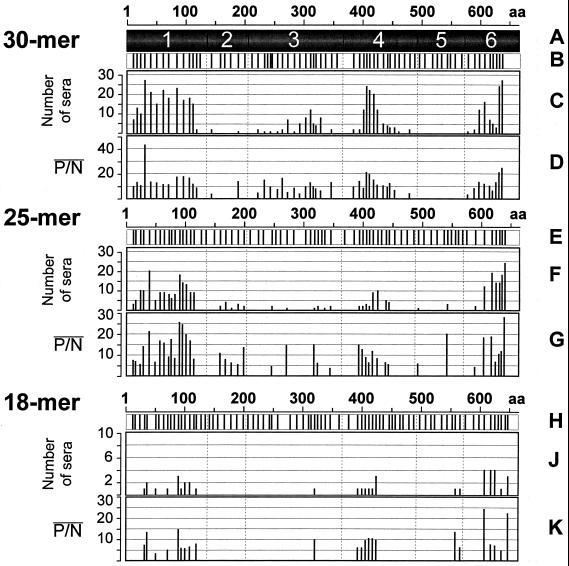
The sequence of the protein encoded by ORF2 of the HEV Burma strain (39) was used to design synthetic peptides. Three sets of overlapping peptides were synthesized. Each set spanned almost the entire ORF2 protein without gaps, with the exception of an 11-aa hydrophobic region at the extreme N terminus (Fig. 1 to 3). One set contained 80 18-mer peptides (Fig. 1H). A second set contained 72 25-mer peptides (Fig. 1E), and a third set contained 72 30-mer peptides (Fig. 1B). Each set spanned the ORF2 protein in such a way that every amino acid of this protein was represented an average of 2.2 times for 18-mers, 2.7 times for 25-mers, and 3.3 times for 30-mer peptides. Within each set, peptides were distributed across the HEV polypeptide chain in an almost uniform manner. However, some regions were spanned with a higher density of peptides. These regions included regions where antigenic epitopes had been found previously (9, 18–20, 43) or where antigenic epitopes could be predicted based on hydropathic plots (23), backbone chain flexibility (17), and protein secondary structure (32). Some other regions, such as a region at aa 360 to 390, where a long hydrophobic alpha-helix was predicted (Fig. 3B and D), were spanned with a lower density of peptides (Fig. 1A).
FIG. 1.
Antigenic reactivity of overlapping synthetic 30-, 25-, and 18-mer peptides with anti-HEV IgG antibodies. (A) Approximate locations of six antigenic domains. (B) Vertical bars show the location of the N-terminal amino acid of each synthesized peptide. (C, F, and J) Charts demonstrating the number of serum specimens that are immunoreactive with each peptide. (D, G, and K) Charts showing the average P/N ratio (see Materials and Methods) calculated for all serum specimens that are immunoreactive with each synthetic peptide.
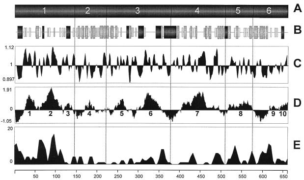
FIG. 3.
Structural properties of the HEV ORF2 antigenic domains. (A) Antigenic domains. (B) Predicted secondary structure (32) with horizontal line representing random coil, open boxes representing beta-turns, shaded boxes representing beta-sheets, and black boxes representing alpha helices. (C) Plot of flexibility of the protein chain (17), in which upward peaks show flexible regions. (D) Hydrophilicity plot (23), in which upward peaks show hydrophilic regions. (E) Profile of amino acid sequence heterogeneity calculated for 10 known ORF2-encoded proteins (1, 2, 3, 5, 11, 15, 29, 30, 38, 39) by using a window of 40 aa sliding along the polypeptide chain with a step of 10 aa; graph values were calculated as the number of positions affected with amino acid substitutions within each window across the entire ORF2 polypeptide.
Immunoreactivity of synthetic peptides with IgG anti-HEV antibodies.
All synthetic peptides were tested for immunoreactivity with IgG anti-HEV antibodies as described in Materials and Methods. In general, 18-mer peptides were less immunoreactive than either 25- or 30-mer peptides. Because of this observation, 18-mer peptides were tested with only 10 anti-HEV-positive serum specimens, whereas 25- and 30-mer peptides were tested with 32 anti-HEV-positive serum specimens. An analysis of the pattern of the antigenic reactivities of all peptides allowed the identification of six antigenic domains (Fig. 1A): domain 1 at aa 12 to 147, domain 2 at aa 143 to 222, domain 3 at aa 221 to 373, domain 4 at aa 381 to 504, domain 5 at aa 490 to 579, and domain 6 at aa 573 to 660 (Table 1).
TABLE 1.
Antigenic domains and regions within the HEV ORF2-encoded protein as modeled with synthetic peptides immunoreactive with IgG anti-HEV antibodies
| Domain | Domain positions (aa) | No. of epitopes within domain | 18-mer peptides
|
25-mer peptides
|
30-mer peptides
|
|||||||||
|---|---|---|---|---|---|---|---|---|---|---|---|---|---|---|
| Total no. of peptides | No. of Aga regions within domain | Positions (aa) | No. of reactive peptides | Total no. of peptides | No. of Ag regions within domain | Positions (aa) | No. of reactive peptides | Total no. of peptides | No. of Ag regions within domain | Positions (aa) | No. of reactive peptides | |||
| 1 | 12–147 | 6 | 18 | 4 | 32–68 | 3 | 16 | 1 | 12–138 | 16 | 13 | 1 | 12–147 | 13 |
| 71–88 | 1 | |||||||||||||
| 89–125 | 4 | |||||||||||||
| 119–136 | 1 | |||||||||||||
| 2 | 143–222 | 2 | 9 | 9 | 1 | 158–222 | 5 | 7 | 2 | 143–172 | 1 | |||
| 188–217 | 1 | |||||||||||||
| 3 | 221–373 | 6 | 18 | 1 | 318–335 | 1 | 14 | 4 | 245–269 | 1 | 19 | 4 | 221–261 | 2 |
| 270–294 | 1 | 242–271 | 1 | |||||||||||
| 316–346 | 2 | 254–355 | 10 | |||||||||||
| 333–367 | 2 | 344–373 | 1 | |||||||||||
| 4 | 381–504 | 5 | 16 | 1 | 390–438 | 6 | 15 | 2 | 391–446 | 6 | 16 | 2 | 381–486 | 12 |
| 436–465 | 2 | 475–504 | 1 | |||||||||||
| 5 | 490–579 | 2 | 9 | 1 | 554–579 | 2 | 10 | 2 | 490–514 | 1 | 7 | |||
| 539–563 | 1 | |||||||||||||
| 6 | 573–660 | 5 | 10 | 3 | 603–638 | 3 | 8 | 1 | 586–660 | 7 | 10 | 1 | 573–660 | 9 |
| 632–649 | 1 | |||||||||||||
| 643–660 | 1 | |||||||||||||
Ag, antigenic.
As can be seen in Fig. 1, the majority of immunoreactive 18-mer peptides could bind IgG anti-HEV antibodies from only one to two serum specimens. Nonetheless, these results allowed the identification of five of the six antigenic domains (Fig. 1J and Table 1). N-terminal domain 1 contains four antigenic regions at aa 32 to 68, 71 to 88, 89 to 125, and 119 to 136. An antigenic region is defined as a region of the protein spanned by one or more overlapping consecutive immunoreactive peptides. Regions 1 and 3 were identified with three and four consecutive overlapping peptides, respectively. Regions 2 and 4 were identified with only one immunoreactive peptide each. Immunoreactive peptides derived from regions 1 and 2 are separated from each other with two consecutive nonimmunoreactive peptides. Regions 3 and 4 are separated with only one nonimmunoreactive peptide. No antigenic epitopes within domain 2 could be modeled with 18-mer peptides, and only one peptide, 18-318 (an 18-mer comprising aa 318 to 335), derived from domain 3, was found to be immunoreactive. Domain 4 was identified with six overlapping peptides spanning aa 390 to 438, and domain 5 was identified with two overlapping peptides spanning aa 554 to 579. Finally, C-terminal antigenic domain 6 contains three antigenic regions at aa 603 to 638, identified with three peptides, aa 632 to 649, identified with 1 peptide, and aa 643 to 660, identified with one peptide. These regions are separated from each other by only one nonimmunoreactive peptide. Synthetic peptides derived from the C-terminal domain were the most immunoreactive among all 18-mer peptides (Fig. 1J and K). Two peptides, 18-603 (aa 603 to 620) and 18-643 (aa 643 to 660), were the most immunoreactive, detecting antibody activity in three to four serum specimens with high P/N values (Fig. 1K).
Analysis of 25-mer peptides revealed all six antigenic domains (Fig. 1F and G; Table 1). N-terminal domain 1 was identified with 16 overlapping immunoreactive peptides spanning the region of aa 12 to 138. Five overlapping immunoreactive peptides spanning aa 158 to 222 were found within the second domain. Thus, antigenic epitopes within this domain were modeled with 25-mer peptides (Fig. 1F). However, none of the 18-mer peptides modeled epitopes within this domain (Fig. 1J) and only two 30-mer peptides were immunoreactive (Fig. 1C). Domain 3 comprises four antigenic regions. Regions 1 and 2 at aa 245 to 269 and 270 to 294, respectively, were identified with only one peptide each. Overlapping regions 3 and 4 at aa 316 to 346 and 333 to 367, respectively, were identified with two peptides each. Regions 1 and 2, 2 and 3, and 3 and 4 were separated by two, three, and one nonimmunoreactive peptide(s), respectively. Domain 4 contains two antigenic regions. One region at aa 391 to 446 was spanned with six consecutive peptides. The second region at aa 436 to 465 was spanned with two peptides, which was separated from the first region by one nonimmunoreactive peptide (Fig. 1F). Domain 5 contains two antigenic regions at aa 490 to 514 and 539 to 563, with each region represented with only one immunoreactive peptide and separated from each other by four nonimmunoreactive peptides. C-terminal domain 6 was spanned with seven overlapping peptides derived from the region of aa 586 to 660. Peptides 25-39 and 25-90 (25-mer peptides comprising aa 39 to 63 and 90 to 114, respectively), derived from N-terminal domain 1, and peptides 25-614 and 25-636 (aa 614 to 638 and 636 to 660, respectively), derived from C-terminal domain 6, were the most immunoreactive among all 25-mer peptides. These peptides immunoreacted with 18 to 24 serum specimens with high P/N values (Fig. 1G). The most immunoreactive 25-mer peptide (25-636) detected IgG anti-HEV IgG activity in 24 of 32 serum specimens.
The set of 30-mer peptides was the most immunoreactive. The pattern of reactivity of these peptides with serum specimens demonstrated the existence of five antigenic domains (Fig. 1C and D; Table 1). N-terminal domain 1 extends from aa 12 to 147. Antigenic epitopes within this domain were modeled with 13 consecutive overlapping synthetic peptides. Also, this domain contains the most immunoreactive peptide, 30-31 (30-mer peptide comprising aa 31 to 60), found in this study. It strongly immunoreacted with 27 of 32 anti-HEV-positive serum specimens (Fig. 1C). As was mentioned above, peptide 25-39, which overlaps peptide 30-31, was one of the most immunoreactive among all 25-mers. The overlapping region between these two peptides (aa 39 to 60) may comprise the immunodominant antigenic epitope within N-terminal domain 1. The other strong antigenic epitope identified with peptide 25-90 (Fig. 1F) was not as well modeled with 30-mer peptides as the epitope located within the region of aa 39 to 60. Peptide 30-85 (aa 85 to 114) immunoreacted with 23 of 32 anti-HEV-positive serum specimens, but with a lower P/N value than peptide 30-31 (Fig. 1D).
Only two 30-mer peptides derived from aa 143 to 172 and 188 to 217 comprised the second antigenic domain, suggesting that epitopes within this domain are more efficiently modeled with 25-mer peptides than with 30-mers. Conversely, antigenic epitopes within the third domain were better modeled with 30-mer synthetic peptides than with 18- or 25-mer peptides (Fig. 1C, F, and J). This finding suggests that antigenic epitopes within this domain are more conformation dependent than epitopes within domains 1, 4, and 6, because the antigenic properties of these domains may be efficiently modeled with peptides of different sizes. The major antigenic region of domain 3 was spanned with 10 overlapping synthetic peptides. None of these 10 peptides were as immunoreactive as the most immunoreactive peptides from the other regions. The most immunoreactive peptide derived from this region is peptide 30-309, comprising the sequence from aa 309 to 338. However, this peptide immunoreacted with only 12 of 32 serum specimens (Fig. 1C). This domain contains three additional weakly immunoreactive antigenic regions (Table 1) at aa 221 to 261 (two peptides), 242 to 271 (one peptide), and 344 to 373 (one peptide).
The fourth antigenic domain contains two antigenic regions (Table 1). The major antigenic region was spanned with 12 overlapping peptides derived from aa 381 to 486 (Fig. 1C). Peptides 30-403 (aa 403 to 432), 30-408 (aa 408 to 437), and 30-415 (aa 415 to 444) derived from this domain were strongly and broadly immunoreactive. These peptides immunoreacted with 20 to 24 serum specimens with high P/N values. The antigenic epitopes within this domain were poorly modeled with 18-mer and 25-mer peptides (Fig. 1F and J); however, 30-mer peptides modeled these antigenic epitopes as efficiently as epitopes from domain 1. This observation suggests that these epitopes as well as epitopes from domain 2 are strongly conformation dependent.
For domain 5, not a single 30-mer synthetic peptide was found to model an antigenic epitope. In contrast, however, epitopes within domain 5 were efficiently modeled with shorter 18- and 25-mer peptides. This finding suggests that these antigenic epitopes are also conformation dependent.
The sixth antigenic domain located within the region of aa 573 to 660 was spanned with nine overlapping synthetic peptides. Peptides 30-626 (aa 626 to 655) and 30-631 (aa 631 to 660) were two of the most immunoreactive peptides derived from this domain. The very C-terminal 25-mer peptide (25-636), which overlapped significantly with peptide 30-631, was the most immunoreactive peptide among all 25-mer peptides. Although very immunoreactive, peptide 30-631 was not the most immunoreactive among the 30-mer peptides. Peptide 30-31 derived from N-terminal domain 1 bound anti-HEV antibodies from the same number of serum specimens (27 of 32 specimens) as peptide 30-631; however, peptide 30-31 was more strongly immunoreactive than peptide 30-631 as evidenced by a comparison of the average P/N values for these two peptides (Fig. 1D).
The number of antigenic epitopes within each domain.
Within N-terminal domain 1, immunoreactive 25- and 30-mer peptides spanned aa 12 to 138 and 12 to 147, respectively. These regions contained 4 to 5 nonoverlapping 25- or 30-mer peptides, with each peptide modeling at least one antigenic epitope. Four antigenic regions were modeled within this domain with 18-mer synthetic peptides (see above). Two of these regions at aa 32 to 68 and 89 to 125 are large enough to be spanned with two nonoverlapping peptides. Therefore, domain 1 may contain at least six antigenic epitopes (Table 1).
Domain 2 was modeled with two nonoverlapping 30-mer peptides (30-143 and 30-188) and five overlapping 25-mer peptides spanning aa 158 to 222, which is of sufficient size to be covered with two nonoverlapping 25-mers. This finding suggests that domain 2 may contain at least two antigenic epitopes.
Within domain 3, only one 18-mer was immunoreactive. 25-mer peptides derived from this domain identified four antigenic regions, each of 25 to 34 aa. Therefore, these peptides model at least four antigenic epitopes. However, 30-mer peptides identified four antigenic regions (Table 1), one of which may be spanned with three nonoverlapping peptides. Thus, at least six antigenic epitopes may be modeled with synthetic peptides within this domain.
The region of aa 390 to 438 within domain 4 was identified with six overlapping immunoreactive 18-mer peptides. The size of this region is sufficient to accommodate three 18-mer peptides overlapped by only 2 aa each, suggesting that this region may contain at least three antigenic epitopes. In addition, the use of 25-mer peptides identified another region at aa 436 to 465 that may contain one antigenic epitope. An additional antigenic epitope was identified with 30-mer peptides within the region of aa 475 to 504. Thus, domain 4 may contain at least five antigenic epitopes.
Domain 5 was identified with only two overlapping 18-mer peptides and two 25-mer peptides representing two separate antigenic regions. Thus, this domain may contain at least two antigenic epitopes. C-terminal domain 6 contains at least five antigenic epitopes because 18-mer peptides identified three antigenic regions, one of which at aa 603 to 638 may be spanned with two nonoverlapping 18-mers, and one immunoreactive 30-mer spans aa 573 to 602 (Table 1), which is not covered with any immunoreactive 18-mer. Collectively, these data suggest that the entire HEV ORF2-encoded protein may contain a minimum of 26 antigenic epitopes.
Immunoreactivity of synthetic peptides with IgM anti-HEV antibodies.
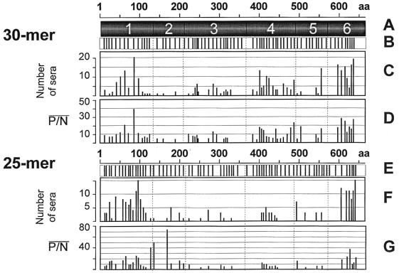
All 25- and 30-mer synthetic peptides were tested for IgM anti-HEV immunoreactivity as described in Materials and Methods. The set of 25-mer peptides was tested with 16 anti-HEV positive serum specimens, whereas 30-mer peptides were tested with 23 anti-HEV-positive serum specimens. An analysis of the pattern of the antigenic reactivities of these peptides allowed for the identification of the very same six antigenic domains that were found when these peptides were tested for IgG immunoreactivity (Fig. 2A and Table 2), although domain 2 was not as readily discernible, especially when 30-mer peptides were tested, an observation similar to that shown for IgG anti-HEV (Fig. 2C). Domain 2 was best identified when 25-mers were tested for IgG immunoreactivity (Fig. 1F). Similarly, this domain was better identified with 25-mer peptides when tested for IgM activity (Fig. 2F).
FIG. 2.
Antigenic reactivity of overlapping synthetic 30- and 25-mer peptides with anti-HEV IgM antibodies (for details see the legend to Fig. 1).
TABLE 2.
Antigenic domains and regions within the HEV ORF2-encoded protein as modeled with synthetic peptides immunoreactive with IgM anti-HEV antibodies
| Domain | Domain positions (aa) | No. of epitopes within domain | 25-mer peptides
|
30-mer peptides
|
||||
|---|---|---|---|---|---|---|---|---|
| No. of Aga regions | Positions (aa) | No. of reactive peptides | No. of Ag regions | Positions (aa) | No. of reactive peptides | |||
| 1 | 12–147 | 5 | 2 | 12–68 | 5 | 2 | 12–41 | 1 |
| 57–138 | 10 | 24–147 | 11 | |||||
| 2 | 143–222 | 3 | 3 | 126–158 | 2 | 2 | 124–187 | 3 |
| 167–201 | 2 | 188–217 | 1 | |||||
| 198–232 | 2 | |||||||
| 3 | 221–373 | 5 | 5 | 222–246 | 1 | 2 | 221–274 | 5 |
| 251–275 | 1 | 271–355 | 8 | |||||
| 270–294 | 1 | |||||||
| 301–333 | 2 | |||||||
| 328–352 | 1 | |||||||
| 4 | 381–504 | 5 | 1 | 403–465 | 7 | 2 | 381–471 | 10 |
| 457–520 | 5 | |||||||
| 5 | 490–579 | 4 | 4 | 490–514 | 1 | 1 | 538–580 | 3 |
| 511–535 | 1 | |||||||
| 539–563 | 1 | |||||||
| 552–576 | 1 | |||||||
| 6 | 573–660 | 2 | 1 | 601–660 | 6 | 1 | 592–660 | 7 |
Ag, antigenic.
An analysis of the immunoreactivity of 30-mer peptides allowed the identification of five of six antigenic domains (Fig. 2C and D). Testing of 25-mers allowed the clear identification of domains 1, 4, and 6, while the other domains were less discernible. Domains 1 and 6 were the most IgM immunoreactive. Domain 1 was spanned with 12 immunoreactive 30-mer peptides and 15 immunoreactive 25-mer peptides. Each set of peptides identified two antigenic regions within this domain. One antigenic region at aa 12 to 41 was revealed with one 30-mer peptide, whereas the second region at aa 24 to 147 was detected with 11 overlapping peptides. The set of 25-mer peptides also identified two antigenic regions that significantly overlap with antigenic regions found with 30-mer peptides (Fig. 2C and F). Five 25-mer peptides spanned the first region at aa 12 to 68, and 10 peptides spanned the second region at aa 57 to 138 (Fig. 2F). An analysis of the number of nonoverlapping 25- or 30-mer peptides that would fit to each antigenic region showed that domain 1 may contain at least five IgM epitopes. The most IgG immunoreactive peptide, 30-31 (see above), was very poorly IgM immunoreactive. This peptide detected only 2 of 23 anti-HEV-positive serum specimens when tested for IgM reactivity, whereas it immunoreacted with 27 of 32 anti-HEV-positive serum specimens when tested for IgG reactivity. Peptide 30-85 was the most IgM-immunoreactive peptide found in this study. It immunoreacted with 20 of 23 anti-HEV-positive serum specimens. This peptide was also very IgG immunoreactive. It detected anti-HEV IgG in 23 of 32 anti-HEV-positive serum specimens.
Domain 2 contains two antigenic regions at aa 124 to 187 and 188 to 217, identified with three 30-mer peptides and one 30-mer peptide, respectively, or three antigenic regions at aa 126 to 158, 167 to 201, and 198 to 232, identified with two 25-mer peptides for each antigenic region. By applying the same criterion as described above, the minimal number of antigenic epitopes within this domain can be estimated as three.
Domain 3 contains two antigenic regions at aa 221 to 274 and 271 to 355, identified with five and eight 30-mer peptides, respectively. When 25-mer peptides were used, five antigenic regions at aa 222 to 246 (one peptide), 251 to 275 (one peptide), 270 to 294 (one peptide), 301 to 333 (two peptides), and 328 to 352 (one peptide) were found. Thus, this domain may contain a minimum of five IgM antigenic epitopes. Domain 4 contains two antigenic regions at aa 381 to 471 and 457 to 520, identified with 10 and five 30-mer peptides, respectively. One antigenic region at aa 403 to 465 was identified with seven overlapping 25-mer peptides. This domain may contain at least five IgM antigenic epitopes. Peptide 30-398 was the most IgM-immunoreactive peptide derived from domain 4. This peptide bound IgM antibodies from 13 of 23 anti-HEV-positive serum specimens (Fig. 2C).
One antigenic region at aa 538 to 580 was identified within domain 5 with three overlapping 30-mer peptides. However, by using 25-mer peptides four antigenic regions at aa 490 to 514, 511 to 535, 539 to 563, and 552 to 576 were identified. Each region was spanned with one peptide. Thus, this domain may contain at least four IgM antigenic epitopes. Peptide 30-551 was the most IgM-immunoreactive peptide derived from domain 5. This peptide immunoreacted with 14 of 23 anti-HEV-positive serum specimens (Fig. 2C). None of the 30-mer peptides derived from domain 5 showed immunoreactivity with IgG anti-HEV antibodies (Fig. 1C), whereas the same peptides were strongly immunoreactive with IgM antibodies (Fig. 2C).
Domain 6 was modeled with six overlapping 25-mer peptides and seven overlapping 30-mer peptides. All peptides derived from this domain were broadly and strongly immunoreactive (Fig. 2C, D, F, and G). The extreme C-terminal peptides (25-636 and 30-631) could efficiently detect IgG and IgM anti-HEV antibodies. These two peptides were among the most immunoreactive peptides found in this study. Domain 6 contains one antigenic region at aa 601 to 660, identified with 25-mer peptides, or at aa 592 to 660, identified with 30-mer peptides. This region is of sufficient size to accommodate two nonoverlapping 25- or 30-mer peptides. Therefore, domain 6 may contain at least two IgM antigenic epitopes.
Antigenic epitopes of the HEV ORF2 protein.
Various investigators have used synthetic peptides (9, 18–20) or recombinant proteins (24, 25, 43) to identify antigenic epitopes within the HEV ORF2-encoded protein. A recombinant protein containing the N-terminal region of approximately 100 aa was strongly immunoreactive (25). An analysis of the immunoreactivities of synthetic peptides derived from this region, however, yielded contradicting results. In one study (19) in which two peptides spanning sequences from aa 54 to 65 (12-mer peptide) and 84 to 101 (18-mer peptide) were tested, no antigenic reactivity was found. Two 18-mer peptides (18-56 and 18-82) described in the present paper, which are most related to the two peptides mentioned above, were also found to be antigenically inactive. On the other hand, a peptide scan using overlapping 10-mer peptides revealed only one antigenic region spanned with two peptides at aa 25 to 38 (18). The sequence from aa 29 to 34, shared by these two peptides, may play an essential role for this antigenic epitope. This sequence is also shared by immunoreactive 25- and 30-mer peptides (Fig. 1 and Table 1). However, the most IgG-immunoreactive peptides identified in this study within N-terminal domain 1 (aa 12 to 147) share sequences from aa 39 to 53 and 90 to 106 (Fig. 1), both of which are outside the region identified by peptide scanning (18). Of these two immunodominant regions within domain 1, only one region (aa 95 to 114) was associated with strong IgM immunoreactivity (Fig. 2). This region was spanned with one 30-mer peptide containing the sequence at aa 85 to 114 and one 25-mer peptide containing the sequence at aa 95 to 119 (Fig. 2).
No antigenic epitopes within domain 2 have been reported. However, within domain 3 two regions of antigenic reactivity were previously identified by using two overlapping 10-mer peptides at aa 341 to 354 (8) and one 20-mer peptide at aa 319 to 340 (20). In the present study, peptides overlapping these two regions were also found to be immunoreactive (Fig. 1 and 2). One of these regions was found to be strongly IgG immunoreactive by using a 30-mer peptide comprising the sequence from aa 309 to 338. This peptide immunoreacted with 12 of 32 serum specimens (Fig. 1C and D). However, two of the most IgM-immunoreactive 30-mer peptides from this domain were located elsewhere, at aa 242 to 271 and 282 to 311 (Fig. 2C).
Domain 4 was previously identified with 20-mer synthetic peptides as one of the most immunodominant regions within the HEV ORF2 protein (20). Surprisingly, a recombinant protein comprising aa 394 to 470, which is a significant part of the domain 4 sequence (aa 381 to 504), was not immunoreactive (25). These contradicting observations suggest that antigenic epitopes within this region are highly conformation dependent and that they can be efficiently modeled only with peptides or protein fragments of specific sizes.
Two antigenic regions within domain 5 at aa 515 to 530 (18, 20) and 546 to 589 (20) have been previously described. One of these regions (aa 515 to 530) was also reported as having nonspecific antigenic reactivity (20). In the present study, none of the synthetic peptides that overlapped this region was either specifically or nonspecifically immunoreactive. IgG antigenic epitopes within this domain could not be efficiently modeled with 30-mer peptides (Fig. 1C). The antigenic reactivity associated with the other region at aa 546 to 589 (20) was confirmed with only two overlapping 18-mer peptides spanning the region at aa 554 to 579 (Fig. 1J). 25- and 30-mer peptides derived from this domain were more immunoreactive with IgM anti-HEV (Fig. 2C and F) than with IgG antibodies (Fig. 1C and F).
Antigenic epitopes derived from the extreme C terminus of the HEV ORF2 protein were found in the very first experiments on the identification of HEV antigenic epitopes (43). Later, this original observation was confirmed by several research groups (9, 19, 24, 25). This further substantiated this observation, namely, that the region from aa 631 to 660 contains strong IgG- and IgM-specific antigenic epitopes that can be efficiently modeled with synthetic peptides of different sizes (Fig. 1 and 2). In addition, synthetic peptides derived from the adjacent region at aa 573 to 630 are also strongly and broadly immunoreactive (Fig. 1 and 2).
Computer-assisted analysis revealed 10 large hydrophilic regions within the HEV ORF2-encoded protein (Fig. 3D). When these hydrophilic regions were aligned with antigenic domains, each one contained at least one hydrophilic region. All domains are separated by broad hydrophobic regions, except at the border between domains 2 and 3 (Fig. 3A and D). Domains 1, 3, and 4 contain highly hydrophilic regions. In addition, domain 1 has a very high predicted backbone chain flexibility (Fig. 3C). Domains 3 and 4 are also quite flexible compared with domains 2 and 6, which have rather rigid backbone structure, and compared with domain 5, which has intermediate flexibility. Domains 1, 4, and 6 were the most immunoreactive domains modeled with synthetic peptides. Although antigenic epitopes tend to be located within highly hydrophilic and flexible regions (16), only domains 1 and 4 have high hydrophilicity and protein chain flexibility. Domain 6 is not very hydrophilic and, except for the extreme C-terminal region, has a very low backbone chain flexibility (Fig. 3C and D). The predicted secondary structure for the HEV ORF2 protein was aligned with the antigenic domains (Fig. 3B). This analysis revealed that (i) antigenic domain 1 is flanked by two alpha-helices and is mainly composed of random coil and beta-turn elements; (ii) domain 2 is a cluster of beta-sheet structures; (iii) domain 3 is alpha-helical with a long stretch of a random coil structure; (iv) domain 4 contains almost an equal mixture of random coil and beta-sheet structures with the centrally located extended random coil structure demonstrating the highest hydrophilicity and flexibility characteristics, which is true for a similar region within domain 3 (Fig. 3B, C, and D); (v) domain 5 is mainly represented with beta-turns and random coils flanked with beta-sheet structures; and (vi) the hydrophilic region of domain 6 comprises a mixture of random coils and beta-turns and contains one alpha-helix. The hydrophobic region of this domain is represented with a cluster of beta-sheets (Fig. 3B). Thus, each antigenic domain has a set of specific structural characteristics as predicted by the computer-assisted analysis.
Finally, sequence heterogeneity along the HEV ORF2 protein was analyzed (Fig. 3E). This analysis revealed that domain 1 was the most heterogeneous. The sequence of the second strongest antigenic region at aa 90 to 106 was identified within this domain (Fig. 1) as very variable among different strains (Fig. 3E). Domain 6, the other strongly antigenic domain, is also variable. However, the most antigenically reactive region located at the extreme C terminus of the protein is rather conserved. Some sequence heterogeneity can also be found within domain 5, whereas sequences of domains 2, 3, and 4 from different HEV strains are conserved. Sequence heterogeneity of antigenic epitopes within domains 1 and 6 may affect the antigenic reactivities of these epitopes. A study of the influence of sequence variation on the antigenic properties of these antigenic regions is warranted.
In conclusion, six antigenic domains within the ORF2-encoded protein were identified by using three sets of overlapping synthetic peptides of different sizes. It was shown that several ORF2 antigenic epitopes can be very efficiently modeled with synthetic peptides. The most diagnostically relevant antigenic regions that were reliably modeled with these peptides were found within domains 1, 4, and 6. The data presented in this paper strongly suggest that recombinant proteins and/or synthetic peptides derived from these domains may be used as diagnostic targets for the development of assays to detect anti-HEV activity in serum specimens obtained from acutely infected patients.
REFERENCES
- 1.Aye T T, Uchida T, Ma X, Iida F, Shikata T, Zhuang H, Win K M. Complete nucleotide sequence of a hepatitis E virus isolated from the Xijiang epidemic (1986–1988) of China. Nucleic Acids Res. 1992;20:3512. doi: 10.1093/nar/20.13.3512. [DOI] [PMC free article] [PubMed] [Google Scholar]
- 2.Aye T T, Uchida T, Ma X Z, Iida F, Shikata T, Zhuang H, Win K M. Sequence comparison of the capsid region of hepatitis E virus isolated from Myanmar and China. Microbiol Immunol. 1992;36:615–621. doi: 10.1111/j.1348-0421.1992.tb02061.x. [DOI] [PubMed] [Google Scholar]
- 3.Aye T T, Uchida T, Ma X, Iida F, Shikata T, Ichikawa M, Rikihisa T, Win K M. Sequence and gene structure of the hepatitis E virus isolated from Myanmar. Virus Genes. 1993;7:95–110. doi: 10.1007/BF01702352. [DOI] [PubMed] [Google Scholar]
- 4.Barany G, Merrifield R B. Solid-phase peptide synthesis. In: Gross E, Meienhoter J, editors. The peptides. Vol. 1. New York, N.Y: Academic Press; 1980. pp. 1–284. [Google Scholar]
- 5.Bi S-L, Purdy M A, McCaustland K A, Margolis H S, Bradley D W. The sequence of hepatitis E virus isolated directly from a single source during an outbreak in China. Virus Res. 1993;28:233–247. doi: 10.1016/0168-1702(93)90024-h. [DOI] [PubMed] [Google Scholar]
- 6.Bradley D W. Hepatitis non-A, non-B viruses become identified as hepatitis C and E viruses. Prog Med Virol. 1990;37:101–135. [PubMed] [Google Scholar]
- 7.Bradley D W. Enterically-transmitted non-A, non-B hepatitis. Br Med Bull. 1990;46:442–461. doi: 10.1093/oxfordjournals.bmb.a072409. [DOI] [PubMed] [Google Scholar]
- 8.Carl M, Isaacs S N, Kaur M, He J, Tam A W, Yarbough P O, Reyes G R. Expression of hepatitis E virus putative structural proteins in recombinant vaccinia viruses. Clin Diagn Lab Immunol. 1994;1:253–256. doi: 10.1128/cdli.1.2.253-256.1994. [DOI] [PMC free article] [PubMed] [Google Scholar]
- 9.Cousaget P, Buisson Y, Depril N, le Cann P, Chabaud M, Molinie C, Roul R. Mapping of linear B cell epitopes on open reading frame 2- and 3-encoded proteins of hepatitis E virus using synthetic peptides. FEMS Microbiol Lett. 1993;109:251–255. doi: 10.1111/j.1574-6968.1993.tb06176.x. [DOI] [PubMed] [Google Scholar]
- 10.Dawson G J, Chau K H, Cabal C M, Yarbough P O, Reyes G R, Mushawar I K. Solid-phase enzyme-linked immunosorbent assay for hepatitis E virus IgG and IgM antibodies utilizing recombinant antigens and synthetic peptides. J Virol Methods. 1992;38:175–186. doi: 10.1016/0166-0934(92)90180-l. [DOI] [PubMed] [Google Scholar]
- 11.Donati M C, Fagan E A, Harrison T J. Sequence analysis of full length HEV clones derived directly from human liver in fulminant hepatitis E. In: Rizzetto M, Purcell R H, Gerin J L, Verme G, editors. Viral hepatitis and liver disease. Turin, Italy: Edizioni Minerva Medica; 1997. pp. 313–316. [Google Scholar]
- 12.Favorov M O, Khudyakov Y E, Mast E E, Yashina T L, Shapiro C N, Khudyakova N S, Jue D L, Onischenko G G, Margolis H S, Fields H A. IgM and IgG antibodies to hepatitis E virus (HEV) detected by an enzyme immunoassay based on an HEV-specific artificial recombinant mosaic protein. J Med Virol. 1996;50:50–58. doi: 10.1002/(SICI)1096-9071(199609)50:1<50::AID-JMV10>3.0.CO;2-1. [DOI] [PubMed] [Google Scholar]
- 13.Goldsmith R, Yarbough P O, Reyes G R, Fry K E, Gabor K A, Kamel M, Zakaria S, Amer S, Gaffar Y. Enzyme-linked immunosorbent assay for diagnosis of acute sporadic hepatitis E in Egyptian children. Lancet. 1992;339:328–331. doi: 10.1016/0140-6736(92)91647-q. [DOI] [PubMed] [Google Scholar]
- 14.He J, Tam A W, Yarbough P O, Reyes G R, Carl M. Expression and diagnostic utility of hepatitis E virus putative structural proteins expressed in insect cells. J Clin Microbiol. 1993;31:2167–2173. doi: 10.1128/jcm.31.8.2167-2173.1993. [DOI] [PMC free article] [PubMed] [Google Scholar]
- 15.Huang C-C, Nguyen D, Fernandez J, Yun K Y, Fry K E, Bradley D W, Tam A W, Reyes G R. Molecular cloning and sequencing of the Mexico isolate of hepatitis E virus (HEV) Virology. 1992;191:550–558. doi: 10.1016/0042-6822(92)90230-m. [DOI] [PubMed] [Google Scholar]
- 16.Jameson B A, Wolf H. The antigenic index: a novel algorithm for predicting antigenic determinants. CABIOS. 1988;4:181–186. doi: 10.1093/bioinformatics/4.1.181. [DOI] [PubMed] [Google Scholar]
- 17.Karplus P A, Schultz G E. Prediction of chain flexibility in protein. Naturwissenschaften. 1985;72:212–213. [Google Scholar]
- 18.Kaur M, Hyams K C, Purdy M A, Krawczynski K, Ching W M, Fry K E, Reyes G R, Bradley D W, Carl M. Human linear B-cell epitopes encoded by the hepatitis E virus include determinants in the RNA-dependent RNA polymerase. Proc Natl Acad Sci USA. 1992;89:3855–3858. doi: 10.1073/pnas.89.9.3855. [DOI] [PMC free article] [PubMed] [Google Scholar]
- 19.Khudyakov Y E, Khudyakova N S, Fields H A, Lue D, Starling C, Favorov M O, Krawczynski K, Polish L, Mast E, Margolis H S. Epitope mapping in proteins of hepatitis E virus. Virology. 1993;194:89–96. doi: 10.1006/viro.1993.1238. [DOI] [PubMed] [Google Scholar]
- 20.Khudyakov Y E, Favorov M O, Jue D L, Hine T K, Fields H A. Immunodominant antigenic regions in a structural protein of the hepatitis E virus. Virology. 1994;198:390–393. doi: 10.1006/viro.1994.1048. [DOI] [PubMed] [Google Scholar]
- 21.Khudyakov Y E, Khudyakova N S, Jue D L, Wels T W, Padhye N, Fields H A. Comparative characterization of antigenic epitopes in the immunodominant region of the protein encoded by open reading frame 3 in Burmese and Mexican strains of hepatitis E virus. J Gen Virol. 1994;75:641–646. doi: 10.1099/0022-1317-75-3-641. [DOI] [PubMed] [Google Scholar]
- 22.Khudyakov Y E, Favorov M O, Khudyakova N S, Cong M E, Holloway B P, Padhye N, Lambert S B, Jue D L, Fields H A. Artificial mosaic protein containing antigenic epitopes of hepatitis E virus. J Virol. 1994;68:7067–7074. doi: 10.1128/jvi.68.11.7067-7074.1994. [DOI] [PMC free article] [PubMed] [Google Scholar]
- 23.Kyte J, Doolittle R F. A simple method for displaying the hydropathic character of a protein. J Mol Biol. 1982;157:105–132. doi: 10.1016/0022-2836(82)90515-0. [DOI] [PubMed] [Google Scholar]
- 24.Li F, Zhuang H, Kolivas S, Locarnini S A, Anderson D A. Persistent and transient antibody responses to hepatitis E virus detected by Western immunoblot using open reading frame 2 and 3 and glutathione S-transferase fusion proteins. J Clin Microbiol. 1994;32:2060–2066. doi: 10.1128/jcm.32.9.2060-2066.1994. [DOI] [PMC free article] [PubMed] [Google Scholar]
- 25.Li F, Torresi J, Locarnini S A, Zhuang H, Zhu W, Guo X, Anderson D A. Amino-terminal epitopes are exposed when full-length open reading frame 2 of hepatitis E virus is expressed in Escherichia coli, but carboxy-terminal epitopes are masked. J Med Virol. 1997;52:289–300. doi: 10.1002/(sici)1096-9071(199707)52:3<289::aid-jmv10>3.0.co;2-e. [DOI] [PubMed] [Google Scholar]
- 26.Linnen J, Wages J, Zhang-Keck Z-Y, Fry K E, Krawczynski K Z, Alter H, Koonin E, Gallagher M, Alter M, Hadziyznnis S, Karayiannis P, Fung K, Nakatsuji Y, Shih J W-K, Young L, Piatak M, Hoover C, Fernandez J, Chen S, Zou J-C, Morris T, Hyams K C, Ismay S, Lison J D, Hess G, Foung S K H, Thomas H, Bradley D, Margolis H, Kim J P. Molecular cloning and disease association of hepatitis G virus: a transfusion-transmissible agent. Science. 1996;271:505–508. doi: 10.1126/science.271.5248.505. [DOI] [PubMed] [Google Scholar]
- 27.Lok A S, Kwan W K, Moeckli R, Yarbough P O, Chan R T, Reyes G R, Lai C L, Chung H T, Lai T S. Seroepidemiological survey of hepatitis E in Hong Kong by recombinant-based enzyme immunoassays. Lancet. 1992;340:1205–1208. doi: 10.1016/0140-6736(92)92901-q. [DOI] [PubMed] [Google Scholar]
- 28.Meng J, Pillot J, Fields H A, Khudyakov Y K. Neutralization of different geographic strains of the hepatitis E virus (HEV) with anti-HEV positive serum samples obtained from different sources. Virology. 1998;249:316–324. doi: 10.1006/viro.1998.9346. [DOI] [PubMed] [Google Scholar]
- 29.Meng X-J, Purcell R H, Halbur P G, Lehman J R, Webb D M, Tsareva T S, Haynes J S, Thacker B J, Emerson S U. A novel virus in swine is closely related to the human hepatitis E virus. Proc Natl Acad Sci USA. 1997;94:9860–9865. doi: 10.1073/pnas.94.18.9860. [DOI] [PMC free article] [PubMed] [Google Scholar]
- 30.Panda S K, Nanda S K, Zafrullah M, Ansari I-ul-H, Ozdener M H, Jameel S. An Indian strain of hepatitis E virus (HEV): cloning, sequence, and expression of structural region and antibody responses in sera from individuals from an area of high-level HEV endemicity. J Clin Microbiol. 1995;33:2653–2659. doi: 10.1128/jcm.33.10.2653-2659.1995. [DOI] [PMC free article] [PubMed] [Google Scholar]
- 31.Paul D A, Knigge M F, Ritter A, Gutierrez R, Pilot M T, Chau K H, Dawson G J. Determination of hepatitis E virus seroprevalence by using recombinant fusion proteins and synthetic peptides. J Infect Dis. 1994;169:801–806. doi: 10.1093/infdis/169.4.801. [DOI] [PubMed] [Google Scholar]
- 32.Ptitsyn O B, Finkelstein A V. Theory of protein secondary structure and algorithm of its prediction. Biopolymers. 1983;22:15–25. doi: 10.1002/bip.360220105. [DOI] [PubMed] [Google Scholar]
- 33.Purdy M A, McCaustland K A, Krawczynski K, Tam A W, Beach M J, Tassopoulos N C, Reyes G R, Bradley D W. Expression of a hepatitis E virus (HEV)-trpE fusion protein containing epitopes recognized by antibodies in sera from human cases and experimentally infected primates. Arch Virol. 1992;123:335–349. doi: 10.1007/BF01317268. [DOI] [PubMed] [Google Scholar]
- 34.Purdy M A, McCaustland K A, Krawczynski K, Spelbring J, Reyes G R, Bradley D W. Preliminary evidence that a trpE-HEV fusion protein protects cynomolgus macaques against challenge with wild-type hepatitis E virus (HEV) J Med Virol. 1993;41:90–94. doi: 10.1002/jmv.1890410118. [DOI] [PubMed] [Google Scholar]
- 35.Reyes G R, Purdy M A, Kim J P, Luk K C, Young L M, Fry K E, Bradley D W. Isolation of a cDNA from the virus responsible for enterically transmitted non-A, non-B hepatitis. Science. 1990;247:1335–1339. doi: 10.1126/science.2107574. [DOI] [PubMed] [Google Scholar]
- 36.Reyes G R, Huang V-C, Yarbough P O, Young L M, Tam A W, Moeckli R A, Yun K Y, Nguyen D, Smith M M, Fernandez J, Guerra M E, Kim J P, Luk K-C, Krawczynski K, Purdy M A, Fry K E, Bradley D W. Hepatitis E virus (HEV): epitope mapping and detection of strain variation. In: Shikata T, Purcell R C, Uchida T, editors. Viral hepatitis C, D, E. New York, N.Y: Elsevier Science Publishers; 1991. pp. 237–245. [Google Scholar]
- 37.Reyes G R, Yarbough P O, Tam A W, Purdy M A, Huang C-C, Kim J P, Bradley D W. Hepatitis E virus (HEV): the novel agent responsible for enterically transmitted non-A, non-B hepatitis. Gastroenterol Jpn. 1991;26(Suppl. 3):142–147. doi: 10.1007/BF02779285. [DOI] [PubMed] [Google Scholar]
- 38.Schlauder G G, Dawson G J, Erker J C, Kwo P Y, Knigge M F, Smalley D L, Rosenblatt J E, Desai S M, Mushawar I K. The sequence and phylogenetic analysis of a novel hepatitis E virus isolated from a patient with acute hepatitis reported in the United States. J Gen Virol. 1998;79:447–456. doi: 10.1099/0022-1317-79-3-447. [DOI] [PubMed] [Google Scholar]
- 39.Tam A W, Smith M M, Guerra M E, Huang C-C, Bradley D W, Fry K E, Reyes G R. Hepatitis E virus (HEV): molecular cloning and sequencing of the full-length viral genome. Virology. 1991;185:120–131. doi: 10.1016/0042-6822(91)90760-9. [DOI] [PMC free article] [PubMed] [Google Scholar]
- 40.Tsarev S A, Tsareva T S, Emerson S U, Kapikian A Z, Ticehurst J, London W, Purcell R H. ELISA for antibody to hepatitis E virus (HEV) based on complete open-reading-frame-2 protein expressed in insect cells: identification of HEV infection in primates. J Infect Dis. 1993;168:369–378. doi: 10.1093/infdis/168.2.369. [DOI] [PubMed] [Google Scholar]
- 41.Tsarev S A, Tsareva T S, Emerson S U, Govindarajan S, Shapiro M, Gerin J L, Purcell R H. Successful passive and active immunization of cynomolgus monkeys against hepatitis E. Proc Natl Acad Sci USA. 1994;91:10198–10202. doi: 10.1073/pnas.91.21.10198. [DOI] [PMC free article] [PubMed] [Google Scholar]
- 42.Wright R. Viral hepatitis comparative epidemiology. Br Med Bull. 1990;46:549–558. doi: 10.1093/oxfordjournals.bmb.a072415. [DOI] [PubMed] [Google Scholar]
- 43.Yarbough P O, Tam A W, Fry K E, Krawczynski K, McCaustland K A, Bradley D W, Reyes G R. Hepatitis E virus: identification of type-common epitopes. J Virol. 1991;65:5790–5797. doi: 10.1128/jvi.65.11.5790-5797.1991. [DOI] [PMC free article] [PubMed] [Google Scholar]


