Abstract
The photocatalytic activity of eco-friendly zinc oxide doped silica nanocomposites, synthesized via a co-precipitation method followed by heat-treatment at 300, 600, and 900 °C is investigated. The samples have been characterized by employing X-ray diffraction method, and further analyzed using the Rietveld Refinement method. The samples show a space group P63mc with hexagonal structure. The prepared composites are tested for their photocatalytic activities for the degradation of methyl orange-based water pollutants under ultra-violet (UV) irradiation using a 125 W mercury lamp. A systematic analysis of parameters such as the irradiation time, pH value, annealing temperatures, and the concentration of sodium hydroxide impacting the degradation of the methyl orange (MO) is carried out using UV-visible spectroscopy. The ZnO.SiO2 nanocomposite annealed at 300 °C at a pH value of seven shows a maximum photo-degradation ability (~98.1%) towards methyl orange, while the photo-degradation ability of ZnO.SiO2 nanocomposites decreases with annealing temperature (i.e., for 600 and 900 °C) due to the aspect ratio. Moreover, it is seen that with increment in the concentration of the NaOH (i.e., from 1 to 3 g), the photo-degradation of the dye component is enhanced from 20.9 to 53.8%, whereas a reverse trend of degradation ability is observed for higher concentrations.
Keywords: water pollutant, Rietveld refinement, Tauc’s plot, photocatalytic activity, methyl orange
1. Introduction
To meet the demand and supply of developing countries, numerous small and large-scale industries are flourishing. The fast-growing industries produce a large number of toxic chemical wastes due to a lack of proper management and knowledge. The chemical wastes contain several non-biodegradable elements and other water pollutants, which are harmful to both aquatic and human life. Major water pollutants involve dye molecules used in pharmaceuticals, paper, leather, plastic, cosmetics, food, and textile industries [1,2]. It is reported that the yearly generation of the synthetic dye is greater than 7 × 10⁵ tons, out of which around 2 × 10³ tons of dyes are discharged in water bodies only as textile waste [3]. Among many dyes, azo dyes like methyl orange (MO) are used in laboratories as pH indicators, water-soluble dye products in textiles, printing, paper, pharmaceuticals, and food industries [4]. MO displays an orange colour in the basic medium and a red colour in the acidic medium [5]. Due to its high solubility, it is difficult to remove MO from solvents using a conventional treatment system [6]. Drinking MO contaminated water may cause vomiting, shock, irregular heart rate, jaundice, and tissue necrosis in humans [7]. Thus, it is important to remove the residual MO content from wastewater produced by industries. Numerous water treatment techniques, such as chemical oxidation, adsorption, precipitation, coagulation, electrolysis, photo-degradation, and many more, are employed for the filtration and purification at the industry level before releasing it into the environment [8,9,10].
In recent times, metal oxides are reported as good photocatalysts due to their excellent photocatalytic activity, high stability in a watery environment, and low cost [11,12], and are the most widely used for wastewater treatment [13,14,15,16]. Lu et al. have reported on the TiO2/biochar composite for photocatalytic degradation of MO, and have illustrated the good catalytic performance of TiO2 nanocomposites [17]. Molkenova et al. examined the photocatalytic performance of hollow CuO microspheres for the degradation of pollutant Rhodamine B dye [18]. With ZnO having remarkable physical, chemical, and optical properties, as well as an easy scale-up and complete mineralization capacity, it is purposefully used in the catalytic processes for the elimination of organic-dye pollutants [19]. Wang et al. used a porous graphene/ZnO nanocomposite to decolor MO and confirmed enhanced efficiency as compared to bare ZnO nanoparticles [20]. Ali et al. reported the photo-degradation of the MO using ZnO/SnO2 nanocomposites. Their studies revealed a decrement in the photo-degradation efficiency of ZnO/SnO2 nanocomposites with an increase in the annealing temperature [21]. Albiss et al. fabricated ZnO nanorods over the activated carbon fiber composites and studied the photocatalytic performance [22]. It is reported that the photocatalytic activities depend on surface area, absorption of UV-light, and charge carrier recombination, and the photocatalytic efficiency can be enhanced by improving these properties.
However, while ZnO is widely used for photocatalytic activities, it has some basic problems such as a high optical band gap (3.37 eV) and a high recombination rate of electron-hole pairs (EHPs), which diminish its photocatalytic efficiency [23,24]. Therefore, it is always preferable to mix doped ZnO with some porous matrix or system, such as silica dioxide (SiO2). The doping of SiO2 leads to high mass transport, which causes higher selectivity and adsorption of the pollutants [25]. Additionally, the core of the silicon dioxide will act as an electron-trapping centre. These trapping centres deal with the electron generated from ZnO due to the process of photon irradiation and excitation, which reduces the recombination rate of EHPs, and in turn, contributes to an enhanced photocatalytic performance [26].
In light of the above, the present work reports on the synthesis of ZnO.SiO2 nanocomposites, the examination of their structure, and photo-degradation of methyl orange pollutant of the nanocomposite. This report explains the correlation between the structure and the parameters such as temperature, pH, irradiation time, and NaOH concentration affecting the photo-degradation performance of nanocomposites.
2. Experimental Details
2.1. Materials Used
For the synthesis of this photocatalyst material, zinc nitrate hexahydrate (Zn(NO3)2·6H2O, ethanol (C2H5OH), silicon oxide (SiO2), sodium hydroxide (NaOH), HCl, and methyl orange (C14H14N3NaO3S) were procured from the Merck, Bengaluru, India. All chemicals were of analytic grade and were used as procured.
2.2. Synthesis
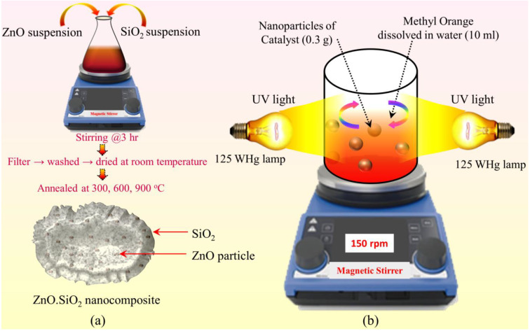
The synthesis of the ZnO.SiO2 nanocomposite involves three steps. The first step involves the preparation of ZnO nano-suspension. For this, 0.1 M NaOH solution was prepared in 50 mL distilled water and was added to 0.1 M of Zn(NO3)2·6H2O dissolved in 100 mL ethanol, following the procedure reported in our previous work [27]. Secondly, the preparation of SiO2 nano-suspension was prepared by mixing 0.3 M SiO2 in 50 mL 0.6 M NaOH, which resulted in Na2SiO3. The prepared solution was drop-wise added (using the piezoelectric nozzle of 0.05 mm and drop rate of 0.02 mL/s) to 50 mL 0.6 M HCl, which resulted in white precipitates of SiO2. The obtained suspension was stirred at 8000 rpm for 3 h at 50 °C. Afterward, NaCl was removed from the suspension by washing it several times using distilled water [28]. For the synthesis of the ZnO.SiO2 nanocomposite, both suspensions were mixed and stirred [28]. The white precipitates were filtered with grade 5 Whatman filter paper and then washed and dried in air. By following the procedure (Scheme 1a) two samples (0.1 M)ZnO.(0.3M)SiO2 (ZS1) and (0.05 M)ZnO.(0.3 M)SiO2 (ZS2) were prepared; furthermore, these samples were annealed at the different temperatures of 300, 600, and 900 °C for 2 h. For ease of understanding, the samples annealed at 300, 600, and 900 °C are named as ZS1300, ZS1600, and ZS1900, respectively.
Scheme 1.
Schematic diagram of (a) preparation of SiO2.ZnO nanoparticles with the expected morphology (ZnO nanoparticles embedded into SiO2 porous structure) and, (b) photocatalytic activity set-up.
2.3. Characterization
For the determination of the crystallite size and lattice constant, the X-ray diffraction (XRD) patterns of the samples were acquired with an X-ray diffractometer (Philips PW/1710), operated at 50 kV and 40 mA in range of 10 to 70 degrees. The XRD data was analyzed through Rietveld refinement for a perfect matching of phases and to determine various lattice parameters, dislocation density, microstrain, Wyckoff positions, bond length, and bond angle, etc. For morphological analysis, transmission electron microscopy was performed on a Hitachi (H-7500) instrument. Fourier transform infrared spectrometer (Bruker compact alpha-II) was used for recording the spectra of nanocomposites in a range of 4000–400 cm−1 using Kbr pallets. The UV-Vis absorption spectra of the prepared samples were acquired with a Lambda 750 spectrophotometer (Perkin) in a wavelength from 300 to 700 nm. The surface area (SBET) was evaluated using N2-adsorption measurements performed on a Quantachrome Instruments at 77.3500 K. Nitrogen and carbon were used as adsorbate and adsorbent, respectively.
2.4. Photocatalytic Activity
The photocatalytic properties of ZnO.SiO2 nanocomposite were evaluated by testing the photo-degradation of methyl orange (MO) in water using UV irradiation (125 WHg lamp). In a typical reaction, 30 mg ZS1 composite and 10 mL of 30 mg/80 mL of methyl orange dye aqueous solution were stirred using a magnetic stirrer for an hour to establish the adsorption/desorption equilibrium of MO on the surface of catalyst before the irradiated process. For this experiment, two mercury lamps were placed 5 cm away from the magnetic stirrer on which the solution of catalyst, as well as dye, were stirred, as shown in Scheme 1b. The effect of pH (at 7 and 12) on photocatalytic activity was analyzed. The pH of methyl orange dye solution (MODS) was controlled by adding diluted Nitric acid and NaOH. For this analysis, solutions of ZS1 (ZS1300, ZS1600 and ZS1900) and MODS were prepared with pH values of 7 and 12. At consequent intervals of time (i.e., 0 min, 20 min, 40 min, 60 min, 100 min) 2 mL of samples were extracted from each solution and analyzed using UV–Vis spectroscopy. The effect of NaOH conc. on the photo-degradation of dye was also evaluated. For this analysis, solutions of ZS1300 and MODS were prepared with different conc. of NaOH (1 g, 2 g, 3 g, and 4 g). Afterward, the slurry was not stirred, and the resulting dye content was determined at the end of the experiment. The photo-degradation percentage of methyl orange is calculated using Equation (1) [29]
| (1) |
where, Ao is the initial concentration of MO and At is the concentration of MO after time t. The experiment has been repeated again to ensure the reproducibility of the results.
2.5. Zero-Point Charge
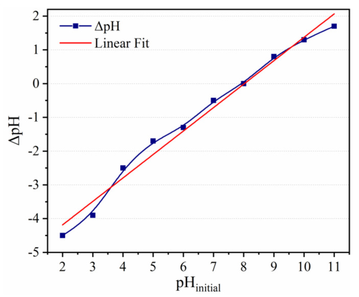
The value of zero-point charge (ZPC) was calculated with the help of salt titration method. The ZPC value of the catalyst was determined using a 10 mol solution of NaClO4. In this method, 0.2 g of catalyst was taken in 10 mL deionized water, and 0.01 mol/L NaOH was added to get the required pH. The suspension was taken in ten different beakers and the pH of the suspensions was initially set to different values from two to 11. This pH value is denoted as pHinitial. The sample of each beaker was stirred briskly for 24 h at room temperature, and the value of pH was recorded. This pH value is denoted as pHfinal. The ZPC is obtained from the plot, between ΔpH (pHfinal–pHinitial) and pHinitial as depicted in Figure 1. For ZPC, ΔpH needs to be zero. The ZPC of ZS1300 is investigated and its value is found as 8 [30].
Figure 1.
Plot of ΔpH versus pHinitial for ZS1300.
The value of ZPC implies that surface of the catalyst is positively charged at pH < pHZPC, and for pH > pHZPC will be negatively charged. Therefore, ZPC is considered an important feature for photocatalytic activity as it affects the surface property of catalyst. Under basic pH conditions, the negatively charged pollutant compounds will be repelled from the negatively charged catalyst surface, which results in to decrease in absorption of the pollutant compound. As a result, the degradation efficiency of the sample falls at a higher pH [31].
3. Results and Discussion
3.1. XRD Analysis
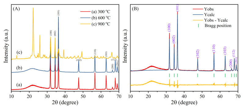
The XRD patterns of ZS1300, ZS1600 and ZS1900 samples are shown in Figure 2A. It can be observed from Figure 2A(a,b) that the XRD patterns of both samples contain various well-defined reflections at 2θ at 31.68°, 34.32°, 36.26°, 47.56°, 56.59°, 62.87°, 66.44°, 67.95°, and 69.08° correspond to Miller planes (100), (002), (101), (102), (110), (103), (200), (112), and (201) respectively, confirming the formation of ZnO [32,33,34]. In both diffraction patterns, a broad hump is present at 2θ~21.98°, which reveals the presence of amorphous silica. The observed peaks in in the diffraction pattern reveal the formation of the hexagonal structure of ZnO embedded in silica matrix, with the space group P63mc (JCPDS card no. 790205).
Figure 2.
(A) XRD patterns of ZS1 annealed at various temperatures and (B) Rietveld Refined XRD pattern of ZS1300 nanocomposite.
However, the XRD pattern of the sample ZS1900 also contained some new peaks (Figure 2A(c)). The peak centred at 2θ (reflection planes) at ~22.06° (101), 28.76° (111), 31.70° (102), 36.14° (200), 45.44° (202), 47.28° (113), 48.72° (212), 62.18° (302), 65.02° (312), and 66° (204), which may be ascribed to Tetragonal Cristobalite (JCPDS File No. 82-1408). In the same pattern, some of the peaks cantered at 2θ (reflection planes) at ~31.70° (113), 34° (410), and 38.80° (223) may be ascribed to Hexagonal Zinc silicate (Zn2SiO4) (JCPDS File No.37-1485).
The value of structural parameters like plane (hkl), crystallite size (D), d-spacing, full width at half maximum (β), intensity, microstrain (ε) and the dislocation density (δ) of ZnO.SiO2 composite are obtained from XRD analysis and are shown in Table S1. The crystallite size (D) is calculated by using Scherrer’s formula as:
| (2) |
The average crystallite size is calculated from the three peaks of highest intensity in the pattern, which was characterized by lattice plane (100), (002) and (101) at 2θ ~ 31.85, 34.55 and 36.35°, respectively inserted in Table S1. Dislocation density (δ) is calculated as [35]:
| (3) |
and microstrain (ε) value of the nanocomposite ZnO.SiO2 is evaluated as,
| (4) |
The Rietveld refined X-ray diffractogram of ZS1300 nanocomposite is shown in Figure 2B. For the quantitative analysis of the phase present in the sample, Rietveld refinement was carried out of the XRD data. The atomic position and isothermal parameters for Zinc and Oxygen are fixed and the other parameters like lattice constants, shape parameters, and scale factors are considered as free parameters during the time of fitting. The parameters, such as background factors and scale factors, are refined in the first step of the Rietveld refinement. The background is fitted using a linear interpolation method and pseudo-voigt is chosen for peak profile. In the succeeding stage, the structural parameters (lattice parameters, width parameter, preferred orientation, asymmetry, and atomic coordinates, etc.) are refined.
Figure 2B approves the hexagonal structure of ZnO embedded in SiO2. The Wyckoff positions parameters of atoms obtained from refinement are tabulated in Table S2. The norm’s factor for the best fit, such as the Reliability factors (R factors), Profile R factor (RB = 13.6), Weighted R Factor (Rwp = 14.3), Expected values (Rexp = 7.17), Bragg R Factor (RB = 6.02), Rf–Factor (RB = 2.78) and Goodness fit factor (χ2 = 3.9) value obtained from Rietveld refinement, confirm the precision of the Rietveld refinement analysis. The value of lattice parameters obtained from the refinement are as follows: ; ; α = 90°; , and ratio of c/a is 1.6022. The bond lengths between different atoms and bond angles between them are evaluated and presented in Table S3.
The type of the grain boundaries may play an important role in the photocatalytic mechanism. The grain boundary specific area (Sa) i.e., the ratio of the area enclosed by the grain boundary and volume of grain, is estimated as [36]
| (5) |
where, D is the crystallite size estimated from XRD studies using Equation (2). The Equation (5) is valid for both single and polycrystalline forms of ZnO system having a random distribution of shapes and grains. The values of Sa are tabulated in Table 1. It is observed that the grain boundary-specific area is decreased with an increase in temperature. The higher value i.e., 4.445 × 10⁷ m2 m−3 of Sa is calculated for the ZS1 sample annealed at 300 °C.
Table 1.
Average crystallite size (D), bandgap energy (Eg) and grain boundary specific area (Sa) w.r.t. to annealing temperatures.
| Annealing Temp (°C) | D (nm) | Sa (m2·m−3) | Eg (eV) |
|---|---|---|---|
| 300 | 36.20 | 4.455 × 10⁷ | 3.26 |
| 600 | 38.81 | 4.251 × 10⁷ | 3.25 |
| 900 | 43.42 | 3.800 × 10⁷ | 3.36 |
With a change in the precursor’s concentration i.e., sample ZS2, it is observed from the XRD patterns of ZS2300 and ZS2600 for the ZnO phase that the crystallite size decreased compared to ZS1300 and ZS1600, respectively. The XRD data for ZS2300 and ZS2600 are provided in the Supplementary File. The diffractogram of ZS2900 was found to have marginal changes compare to ZS1900. The phase structure observed for ZS2 samples is the same as observed in the case of ZS1 samples.
3.2. TEM Analysis
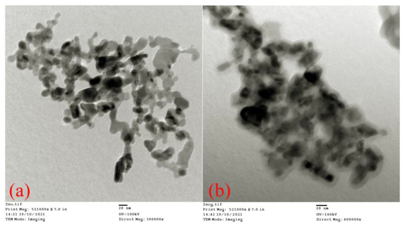
Transmission electron microscopy (TEM) is a utile technique for the analysis of particle size and the morphology of materials. The microscopic images of ZnO.SiO2 nanocomposites annealed at 300 °C are depicted in Figure 3. The average particles size is found to vary between 20 to 30 nm, which is nearly analogous to the crystallite size calculated from the XRD results. From the figure, a uniform morphology has been observed in which the particles are appearing roughly as spherical shapes. The micrograph shows the weak aggregation of ZnO particles that confirm the presence of particles in the silica matrix. Some particles are observed to possess sharp grain boundaries and a clear morphology in the silica matrix.
Figure 3.
Transmission electron microscopy images of ZS1300 (a) lower magnification and (b) higher magnification.
3.3. FTIR Analysis
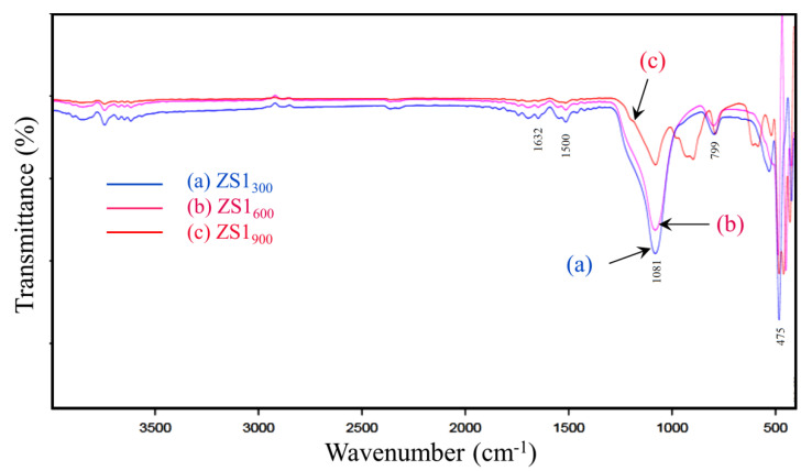
For the further investigation of the molecular structures of ZS1300, ZS1600 and ZS1900 nanocomposites, Fourier-transform infrared (FTIR) spectroscopy studies are carried out and the FTIR spectra of these samples are shown in Figure 3.
Characteristic peaks of these samples are observed at 1081 and 799 cm−1, which represent the stretching vibration mode and symmetric stretching of the Si-O-Si group, respectively [26]. The peak at around 475 cm−1 in the spectra is due to the vibration of the Zn, and the O bond confirms the presence of ZnO in the samples [37]. The presence of bonds at 1500 cm−1 and 1632 cm−1 are attributed to the stretching vibration of the (−OH) group attached with Si and ZnO, respectively [38]. The hydroxyl group results from the hygroscopic nature of ZnO and Si that come during the synthesis. It is also observed that the content of hydroxyl group in the sample decreased with an increase in temperature, suggesting the presence of impurities mostly near the surfaces of ZnO and Si. Moreover, it can be found that the peak at 1081.98 of ZS1300 sample had a strong intensity. However, for ZS1600 and ZS1900, the peak intensity decreased due to the decomposition of hydroxyl group Figure 4a,b. In the spectrum of ZS1900 sample (Figure 4c), some new peaks are also observed, which confirm the formation of some intermediate phases of ZnO and cristobalite (crystallite form of silica). The observed changes in the FTIR spectra of the nanocomposites samples are well-matched with the findings of XRD analysis.
Figure 4.
Fourier transforms infrared spectra of (a) ZS1300, (b) ZS1600, and (c) ZS1900 nanocomposite.
3.4. UV-Visible Analysis
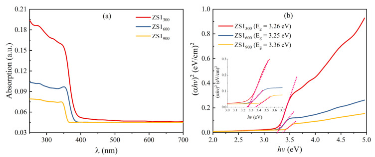
For the examination of the optical band gap of the ZS1300, ZS1600 and ZS1900 samples, UV-Visible spectroscopy is performed, as shown in Figure 5. It is seen that band edge shifted to a lower wavelength edge (i.e., blue shift), suggesting an alteration in the optical band gap. Thereby, to estimate the energy band gap (Eg) of the catalyst samples, Tauc’s relation the spectra (250–700 nm) is used, which is given by
| (6) |
where , , and are the absorption coefficients, Planks constant, power factor for the transition mode, and constant, respectively. For the allowed direct transitions, is taken as 1/2. The band gap values for the nanocomposites samples are determined by plotting versus , as shown in Figure 5.
Figure 5.
(a) UV plots (b) Tauc’s plot of ZS1300, ZS1600 and ZS1900.
The estimated values of Eg for the ZS1300, ZS1600, and ZS1900 nanocomposites are 3.26 eV, 3.25 eV, and 3.36 eV, respectively. The optical band gap of ZS1300 is found to be smaller than the characteristic value of ZnO, i.e., 3.37 eV at room temperature. This may be due to the occurrence of intrinsic defects like oxygen vacancies and zinc interstitials in the ZnO [36,39]. The ZS1900 sample has a higher optical band gap (very near to 3.37 eV) which attributes to an increase in crystallite size, as can be seen from XRD studies. The observed values of Eg w.r.t. crystallite size are tabulated in Table 1.
3.5. BET Analysis
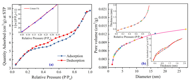
Surface area is an important parameter that affects photocatalytic activity due to the surface dependency of the process. With a larger surface area, the active sites are larger and with better photocatalytic activities. Therefore, to measure the surface and porosity of the ZS1300 and ZS1900 nanocomposite samples, the Brunauer-Emmett-Teller (BET) measurements are performed in a nitrogen environment. The nitrogen (N2) adsorption-desorption isotherm curves of the ZS1300 sample are shown in Figure 6a. The shape of adsorption-desorption curves depicts the features of isotherm of type IV (as per IUPAC classification) with H4 hysteresis, which reflects the mesoporous kind nature of the samples under discussion. The BET curve having three main segments, i.e., (i) concave-shaped for relative pressures from 0.05 to 0.1, (ii) linear-shaped up to 0.3, and (iii) convex-shaped for above 0.3, the (ii) or linear-shaped segment, as shown in inset of Figure 6a, denotes a realization of the first monolayer of the gas molecules and commencement of the second layer. The linear segment of the BET curve is known as the ‘B’ point, which is further used to calculate the specific surface area. From the BET equation:
| (7) |
whereas, denotes the volume of adsorbed gas at normal temperature (T) and pressure (P); represents the volume of adsorbed gas for monolayer per gram of adsorbent; P° denotes saturated vapor pressure of N2 at normal temperature; C refers to a constant depending on the Ha (heat absorbed by the monolayer of N2); and HL (liquefaction parameter of the gas) in the range of 0.05–0.3 of relative pressure, given by flowing relation
| (8) |
Figure 6.
BET isotherm of ZS1300 (a) adsorption and desorption curve and (b) pore size distribution, the thickness of deposited layer (inset) & adsorption quantity versus size of the pore (inset).
The Equation (7) represents an equation of a straight line in between vs. with slope and intercept of having the values of 206.87578 g/cm3 and 0.0088 g/cm3, respectively. The volume of the gas molecules monolayer () can be obtained using the values of slope and intercept of the curve in the straight-line portion of the isotherm. The specific surface area can be calculated using the relation, , where N is the Avogadro number and Am is the molecular cross-sectional area (for nitrogen; Am = 0.162 nm2). The measured values of the specific surface areas (SBET) for the samples ZS1300 & ZS1900 are found to be 16.8 m2/g and 0.001 m2/g, respectively. The decrease in specific surface area with annealing temperature can be attributed to the penetration of ZnO nanocrystallites into the porous structure of SiO2. The filling of the closed pores of SiO2 with ZnO resulted in the decrease in porosity of the nanocomposite samples. This can easily be predicted and corroborated with the change in XRD pattern from ZS1300 to ZS1900, wherein the XRD pattern of ZS1300 is vastly matched with the pure ZnO hexagonal structure. On the flip side, the XRD pattern of ZS1900 consists of the sharps reflections of ZnO along with the well-visualized reflection of SiO2, which points to the mixed phase of the nanocomposite.
The respective pore size distribution curve for the sample under discussion is obtained with the adsorption particulars employing the Barrett-Joyner-Halenda (BJH) method and depicted in Figure 6b. This figure confirms the mesoporous nature of the prepared composite. The insets depict the change in quantity adsorbed as a function of the deposited layer thickness and the change in layer thickness with relative pressure. It can be seen that the composite contains a total pore volume of 0.01667 cm3/g and an average pore diameter of 3.838 nm. The ZS1300 nanocomposite shows a higher specific surface area and it is reported that a higher surface area is advantageous for superior photocatalytic performance [40].
3.6. Photocatalytic Studies
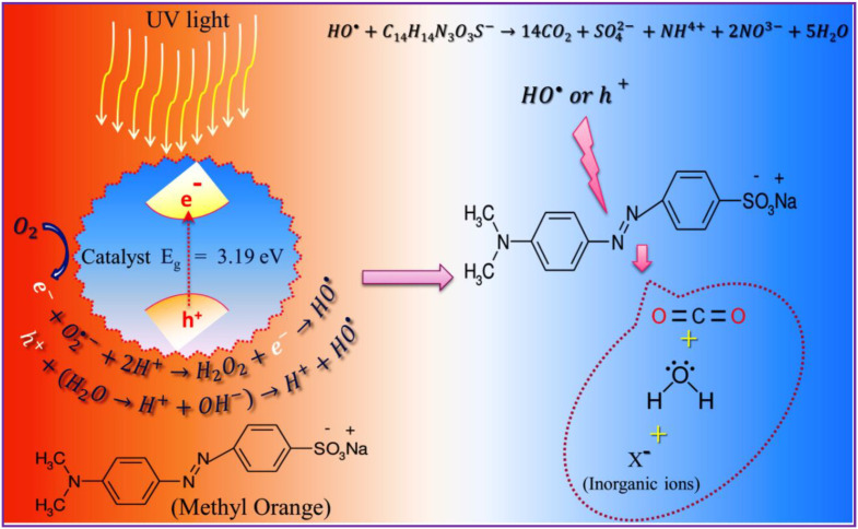
Absorption of UV light with energy greater than the band gap of the catalyst material causes generation of electron-hole pairs in ZnO particles i.e., electrons (e−) in conduction band and holes (h+) in the valence band holes. The holes react with H2O or hydroxide ions, which are adsorbed on the surface of the ZnO particles and produce •OH. Conversely, the electron reduces O2 and produces O2•− as well as other oxygen species (H2O2 and •OH) [32,33]. Both the holes and the •OH are super reactive to organic compounds that are in contact with these radicals [33]. The oxidizing tendency of the •OH radicals is high enough to split C-H as well as C-C bonds of MO sitting on the outer surface of ZnO.SiO2 nanocomposite leading to CO2 and H2O production as shown in Scheme 2.
Scheme 2.
Schematic illustration of the photocatalytic mechanism of ZnO.SiO2 photocatalyst for degradation of MO.
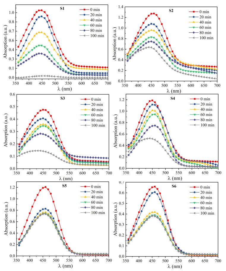
The ability of photocatalyst to decompose the pollutant compound MO, depends upon the different factors, such as the annealing temperature of the photocatalyst, the pH value of solution containing MO dye, and the time of irradiation and different conc. of NaOH. The effect of these factors on the catalytic performance of the material is examined. For an investigation of the effect of pH conc. and annealing temperatures of catalytic material on MO dye, samples of MO and ZS1 (annealed at 300, 600 & 900 °C) were prepared distinctly with different pH concentrations. The samples’ names are abbreviated as S1 (ZS1300, pH7), S2 (ZS1300, pH12), S3 (ZS1600, pH7), S4 (ZS1600, pH12), S5 (ZS1900, pH7), and S6 (ZS1900, pH12). The UV–visible spectra of each sample showing degradation of MO w.r.t. irradiation time, depicted in Figure 7.
Figure 7.
UV–visible spectra of S1, S2, S3, S4, S5, and S6 samples showing degradation of MO.
3.6.1. Effect of pH and Irradiation Time
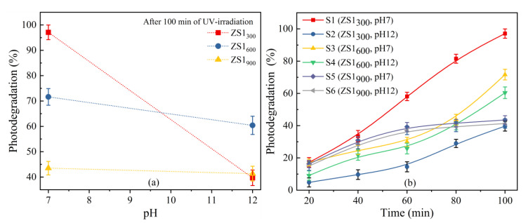
Using Equation (1), the photo-degradation percentages of samples S1, S2, S3, S4, S5, and S6 irradiated under the UV light for 100 min are calculated and the values of photo-degradation (%) are found to be 98.1, 39.7, 71.63, 60.39, 43.51, and 41.34%, respectively (Figure 8a). Interestingly, it is observed that the photocatalytic activity of the S1 sample is relatively higher than the other samples. As the value of pH is increased, the degradation response is decreased and only 39.7 % of MO degradation response is observed for sample S2 (i.e., at pH = 12). The photo-degradation activity depends on the surface properties of the catalyst. Generally, pH changes the surface properties of catalysts like surface charge properties that oversee the absorption mechanism of MO on catalytic surface. At a pH 7 (below ZPC pH i.e., 8), the surface of ZnO is positively charged, which can absorb negatively charged MO due to electrostatic attraction, facilitating the degradation reaction. In contrast, at a higher pH (=12), absorption is decreased due to electrostatic repulsive forces among the catalytic surface and the dyed surface. As a result, photo-degradation response becomes minuscule; this is firstly because of the decrease in adsorption of the dye molecules on surface of the photocatalyst surface at higher pH [41,42], and secondly due to rapid scavenging of the hydroxyl radicals produced via reaction between holes and OH−, which causes a decrease in free radicals [42,43]. It is reported that pH affects the production of these free radicals that, in turn, lower the degradation rate of dye, or the termination of degradation [31].
Figure 8.
Effect of (a) irradiation time and (b) pH on photodegradation rate (%) of methyl orange (UV irradiation = (2 × 125) W).
It is observed that on increasing the irradiation time under light irradiating lamps, the photo-degradation percentage of methyl orange also increased, as shown in Figure 8b. It is observed from Figure 8b that all the samples attained maximum photo-degradation (%) in about 100 min. This may be due to the production of more and more e-h pairs and the corresponding excitation of an electron from the valence band to the conduction band [44]. Accordingly, the production of hydroxyl radicals is rose, which resulted in an enhanced photodegradation (%). The higher degradation efficiency of the ZS1300 nanocomposite sample, as compared to other samples, can be attributed to its small band gap and larger surface area.
3.6.2. Effect of Annealing Temperatures
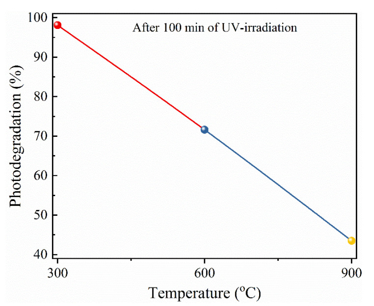
The photocatalytic efficiency of the ZS1 nanocomposite at different annealing temperatures has also been assessed for the photo-degradation of MO. Figure 9 shows the correlation between photo-degradation (%) of MO with annealing temperatures of ZS1 nanocomposite. It is observed that the photocatalytic degradation of MO decreases from 98.1% to about 39% with an increase in the annealing temperature of the sample ZS1 (i.e., sample ZS1300 to ZS1900. The highest activity observed for the ZS1300 nanocomposite may be ascribed to the higher specific surface area, lower band gap value, and efficient charge separation ability of the sample through the charge exchange process in the existence of UV light. The lowest photo-degradation ability of the sample ZS1900 can be due to the decrease in specific surface area due to the filling of the pore by the penetration of ZnO into the SiO2 porous matrix, as observed from the XRD and BET studies. The lower reactivity of SiO2 towards photo-degradation and the presence of higher content of SiO2 on the upper surface as expected from XRD pattern could be another reason for the lower efficiency of ZS1900 sample.
Figure 9.
Variation in photo-degradation performance of ZS1 catalyst w.r.t. annealing temperature at pH 7 after 100 min of UV exposure.
It is reported that in the photocatalytic mechanism, the production of •OH is expected to occur mainly on an internal surface i.e., the ZnO surface [45,46]. In addition, MO approaches the ZnO surface via the pores present in the surface area of sample ZS1. Thus, the photonic degradation efficiency is being limited by the absorption or diffusion rate of the MO molecule into the photo-catalytically active surface. As a result of the difference in the size and the number of pores of the ZS1 samples (ZS1300, ZS1600 & ZS1900,), the photo-degradation (%) is changed accordingly. Therefore, it is concluded that the ZS1300 assists the transport properties of all reactants involved in the photocatalytic process, which enhanced the overall photocatalytic activity.
3.6.3. Effect of NaOH Concentration
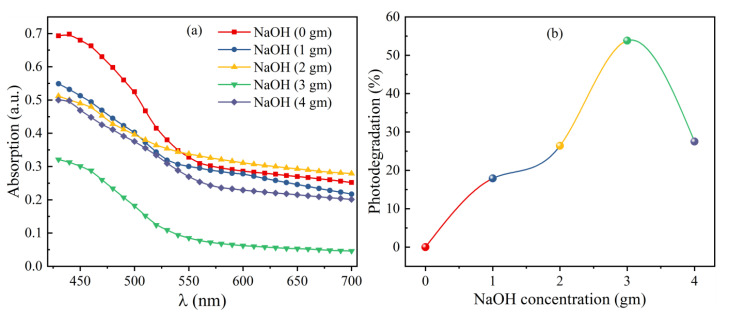
To investigate the effect of NaOH concentration on the photo-degradation ability of the nanocomposite samples, four different concentrations (1, 2, 3 and 4 g) of NaOH are mixed with the photocatalyst and MO solution. In a typical process, 0.38 g of catalyst powder was dispersed in 100 mL deionized water and then 10 mL of methyl orange dye solution was added to catalyst solution and stirred to adsorb the dye molecules on surface of the photocatalyst. After that, 1 g of NaOH powder was mixed with the above solution containing methyl orange and photocatalyst, then the solution was stirred in absence of light to achieve equilibrium between adsorption and desorption and then irradiated with visible irradiation light with continuous stirring [47]. After the decoloration of methyl orange, a part of the solution was used for recording the absorbance spectrum using a UV-Visible spectrophotometer and photo-degradation percentage was calculated using Equation (1). The same steps are followed for the degradation of MO with different weights (2, 3 and 4 g) of NaOH and the recorded absorption spectra are shown in Figure 10a.
Figure 10.
Effect of NaOH on catalyst; (a) UV–visible spectra for different concentrations of NaOH and (b) photo-degradation efficiency w.r.t. concentration NaOH.
It is observed that the decoloration time of methyl orange decreases from 275 through 95 to 40 min with an increase in NaOH concentration from 1 g through 2 g to 3 g. However, for the 4 g of NaOH, again the decoloration time increases to 80 min. It is observed that the sample containing 3 g concentration of NaOH shows a higher photo-degradation of 53.8 %, and beyond 3 g NaOH concentration again the photo-degradation (%) decreases to 27.5%, with 4 g of NaOH (Figure 10b). The results showed that the photo-degradation rate increased with an increase in NaOH concentration up to 3 g of NaOH, which may be due to OH− ions produced by NaOH acting as a strong hole scavenger and generating hydroxyl radicals, thus reducing the e-h pair recombination [45]. Moreover, with a further increase in concentration (>3 g) of NaOH, the degradation rate became poor, since a greater amount of hydroxyl radicals may oxidize the catalyst [45].
Compared to other metal oxide composites tabulated in Table 2, the present ZnO.SiO2 prepared through the co-precipitation method shows enhanced photocatalytic efficiency for the degradation of MO.
Table 2.
Photocatalytic activity of various ZnO composites.
| Catalyst Composite | Method of Synthesis | Light Used for Irradiation | Irradiation Time (min) | Photo-Degradation (%) | Refs. |
|---|---|---|---|---|---|
| ZnO nano-needles on Si substrates | Hydrothermal | UV light | 140 | 95.4 | [48] |
| ZnO quantum dots | Flame spray pyrolysis | UV light | 160 | 97 | [49] |
| TiO2/biochar | Hydrolysis | UV light | 160 | 96.88 | [17] |
| Xanthan gum/TiO2 | Hydro-gel | Visible light | 90 | 89 | [15] |
| Anatase TiO2 | Sol-gel | Visible light | 240 | 67.12 | [50] |
| ZnO catalyst | Hydrothermal | UV light | 180 | 99.7 | [51] |
| Rare earth metal doped ZnO | Sol-gel | UV light | 180 | 98 | [32] |
| ZnO.SiO2 | Co-precipitation | UV light | 100 | 98.1 | Present work |
4. Conclusions
ZnO.SiO2 nanocomposites are successfully synthesized via the co-precipitation method. The specific surface area decreases from 16.8 m2/g and 0.001 m2/g on increasing the annealing temperature from 300 to 900 °C due to blocking of SiO2 pores via the penetration of ZnO into these pores. It is found that the specific surface area of the nanocomposite plays an important role in photocatalytic activities and the degradation of methyl orange dye. The effect of various parameters, such as the pH value of the solution, irradiation time, annealing temperature, and NaOH concentration on photocatalytic performance of ZS1 nanocomposite have been investigated. The sample ZS1300 shows a higher photo-degradation efficiency of 98.1% at pH 7 for 100 min irradiation times. The high efficiency of ZS1300 can be governed by the following mechanisms: (i) a higher specific area that assures the maximum interaction with UV irradiation; (ii) an optimized bandgap resulting in enhanced absorption efficiency of UV irradiation where more electron-hole pairs are produced; (iii) more irradiation time proportional to more generation electron-hole pairs; and (iv) at lower pH below the ZPC pH, which increases the absorption of pollutant dye that resulting higher efficiency. OH− radicals act as active species for the degradation of the pollutant. For the degradation, the formation of hole and hydroxyl radicals is proven to be the crucial factor. The results revealed that ZnO.SiO2 nanocomposite is an effective, inexpensive, and eco-friendly catalyst for the degradation of methyl organic pollutants.
Supplementary Materials
The following are available online at www.mdpi.com/article/10.3390/nano11102548/s1, Table S1: Structural parameters of ZS1300 achieved from X-ray Diffraction Pattern, Table S2: Fractional atomic coordinates and isothermal parameter of different atoms obtained from the Rietveld anal-ysis of XRD patterns for the sample ZS1300, Table S3: Bond Length and bond angles of sample ZS1300, Table S4: Structural parameters of ZS1600 achieved from X-ray Diffraction Pattern, Table S5: Structural parameters of ZS1900 achieved from X-ray Diffraction Pattern, Table S6: Structural parameters of ZS2300 achieved from X-ray Diffraction, Table S7: Structural parameters of ZS2600 achieved from X-ray Diffraction Pattern.
Author Contributions
Conceptualization & Methodology, S.R. and A.G.; Sample characterization, A.G. and S.K.; Validation, V.K. and N.A.; Formal analysis, J.D., S.R.; Investigation, J.D. and M.P.; Data curation, J.D. and M.T.N.; Writing—original draft preparation, S.R.; Writing—review and editing, J.D.; Funding acquisition, M.P. and M.T.N. All authors have read and agreed to the published version of the manuscript.
Funding
This work was supported by the Ministry of Education, Youth and Sports of the Czech Republic and the European Union (European Structural and Investment Funds-Operational Programme Research, Development and Education) in the frames of the project “Modular platform for autonomous chassis of specialised electric vehicles for freight and equipment transportation”, Reg. No. CZ.02.1.01/0.0/0.0/16_025/0007293.
Conflicts of Interest
The authors declare no conflict of interest.
Footnotes
Publisher’s Note: MDPI stays neutral with regard to jurisdictional claims in published maps and institutional affiliations.
References
- 1.Karimi A., Kazeminezhad I., Azizi S. Ag/αFe2O3-rGO novel ternary nanocomposites: Synthesis, characterization, and pho-tocatalytic activity. Ceram. Int. 2019;45:3441–3448. doi: 10.1016/j.ceramint.2018.10.259. [DOI] [Google Scholar]
- 2.Robinson T., McMullan G., Marchant R., Nigam P. Remediation of dyes in textile effluent: A critical review on current treatment technologies with a proposed alternative. Bioresour. Technol. 2001;77:247–255. doi: 10.1016/S0960-8524(00)00080-8. [DOI] [PubMed] [Google Scholar]
- 3.Ajmal A., Majeed I., Malik R.N., Idriss H., Nadeem M.A. Principles and mechanisms of photocatalytic dye degradation on TiO2 based photocatalysts: A comparative overview. RSC Adv. 2014;4:37003–37026. doi: 10.1039/C4RA06658H. [DOI] [Google Scholar]
- 4.Rakhshaee R., Giahi M., Pourahmad A. Removal of methyl orange from aqueous solution by Azolla filicoloides: Synthesis of Fe3O4 nano-particles and its surface modification by the extracted pectin of Azolla. Chin. Chem. Lett. 2011;22:501–504. doi: 10.1016/j.cclet.2010.10.041. [DOI] [Google Scholar]
- 5.Al-Qaradawi S., Salman S.R. Photocatalytic degradation of methyl orange as a model compound. J. Photochem. Photobiol. A Chem. 2002;148:161–168. doi: 10.1016/S1010-6030(02)00086-2. [DOI] [Google Scholar]
- 6.Katsuda T., Ooshima H., Azuma M., Kato J. New detection method for hydrogen gas for screening hydrogen-producing microorganisms using water-soluble wilkinson’s catalyst derivative. J. Biosci. Bioeng. 2006;102:220–226. doi: 10.1263/jbb.102.220. [DOI] [PubMed] [Google Scholar]
- 7.Chung K.-T. Azo dyes and human health: A review. J. Environ. Sci. Heal. Part C. 2016;34:233–261. doi: 10.1080/10590501.2016.1236602. [DOI] [PubMed] [Google Scholar]
- 8.Sha Y., Mathew I., Cui Q., Clay M., Gao F., Zhang X.J., Gu Z. Rapid degradation of azo dye methyl orange using hollow cobalt nanoparticles. Chemosphere. 2016;144:1530–1535. doi: 10.1016/j.chemosphere.2015.10.040. [DOI] [PubMed] [Google Scholar]
- 9.Chamjangali M.A., Bagherian G., Javid A., Boroumand S., Farzaneh N. Synthesis of Ag-ZnO with multiple rods (multipods) morphology and its application in the simultaneous photo-catalytic degradation of methyl orange and methylene blue. Spectrochim. Acta Part A Mol. Biomol. Spectrosc. 2015;150:230–237. doi: 10.1016/j.saa.2015.05.067. [DOI] [PubMed] [Google Scholar]
- 10.Paul K.K., Ghosh R., Giri P.K. Mechanism of strong visible light photocatalysis by Ag2O-nanoparticle-decorated monoclinic TiO2(B) porous nanorods. Nanotechnology. 2016;27:315703. doi: 10.1088/0957-4484/27/31/315703. [DOI] [PubMed] [Google Scholar]
- 11.Theerthagiri J., Chandrasekaran S., Salla S., Elakkiya V., Senthil R.A., Nithyadharseni P., Maiyalagan T., Micheal K., Ayeshamariam A., Arasu A.M., et al. Recent developments of metal oxide based heterostructures for photocatalytic applica-tions towards environmental remediation. J. Solid State Chem. 2018;267:35–52. doi: 10.1016/j.jssc.2018.08.006. [DOI] [Google Scholar]
- 12.Zaleska-Medynska A. Metal Oxide-Based Photocatalysis: Fundamentals and Prospects for Application. Elsevier; Amsterdam, The Netherlands: 2018. [Google Scholar]
- 13.Lee K.M., Lai C.W., Ngai K.S., Juan J.C. Recent developments of zinc oxide based photocatalyst in water treatment technology: A review. Water Res. 2016;88:428–448. doi: 10.1016/j.watres.2015.09.045. [DOI] [PubMed] [Google Scholar]
- 14.Siddiquey I.A., Furusawa T., Sato M., Bahadur N.M., Alam M., Suzuki N. Sonochemical synthesis, photocatalytic activity and optical properties of silica coated ZnO nanoparticles. Ultrason. Sonochem. 2012;19:750–755. doi: 10.1016/j.ultsonch.2011.12.011. [DOI] [PubMed] [Google Scholar]
- 15.Inamuddin Xanthan gum/titanium dioxide nanocomposite for photocatalytic degradation of methyl orange dye. Int. J. Biol. Macromol. 2019;121:1046–1053. doi: 10.1016/j.ijbiomac.2018.10.064. [DOI] [PubMed] [Google Scholar]
- 16.Kumar A., Dalal J., Dahiya S., Chowdhury A., Khandual A., Ohlan A., Punia R., Maan A.S. Coating of multi-walled carbon nanotubes on cotton fabric via conventional dyeing for enhanced electrical and mechanical properties. AIP Conf. Proc. 2019;2142:140019. doi: 10.1063/1.5122532. [DOI] [Google Scholar]
- 17.Lu L., Shan R., Shi Y., Wang S., Yuan H. A novel TiO2/biochar composite catalysts for photocatalytic degradation of methyl orange. Chemosphere. 2019;222:391–398. doi: 10.1016/j.chemosphere.2019.01.132. [DOI] [PubMed] [Google Scholar]
- 18.Molkenova A., Sarsenov S., Atabaev S., Khamkhash L., Atabaev T.S. Hierarchically-structured hollow CuO microparticles for efficient photo-degradation of a model pollutant dye under the solar light illumination. Environ. Nanotechnol. Monit. Manag. 2021;16:100507. doi: 10.1016/j.enmm.2021.100507. [DOI] [Google Scholar]
- 19.Qu Y., Huang R., Qi W., Shi M., Su R., He Z. Controllable synthesis of ZnO nanoflowers with structure-dependent photo-catalytic activity. Catal. Today. 2020;355:397–407. doi: 10.1016/j.cattod.2019.07.056. [DOI] [Google Scholar]
- 20.Wang L., Li Z., Chen J., Huang Y., Zhang H., Qiu H. Enhanced photocatalytic degradation of methyl orange by porous graphene/ZnO nanocomposite. Environ. Pollut. 2019;249:801–811. doi: 10.1016/j.envpol.2019.03.071. [DOI] [PubMed] [Google Scholar]
- 21.Ali W., Ullah H., Zada A., Alamgir M.K., Muhammad W., Ahmad M.J., Nadhman A. Effect of calcination temperature on the photoactivities of ZnO/SnO2 nanocomposites for the degradation of methyl orange. Mater. Chem. Phys. 2018;213:259–266. doi: 10.1016/j.matchemphys.2018.04.015. [DOI] [Google Scholar]
- 22.Albiss B., Abu-Dalo M. Photocatalytic Degradation of Methylene Blue Using Zinc Oxide Nanorods Grown on Activated Carbon Fibers. Sustainability. 2021;13:4729. doi: 10.3390/su13094729. [DOI] [Google Scholar]
- 23.Mohamed R.M., Barakat M.A. Enhancement of photocatalytic activity of ZnO/SiO2 by nanosized Pt for photocatalytic degradation of phenol in wastewater. Int. J. Photoenergy. 2012;2012:103672. doi: 10.1155/2012/103672. [DOI] [Google Scholar]
- 24.Kumari S., Malik S., Kumar S., Dalal J., Dahiya S., Ohlan A., Punia R., Maan A.S. Excellent photoelectrical properties of ZnO thin film based on ZnO/epoxy-resin ink for UV-light detectors. AIP Conf. Proc. 2019;2142:120004. [Google Scholar]
- 25.Feng Q., Chen K., Ma D., Lin H., Liu Z., Qin S., Luo Y. Synthesis of high specific surface area silica aerogel from rice husk ash via ambient pressure drying. Colloids Surf. A Physicochem. Eng. Asp. 2018;539:399–406. doi: 10.1016/j.colsurfa.2017.12.025. [DOI] [Google Scholar]
- 26.Galedari N.A., Rahmani M., Tasbihi M. Preparation, characterization, and application of ZnO@SiO2 core-shell structured catalyst for photocatalytic degradation of phenol. Environ. Sci. Pollut. Res. 2017;24:12655–12663. doi: 10.1007/s11356-016-7888-2. [DOI] [PubMed] [Google Scholar]
- 27.Rohilla S., Lal B., Sunder S., Aghamkar P., Kumar S., Aggarwal A. Synthesis of Fe4[Fe(CN)6]3·14H2O Nanopowder by Co-Precipitation Technique and Effect of Heat Treatment. Acta Phys. Pol. A. 2010;118:696. doi: 10.12693/APhysPolA.118.696. [DOI] [Google Scholar]
- 28.Rohilla S., Kumar S., Aghamkar P., Sunder S., Agarwal A. Investigations on structural and magnetic properties of cobalt ferrite/silica nanocomposites prepared by the coprecipitation method. J. Magn. Magn. Mater. 2011;323:897–902. doi: 10.1016/j.jmmm.2010.11.001. [DOI] [Google Scholar]
- 29.Li N., Yang B., Xu L., Xu G., Sun W., Yu S. Simple synthesis of Cu2O/Na-bentonite composites and their excellent photocatalytic properties in treating methyl orange solution. Ceram. Int. 2016;42:5979–5984. doi: 10.1016/j.ceramint.2015.12.145. [DOI] [Google Scholar]
- 30.Mahmood T., Saddique M.T., Naeem A., Westerhoff P., Mustafa S., Alum A. Comparison of Different Methods for the Point of Zero Charge Determination of NiO. Ind. Eng. Chem. Res. 2011;50:10017–10023. doi: 10.1021/ie200271d. [DOI] [Google Scholar]
- 31.Benhebal H., Chaib M., Salmon T., Geens J., Leonard A., Lambert S.D., Crine M., Heinrichs B. Photocatalytic degradation of phenol and benzoic acid using zinc oxide powders prepared by the sol–gel process. Alex. Eng. J. 2013;52:517–523. doi: 10.1016/j.aej.2013.04.005. [DOI] [Google Scholar]
- 32.Alam U., Khan A., Ali D., Bahnemann D., Muneer M. Comparative photocatalytic activity of sol–gel derived rare earth metal (La, Nd, Sm and Dy)-doped ZnO photocatalysts for degradation of dyes. RSC Adv. 2018;8:17582–17594. doi: 10.1039/C8RA01638K. [DOI] [PMC free article] [PubMed] [Google Scholar]
- 33.Ali A.M., Ismail A.A., Najmy R., Al-Hajry A. Preparation and characterization of ZnO-SiO2 thin films as highly efficient photocatalyst. J. Photochem. Photobiol. A Chem. 2014;275:37–46. doi: 10.1016/j.jphotochem.2013.11.002. [DOI] [Google Scholar]
- 34.Muthulingam S., Bin Bae K., Khan R., Lee I.-H., Uthirakumar P. Improved daylight-induced photocatalytic performance and suppressed photocorrosion of N-doped ZnO decorated with carbon quantum dots. RSC Adv. 2015;5:46247–46251. doi: 10.1039/C5RA07811C. [DOI] [Google Scholar]
- 35.Nogueira I.C., Cavalcante L.S., Pereira P.F.S., De Jesus M.M., Rivas Mercury J.M., Batista N.C., Li M.S., Longo E. Rietveld refinement, morphology and optical properties of (Ba1−xSrx).MoO4 crystals. J. Appl. Crystallogr. 2013;46:1434–1446. doi: 10.1107/S0021889813020335. [DOI] [Google Scholar]
- 36.Singh K., Rawal I., Gautam P., Sharma N., Dhar R. Diluted magnetic semiconducting properties of nanocrystalline Zn0.98X0.02O (X = Fe, Ga, Ni) thin films deposited by PLD technique for spintronic applications. J. Magn. Magn. Mater. 2018;468:259–268. doi: 10.1016/j.jmmm.2018.08.024. [DOI] [Google Scholar]
- 37.Zeng X., Yu S., Sun R., Xu J. Mechanical reinforcement while remaining electrical insulation of glass fibre/polymer composites using core-shell CNT@SiO2 hybrids as fillers. Compos. Part A Appl. Sci. Manuf. 2015;73:260–268. doi: 10.1016/j.compositesa.2015.03.015. [DOI] [Google Scholar]
- 38.Tinio J.V.G., Simfroso K.T., Peguit A.D.M.V., Candidato R.T. Influence of OH-ion concentration on the surface morphology of zno-SiO2 nanostructure. J. Nanotechnol. 2015;2015:686021. doi: 10.1155/2015/686021. [DOI] [Google Scholar]
- 39.Sharma D., Jha R. Analysis of structural, optical and magnetic properties of Fe/Co co-doped ZnO nanocrystals. Ceram. Int. 2017;43:8488–8496. doi: 10.1016/j.ceramint.2017.03.201. [DOI] [Google Scholar]
- 40.Zhong J.B., Li J.Z., He X.Y., Zeng J., Lu Y., He J.J., Zhong F. Fabrication and Catalytic Performance of SiO2-ZnO Composite Photocatalyst. Synth. React. Inorg. Met. Nano-Met. Chem. 2014;44:1203–1207. doi: 10.1080/15533174.2013.799208. [DOI] [Google Scholar]
- 41.Wang W.-Y., Ku Y. Effect of solution pH on the adsorption and photocatalytic reaction behaviors of dyes using TiO2 and Nafion-coated TiO2. Colloids Surf. A Physicochem. Eng. Asp. 2007;302:261–268. doi: 10.1016/j.colsurfa.2007.02.037. [DOI] [Google Scholar]
- 42.Huang M., Xu C., Wu Z., Huang Y., Lin J., Wu J. Photocatalytic discolorization of methyl orange solution by Pt modified TiO2 loaded on natural zeolite. Dye. Pigment. 2008;77:327–334. doi: 10.1016/j.dyepig.2007.01.026. [DOI] [Google Scholar]
- 43.Moignard M., James R., Healy T. Adsorption of calcium at the zinc sulphide-water interface. Aust. J. Chem. 1977;30:733–740. doi: 10.1071/CH9770733. [DOI] [Google Scholar]
- 44.Ghaderi A., Abbasi S., Farahbod F. Synthesis, characterization and photocatalytic performance of modified ZnO nanoparticles with SnO2 nanoparticles. Mater. Res. Express. 2018;5:065908. doi: 10.1088/2053-1591/aacd40. [DOI] [Google Scholar]
- 45.Liu S., Zhao Z., Wang Z. Photocatalytic reduction of carbon dioxide using sol–gel derived titania-supported CoPc catalysts. Photochem. Photobiol. Sci. 2007;6:695–700. doi: 10.1039/B613098D. [DOI] [PubMed] [Google Scholar]
- 46.Reedijk J. Reference Module in Chemistry, Molecular Sciences and Chemical Engineering. Elsevier BV; Amsterdam, The Netherlands: 2013. [Google Scholar]
- 47.Ouyang K., Xie S. Effect of key operational factors on decolorization of methyl orange by multi-walled carbon nanotubes (MWCNTs)/TiO2/CdS composite under simulated solar light irradiation. Ceram. Int. 2013;39:8035–8042. doi: 10.1016/j.ceramint.2013.03.073. [DOI] [Google Scholar]
- 48.Tripathy N., Ahmad R., Song J.E., Ko H.A., Hahn Y.-B., Khang G. Photocatalytic degradation of methyl orange dye by ZnO nanoneedle under UV irradiation. Mater. Lett. 2014;136:171–174. doi: 10.1016/j.matlet.2014.08.064. [DOI] [Google Scholar]
- 49.Khan R., Hassan M.S., Jang L.-W., Yun J.H., Ahn H.-K., Khil M.-S., Lee I.-H. Low-temperature synthesis of ZnO quantum dots for photocatalytic degradation of methyl orange dye under UV irradiation. Ceram. Int. 2014;40:14827–14831. doi: 10.1016/j.ceramint.2014.06.076. [DOI] [Google Scholar]
- 50.Dhanalakshmi J., Padiyan D.P. Photocatalytic degradation of methyl orange and bromophenol blue dyes in water using sol–gel synthesized TiO2 nanoparticles. Mater. Res. Express. 2017;4:095020. doi: 10.1088/2053-1591/aa85fd. [DOI] [Google Scholar]
- 51.Chowdhury M.I.H., Hossain M.S., Azad M.A.S., Islam M.Z., Dewan M.A. Photocatalytic degradation of methyl orange under UV using ZnO as catalyst. Int. J. Sci. Eng. Res. 2018;9:1646–1649. [Google Scholar]
Associated Data
This section collects any data citations, data availability statements, or supplementary materials included in this article.


