Figure 1.
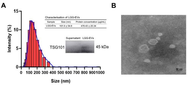
Characterization of L. rhamnosus GG-derived EVs. (A) Size distribution of LGG-EVs (Lactobacillus rhamnosus GG derived extracellular vesicles) was measured by DLS (Dynamic Light Scattering). The peak diameter was about 150 nm. Immunoblot bands demonstrating the presence of TSG101 (Tumor susceptibility gene 101) marker in LGG-EVs. (B) Transmission electron microscopy images of isolated LGG-EVs. Scale bar, 50 nm.

