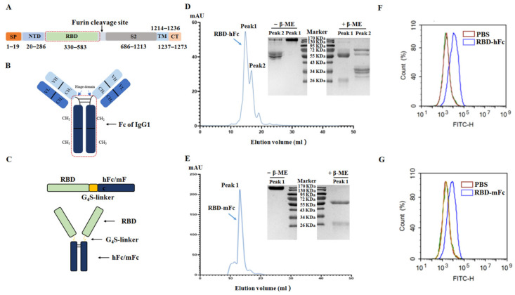
Figure 2.
Construction and characterization of SARS-CoV-2 RBD recombinant proteins. (A) A schematic view of the SARS-CoV-2 S protein. RBD ranges from 330 to 583 aa of the SARS-CoV-2 S protein. SP, signal peptide; NTD, N-terminal domain; TM, transmembrane; CT, C-terminal domain. (B) Structure diagram of IgG1. Fc contains the hinge domain, CH2 and CH3. (C) A schematic view of the SARS-CoV-2 recombinant protein. Residues 330–583 aa of the SARS-CoV-2 S protein were fused with a modified Fc fragment of human IgG1 or mouse IgG1 (hFc or mFc) via a G4S (Gly-Gly-Gly-Gly-Ser) flexible linker and engineered in a mammalian cell expression system. The purification results of RBD-hFc recombinant protein (D) and RBD-mFc recombinant protein (E) after purified by the protein A/G chromatographic column and a Superdex 200 increase column. Each elution peak was analyzed by SDS-PAGE in the presence of β-mercaptoethanol (β-ME) or not. The elution peaks of both recombinant proteins were noted with blue arrows. The cell binding ability of the RBD-hFc recombination protein (F) and RBD-mFc recombinant protein (G) were measured by flow cytometry using the Vero E6 cell line.

