Abstract
DNA interstrand cross-links are induced by many carcinogens and anticancer drugs. It was previously shown that mammalian DNA excision repair nuclease makes dual incisions 5′ to the cross-linked base of a psoralen cross-link, generating a gap of 22 to 28 nucleotides adjacent to the cross-link. We wished to find the fates of the gap and the cross-link in this complex structure under conditions conducive to repair synthesis, using cell extracts from wild-type and cross-linker-sensitive mutant cell lines. We found that the extracts from both types of strains filled in the gap but were severely defective in ligating the resulting nick and incapable of removing the cross-link. The net result was a futile damage-induced DNA synthesis which converted a gap into a nick without removing the damage. In addition, in this study, we showed that the structure-specific endonuclease, the XPF-ERCC1 heterodimer, acted as a 3′-to-5′ exonuclease on cross-linked DNA in the presence of RPA. Collectively, these observations shed some light on the cellular processing of DNA cross-links and reveal that cross-links induce a futile DNA synthesis cycle that may constitute a signal for specific cellular responses to cross-linked DNA.
Interstrand cross-links are common lesions introduced into DNA by drugs such as psoralen, cisplatin, mitomycin C, and melphalan (17). These lesions are eliminated from DNA by a mechanism involving excision repair and recombination in Escherichia coli (3, 5, 32) and in yeast (15, 20). In mammalian cells, the precise role of excision repair in eliminating cross-links is not known (35, 36). Although mutations in any of the genes required for the dual-incision step of excision repair cause sensitivity to cross-linking chemicals, the XPF and ERCC1 mutant cell lines, in addition to being defective in excision repair, are particularly sensitive to cross-linking agents and hence have been presumed to play a special role in cross-link repair (13). Similarly, mutations in the XRCC2 and XRCC3 genes, encoding proteins with sequence homology to the human RAD51 protein (19), confer sensitivity to cross-linking agents without affecting the excision repair system and hence are thought to play a unique role in processing of cross-links (35). To understand the mechanism of cross-link repair, it appears that the actions of the excision repair system, the XPF-ERCC1 complex, and XRCC2 and XRCC3 on cross-links must be investigated.
The human nucleotide excision repair system removes base monoadducts and intrastrand diadducts by making a dual incision bracketing the lesion (14). Recently, we reported the surprising finding that with a cross-linked substrate, the human excision nuclease makes both incisions 5′ to the cross-linked base, excising a damage-free oligomer and generating a gap of 22 to 28 nucleotides (nt) 5′ to either the furan-side or the pyrone-side adducted thymine of a psoralen cross-link (1). We proposed that the gap generated by this unusual type of dual incision may initiate at least one pathway of cross-link repair. In the present study, we have investigated the fate of the 5′ gap by using cell extracts from wild-type, excision repair-defective, and recombination repair-defective cell lines (16, 29). We found that the 5′ gap was filled efficiently but remained mostly unligated both in wild-type and in cross-link-sensitive XRCC3 mutant cells and that “repair patch” formation was dependent on an intact excision repair nuclease. Ligation occurred in a small fraction of molecules with repair patches and thus regenerated the original cross-linked substrate. Furthermore, we found that the XPF-ERCC1 complex in the presence of replication protein A (RPA) could hydrolyze a linear cross-linked DNA past the cross-link by a 3′-to-5′ exonuclease action, thus converting an interstrand cross-link to a single-stranded DNA with a dinucleotide adduct.
MATERIALS AND METHODS
Cell lines and proteins.
The CHO cell lines used in this study were obtained from the American Type Culture Collection Repository (Rockville, Md.): CRL1589 (AA8 wild type), CRL1867 (UV135, XPG mutant), and irs1SF (XRCC3 mutant). Cell extracts were prepared as described previously (30). Recombinant XPF-ERCC1 was purified as described previously (2). Recombinant human RPA was expressed in E. coli BL21 cells and purified by the method of Henricksen et al. (12).
DNA substrates.
Plasmid substrates containing a single psoralen monoadduct or cross-link at a unique position were prepared as described elsewhere (1, 34). Briefly, the oligonucleotide (5′-GCTCGGTACCCCG) with a furan-side monoadduct of 4′-hydroxymethyl-4,5′,8-trimethylpsoralen at the central T residue (33) was annealed to a single-stranded M13mp19(+) circular DNA and was converted to a duplex by T4 DNA polymerase and ligated with T4 DNA ligase. The covalently closed circles were purified by CsCl-ethidium bromide equilibrium density gradient centrifugation. To convert the monoadduct to a cross-link, the DNA was irradiated with 366-nm light as described previously (1).
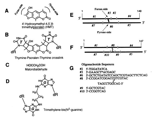
Linear substrates containing a psoralen or a 1,3-bis(2′-deoxyguanosine-N2-yl)propane or trimethylene-bis(N2-guanine) (TBG) interstrand cross-link were synthesized by annealing and ligating seven partially overlapping complementary oligonucleotides (see Fig. 5E and F). The 5′ or 3′ termini of these substrates were radiolabeled by standard procedures. To prepare a psoralen-cross-linked linear duplex, oligonucleotide 3, which contains a furan-side adducted thymine (33), was mixed with the other oligomers and ligated to obtain a 149-mer duplex, which was separated from the partial ligation products by sequential purification on 7% denaturing and 5% nondenaturing polyacrylamide gels. The monoadduct was then converted into a cross-link by irradiating the duplex with 366-nm light at a fluence rate of 2 mW/cm2 for 20 min at 0°C. Subsequently, the cross-linked material was purified from the non-cross-linked duplex by electrophoresis on a 7% denaturing polyacrylamide gel. The TBG cross-link-containing 147-bp duplex was derived from 19-mer and 11-mer oligomers connected by a trimethylene group through two guanines on both strands (8) (see Fig. 5A). The 147-mer with the TBG cross-link was assembled from six oligomers and purified as described for the psoralen-cross-linked substrate.
FIG. 5.
Linear cross-linked substrates for the XPF-ERCC1 nuclease. (A) Structure of the psoralen used in the present study. Both the furan side and pyrone side of the psoralen molecule can be adducted to thymine through a cyclobutane ring. (B) Structure of an interstrand psoralen cross-link. Only the two adducted thymines are shown. dR, deoxyribose. (C) Malondialdehyde. (D) TBG interstrand cross-link. This is a synthetic analog of a malondialdehyde-induced interstrand cross-link. The trimethylene group is linked to N2 of guanines on both strands. (E) Linear psoralen cross-link substrate. The oligomers used to assemble the duplex and the side of the cross-link are indicated. Both strands have a 1-base protruding 5′ end and are 149 nt long. (F) Linear TBG cross-link substrate. The oligomers used to assemble the duplex and the position of the TBG cross-link are shown. Both strands have a 1-base protruding 5′ terminus and are 147 nt long. (G) Nucleotide sequences of some of the oligomers used to make linear cross-link substrates. The thymine base of oligomer 3 linked to the furan side of a psoralen molecule is indicated by an asterisk. Oligomer 8 contains a TBG moiety cross-linking a 19-mer and an 11-mer, as shown. The sequences of oligomers 1, 4, 5, and 7 have been published (23).
Repair synthesis.
The repair synthesis assay was conducted in 25 μl of reaction buffer containing 20 mM HEPES-KOH (pH 7.9), 50 mM KCl, 5 mM MgCl2, 2 mM ATP, 100 μM each deoxynucleoside triphosphate except dCTP, 4 μM [α-32P]dCTP (7,000 Ci/mmol), 0.2 mM EDTA, 50 μg of cell extract. The reaction was conducted at 30°C for 1 h unless indicated otherwise. Following phenol-chloroform extraction, the DNA was digested with the indicated restriction enzymes and analyzed on 5% denaturing polyacrylamide gels.
Exonuclease assay.
Linear monoadducted or cross-linked substrates (25 fmol) were incubated with the indicated amounts of XPF-ERCC1 and RPA in 25 μl of reaction buffer containing 30 mM HEPES-KOH (pH 7.9), 40 mM KCl, 3 mM MgCl2, 5% (vol/vol) glycerol. After incubation at 30°C for 15 min, the DNA was deproteinized with proteinase K-phenol, precipitated with ethanol, and analyzed on 7% denaturing polyacrylamide gels.
Immunodepletion.
Polyclonal anti-ERCC1 or anti-XPB antibodies (26) (30 μl each) were incubated with 20 μl of protein A-agarose beads in 200 μl of buffer A (0.1 M KCl, 50 mM Tris-HCl [pH 7.5], 0.05% Nonidet P-40). After gentle mixing at 4°C for 30 min, the beads were collected by centrifugation and washed three times with 200 μl of buffer A. Subsequently, the beads containing either anti-ERCC1 or anti-XPB antibodies were mixed with XPF-ERCC1 (50 ng) in 60 μl of buffer B (30 mM HEPES-KOH [pH 7.9], 40 mM KCl, 3 mM MgCl2, 20 μM dithiothreitol) and incubated at 4°C for 3 h with rocking. After centrifugation to remove the proteins bound to the antibodies, the supernatant was supplemented with RPA (60 ng), cross-linked substrate (60 fmol), and buffer B to a volume of 100 μl. The reaction mixture was incubated at 30°C for 20 min, and then the DNA was deproteinized and analyzed on a 7% denaturing polyacrylamide gel.
RESULTS
Futile repair synthesis with cross-linked substrate.
A previous study on processing of psoralen interstrand cross-links by mammalian cell extract showed that the mammalian excision repair nuclease makes dual incisions 5′ to the cross-linked base, releases a 22- to 28-nt fragment free of damage, and generates a gap of equal size immediately 5′ to the cross-linked base (1). To investigate the fate of this excision gap, we conducted a repair synthesis assay with a plasmid containing a single cross-link and analyzed the reaction product following digestion of DNA with restriction endonucleases which cut the DNA in the vicinity of the cross-link. The restriction enzyme digestion scheme and the predicted product sizes are shown schematically in Fig. 1.
FIG. 1.
Schematic drawing of the covalently closed circular DNA substrate containing a psoralen interstrand cross-link. Restriction sites around the interstrand cross-link are indicated, and the sizes of the corresponding restriction fragments are shown.
Single-enzyme digestion (HindIII or PvuI), which incises the substrate either 5′ to the pyrone-side or 5′ to the furan-side adducted thymines of the cross-link, yielded results consistent with filling in of the gap adjacent to the cross-link but with minimal ligation to parental DNA (Fig. 2, lanes 2 and 3). Thus, it appears that the cross-link induces a “futile” repair synthesis reaction which is not accompanied by damage removal. Interestingly, there is a striking difference between the “repair synthesis” signal induced by the pyrone-side adducted thymine and the furan-side adducted thymine, with the repair synthesis induced by the former (HindIII, lane 3) being 10-fold stronger than that induced by the latter (PvuI, lane 2).
FIG. 2.
Interstrand psoralen cross-link induces futile DNA synthesis. The covalently closed circular DNA containing a site-specific psoralen interstrand cross-link (XL) was incubated with wild-type rodent extract (AA8) in the presence of [α-32P]dCTP under repair synthesis conditions. After incubation at 30°C for 60 min, the DNA was digested with HindIII (lane 2), PvuI (lane 3), or both (lane 4) and examined on a 7% denaturing polyacrylamide gel. W indicates the signal representing the ligated fraction of DNA that underwent futile repair synthesis. The unligated 130-nt fragment arose from the repair synthesis to fill the gap adjacent to the furan-side adducted thymine, and the 40-nt fragment was generated by filling in the gap adjacent to the pyrone-side adducted thymine. Lane 4 contains psoralen furan-side monoadducted DNA (MA) digested with HindIII and PvuI following the repair synthesis reaction. The 168-nt fragment carrying the 26-nt repair patch (1) is indicated by an arrow. The high-molecular-weight DNA near the origin represents nonspecific incorporation of radiolabel randomly throughout the plasmid. When normalized for fragment length, the background synthesis is about 20% of the damage-induced synthesis.
Futile repair synthesis and ligation.
Digestion of cross-linked DNA which had been subjected to the repair reaction with both PvuI and HindIII was expected to generate two fragments of 168 and 174 nt if the cross-linked-induced DNA synthesis were true repair synthesis which followed the removal of the cross-link (Fig. 1). Instead, the double digestion revealed three bands (Fig. 2, lane 4), none of which corresponded to these sizes. The fragments of 130 and 40 nt corresponded to the sizes expected from filling in the gaps 5′ to the cross-linked bases without ligation of the newly synthesized DNA as described above. The larger one (Fig. 2, lane 4, band W) migrated like a fragment twice the size of the distance between the two restriction sites (Fig. 1). Upon photoreversal, the fragment disappeared and a new species whose size was equal to the distance between the two restriction sites appeared, indicating that this fragment contains the cross-link (data not shown; see below). In contrast to these unusual results with cross-linked DNA, when the psoralen monoadduct was the substrate, true repair synthesis occurred, as evidenced by the appearance of a 168-nt radiolabeled fragment following the HindIII-PvuI digestion (Fig. 2, lane 5).
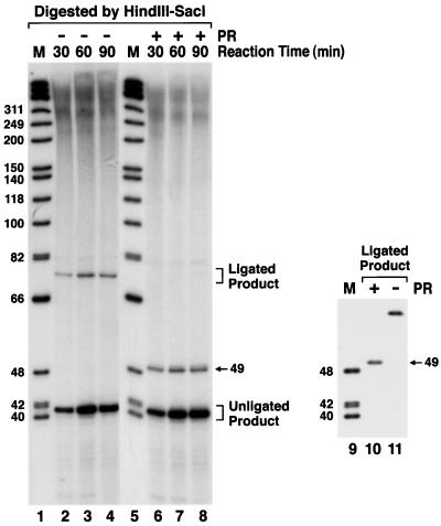
To confirm our interpretation of the fate of the “repair synthesis” patch induced by cross-linked DNA, i.e., that cross-link-induced DNA synthesis was mostly terminated at a nick and that the repair patch was ligated only in a small fraction of the molecules, we carried out a time course experiment of repair synthesis followed by double digestion with HindIII and SacI (Fig. 3). The two restriction incision sites are separated by 41 nt in the furan-side strand and by 49 nt in the pyrone-side strand (Fig. 1). At 60 min, the filling in of the excision gap at the pyrone side (40 nt in Fig. 3, lanes 3 and 7) was complete; further incubation did not promote more synthesis. Similarly, a larger fragment of about 80 nt was detected after 30 min (lane 2) and also reached a plateau level after 60 min (lanes 2 to 4). This was assigned to the small fraction of repair patches ligated to parental DNA. Indeed, photoreversal of the psoralen cross-link by short-wavelength UV light caused the disappearance of the 80-nt fragment and the appearance of a 49-nt species (compare lanes 2 to 4 with lanes 6 to 8), consistent with filling in of the pyrone-side gap followed by ligation. This interpretation was confirmed by isolating the 80-mer and subjecting it to photoreversal (lanes 10 and 11). These results show that the 80-mer represents a species generated by filling in and ligating the gap 5′ to the pyrone-side adducted thymine without removing the cross-link. Thus, in a small fraction (<10%) of the molecules containing an excision gap 5′ to the cross-link, the gap was filled and ligated to generate the structure existing before the excision-resynthesis-ligation had taken place.
FIG. 3.
Cross-link-induced DNA synthesis and ligation without cross-link repair. After the “repair synthesis” reaction, the DNA was digested with HindIII and SacI and analyzed on a 7% denaturing polyacrylamide gel (lanes 2 to 4). The samples in lanes 6 to 8 were subjected to photoreversal (PR) treatment to convert interstrand cross-links to monoadducts prior to gel electrophoresis. To confirm that the fragment marked “ligated product” observed in lanes 2 to 4 contains a psoralen cross-link, the DNA was excised from the gel, purified, and subjected to photoreversal (lanes 10 and 11). Note that because of the pyrone-side preference of excision and synthesis, the 49-nt strand containing the pyrone-side adducted thymine is radiolabeled whereas the 41-nt complementary fragment with the furan-side adducted thymine (Fig. 1) is undetectable. DNA size markers (φX174 digested with HinfI) are shown in lanes 1, 5, and 9 (M).
Requirement for XP but not XRCC genes for cross-link-induced DNA synthesis.
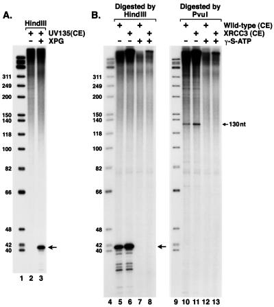
Repair synthesis reactions with whole-cell extracts can give rise to artifacts due to synthesis initiated at nicks and gaps created by nonspecific nucleases. Indeed, as seen in Fig. 2 and 3, in our study there was considerable incorporation of radiolabel into the plasmid substrate in regions far from the cross-link site. Digestion of DNA by several restriction enzymes revealed that this incorporation is evenly distributed throughout the plasmid and is not dependent on the presence of damage (monoadduct or cross-link) in the plasmid (reference 14 and data not shown). Hence, to examine the requirements for and the specificity of the repair synthesis in the vicinity of the cross-link which we have observed in our experiments, we conducted repair synthesis reactions with mutant cell extracts. Figure 4A shows that the cross-link-induced repair synthesis required XPG, one of the six excision nuclease factors (26). Since generation of the gap requires the entire set of excision repair factors (1), we conclude that the nicking as well as the futile DNA synthesis is initiated by the excision repair nuclease system.
FIG. 4.
(A) Futile DNA synthesis is dependent on the nucleotide excision repair nuclease. XPG mutant cell extract (UV135) was incubated with the cross-linked substrate in the presence (lane 3) or absence (lane 2) of purified XPG protein (40 ng) under repair synthesis conditions. After incubation at 30°C for 90 min, the DNA was digested by HindIII and analyzed on a 7% denaturing polyacrylamide gel. Lane 1 contains DNA size markers. The 40-nt fragment arising from futile DNA synthesis is indicated by an arrow. (B) Futile DNA synthesis is independent of the XRCC3 function. The cross-link-containing plasmid was incubated with either wild-type (AA8) or XRCC3 mutant (irs1SF) cell extracts (CE) with or without the nonhydrolyzable ATP analog γ-S-ATP (2 mM), as indicated. Following incubation at 30°C for 60 min, the DNA was digested by either HindIII or PvuI and analyzed on 7% denaturing polyacrylamide gels. Lanes 1 and 6 contain size markers. The bands arising from futile DNA synthesis are indicated by arrows.
It is known that XRCC2 and XRCC3 mutant CHO cell lines are extremely sensitive to killing by cross-linking agents (6, 29, 35, 36). Hence, we wished to investigate if the products of these genes affected the unusual repair synthesis observed in our system. Figure 4B shows that wild-type and XRCC3 mutant cell extracts carried out the cross-link-specific DNA synthesis identically. Similar results were obtained with an XRCC2 mutant extract (data not shown). Therefore, we conclude that XRCC2 and XRCC3 proteins do not play a role at this step of cross-link repair. Figure 4B also shows that ATP hydrolysis was required for the cross-link-induced DNA synthesis since the futile repair synthesis was inhibited by ATP-γ-S (lanes 4, 5, 9, and 10), as is the case for the excision nuclease-initiated “authentic” repair synthesis, which fills in gaps generated by intrastrand monoadduct or diadduct removal (1, 26).
Effect of XPF-ERCC1 on cross-links.
In nucleotide excision repair, the heterodimeric protein complex XPF-ERCC1 is responsible for the 5′ incision of the human excinuclease (21, 22). XPF alone has an endonuclease activity (23), and the XPF-ERCC1 complex has a structure-specific endonuclease activity which incises bubble structures at the 5′ junction (2, 22). Because rodent cell mutants defective in either of these two proteins are much more sensitive (ca. 30-fold) to cross-linking agents than are mutants defective in any of the other excision repair genes (13, 35), it is thought that these proteins play a crucial role in cross-link repair outside the context of their role in excision repair nuclease. Indeed, it has been found that the yeast homologs of XPF (Rad1) and ERCC1 (Rad10) are required for trimming the 3′ overhanging nonhomologous DNA tails during single-strand annealings in certain repair reactions (10). Hence, we investigated the effect of XPF-ERCC1 on DNA containing a cross-link.
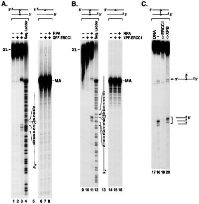
Under a variety of experimental conditions, we failed to see an effect of XPF-ERCC1 on covalently closed or nicked circular plasmid DNA containing a psoralen cross-link (reference 1 and data not shown). Similarly, the heterodimer failed to nick or degrade a cross-linked linear duplex (see below). Since it is known that RPA stimulates the nuclease activity of XPF-ERCC1 (2, 22), RPA was included in a reaction mixture containing XPF-ERCC1 and linear cross-linked substrates. Schematic diagrams of cross-linked substrates and the structures of the cross-links are shown in Fig. 5. The experimental results are shown in Fig. 6. With psoralen-cross-linked DNA, whether the DNA is terminally labeled at the furan-side adducted strand or the pyrone-side adducted strand, inclusion of RPA in the reaction mixture gave rise to two unique products; one corresponded to a fragment extending from the 5′ terminus of the radiolabeled strand to 1 to 5 nt 5′ to the cross-link (the short product), and the other corresponded to a fragment migrating slightly (ca. 1 base) slower than the full-length single-stranded substrate DNA (Fig. 6A and B, lanes 3 and 11). No such products were detected with a duplex with a psoralen monoadduct (lanes 8 and 16). Importantly, both the long and short products were generated by XPF-ERCC1 and not a contaminating nuclease, since both products essentially disappeared upon immunodepletion of the reaction mixture with anti-ERCC1 antibodies and since an unrelated anti-XPB antibody had no effect on the activity (Fig. 6C, lanes 19 and 20).
FIG. 6.
Specific degradation of a linear duplex containing a psoralen cross-link by XPF-ERCC1 and RPA. (A) Reactions performed with a substrate containing 5′ label on the furan-side adducted strand. The triangle and circle represent pyrone-side and furan-side adducted thymines of the cross-link (XL), respectively. Asterisks indicate 5′-terminally labeled strands. The reactions in lanes 2 and 3 were carried out with XPF-ERCC1 (30 ng) alone or in combination with RPA (60 ng) as indicated. Lane 4 shows the Maxam-Gilbert purine sequencing ladder of the same DNA. The nucleotide sequence around the cross-linked thymine is indicated to the right of lane 4. The psoralen-adducted thymine is circled. In the sequence ladder, a significant portion of the cross-linked substrate was converted to monoadduct by the hot alkali used in the sequencing reaction (4). Control experiments using a monoadducted psoralen substrate (MA) are shown in lanes 7 and 8. (B) Cleavage reactions of the same substrate radiolabeled at the 5′ terminus of the pyrone-side adducted strand. The experiments were carried out as described in panel A. Lanes 14 to 16 contain control reactions with monoadducted DNA. (C) The exonucleolytic activity on cross-linked DNA is intrinsic to XPF-ERCC1. The substrate, radiolabeled at the 5′ end of the pyrone-adducted strand, is shown schematically at the top. Where indicated, XPF-ERCC1 (50 ng) was mixed with either anti-ERCC1 (α-ERCC1) or anti-XPB (α-XPB) antibodies linked to protein A-agarose beads, and following removal of the beads by centrifugation, the supernatant was incubated with the substrate. Schematic drawings to the right of lane 4 show the long and short cleavage products generated by XPF-ERCC1.
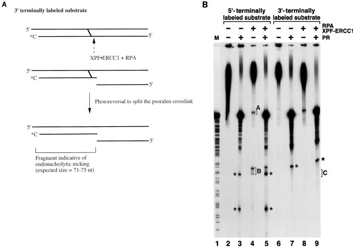
The reaction products of cross-linked DNA were consistent with a 3′-to-5′ exonuclease action of high processivity which was attenuated at the site of the cross-link. The size of the ≈73-nt species (i.e., the short product) indicates that it may arise from a processive digestion beginning at the 3′ end that stops immediately past the cross-link or from endonucleolytic nicking immediately 5′ to the cross-link (Fig. 7A). Experiments with 3′-terminally radiolabeled cross-linked DNA ruled out the second possibility (Fig. 7B). Cross-linked DNA, labeled at the 3′ end of the pyrone-side adducted strand or the corresponding strand in the control DNA, was subjected to cleavage by XPF-ERCC1 in the presence of RPA. Prior to the analysis by denaturing polyacrylamide gels, the DNA was irradiated with 254-nm UV light to photoreverse the psoralen cross-link. If the cleavage reaction were carried out endonucleolytically, only after photoreversal would fragments of 71 to 75 nt be observed on the denaturing polyacrylamide gels (Fig. 7A). Positive control experiments with a 5′-end-labeled substrate yielded large and small products (Fig. 7B, lane 4). In the experiments with 3′-terminally radiolabeled substrate, the fragments that would arise from endonucleolytic incisions were not observed (Fig. 7B, lane 9), indicating that the short product observed with 5′-labeled DNA is generated exonucleolytically by XPF-ERCC1 and RPA. Furthermore, as discussed below, the analysis of the long product seen in lane 11 of Fig. 6B and lane 4 of Fig. 7B lends further support to the notion that XPF-ERCC1, assisted by RPA, acts on cross-linked DNA as a 3′-to-5′ exonuclease with high processivity.
FIG. 7.
(A) Experimental design to determine whether XPF-ERCC1 nicks DNA 5′ to a cross-link. A 3′-terminally labeled psoralen cross-linked substrate is treated with XPF-ERCC1 plus RPA and then irradiated with 254-nm light to reverse the cross-link. A specific nick 5′ to the cross-link would cause the release of a 71- to 75-nt oligomer upon photoreversal. (B) Lack of specific nicking 5′ to the cross-link by XPF-ERCC1. The reactions in lanes 2 to 5 are control experiments with 5′-terminally labeled substrate. Brackets A and B (lane 4) indicate the short and long products, respectively, as seen in lane 3 of Fig. 6. Bracket C (lane 9) defines the area where, with 3′-labeled substrate, the products indicative of endonucleolytic cutting 5′ to the cross-link would have migrated. The bands marked with asterisks are background fragments arising from partial ligation during the construction of the cross-linked substrates by ligating seven oligomers (Fig. 5E and F).
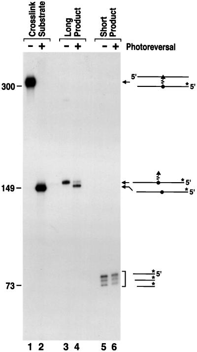
Two possibilities were considered as the source of the larger species of 150 nt. The first was that an exonuclease acting 3′ to 5′ on both strands and stopping at the cross-link would generate a “Z-shaped” molecule in which the horizontal parts consisting of a 70-nt oligomer and a 80-nt oligomer are joined via the crosslink. The second possibility was that the 150-nt species arose from 3′-to-5′ exonucleolytic degradation of the unlabeled strand in a processive manner such that the exonuclease digests the unlabeled strand past the cross-link and all the way to the 5′ terminus, leaving behind the radiolabeled strand attached to a single nucleotide through the psoralen molecule. To differentiate between these two possibilities, we purified the 150-nt species and subjected it to short-wavelength photoreversal. A Z-shaped molecule would be expected to yield two fragments of 70 and 80 nt. In contrast, a 149-mer attached to a thymine via a psoralen cross-link would be shortened by approximately 1 nt after photoreversal (Fig. 7A). Figure 8 shows the result of this experiment. Under photoreversal conditions which reverted the control cross-linked substrate quantitatively (compare lanes 1 and 2), the 150-nt species (the long product) became shorter by 1 nt (compare lanes 3 and 4). This is consistent with the long (150-nt) cleavage product being a single-stranded full-length substrate of 149 nt attached to a thymine via a psoralen cross-link. In contrast, the 71- to 74-mer species were not affected by the photoreversal treatment (compare lanes 5 and 6), in line with our interpretation that these species are free of cross-links and are generated by exonucleolytic degradation of the labeled strand to a point immediately past the cross-link.
FIG. 8.
Analysis of XPF-ERCC1 reaction products from linear substrates with a psoralen cross-link. In lanes 1 and 2, the conversion of the control substrate to the monoadducted form with 254-nm light at 10 kJ/m2 for 3 min is shown. The long product (lane 3) and the short product (lane 5) generated from this substrate by XPF-ERCC1 plus RPA were gel purified from band A and band B in Fig. 7B, lane 4, and subjected to photoreversal under the same conditions. The photoreversed products are shown in lanes 4 and 6, respectively. DNA fragment sizes in nucleotides are indicated to the left of the figure. Drawings illustrating the corresponding species are shown to the right of lane 6.
Effect of XPF-ERCC1 on the malondialdehyde cross-link.
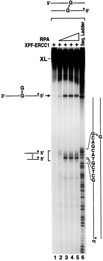
To explore the generality of the unusual cross-link-cleaving activity of XPF-ERCC1 in the presence of RPA, we performed the same type of experiments using a different substrate containing a lesion mimicking a malondialdehyde-directed interstrand cross-link. Malondialdehyde is a known mutagen and suspected endogenous carcinogen generated during lipid peroxidation and eicosanoid synthesis (9, 28). A malondialdehyde interstrand cross-link contains two quite unstable imino attachments to exocyclic amine groups of DNA. To mimic such a lesion, a stable trimethylene linkage was joined to two guanines through N-2 of guanine (Fig. 5D) on two complementary oligonucleotides (oligomer 8, Fig. 5G). When a 5′-terminally radiolabeled duplex containing this cross-link (Fig. 5F) was used as a substrate, XPF-ERCC1, in combination with RPA, digested the DNA and produced both long and short products as for psoralen cross-links (Fig. 9). The short products (63 to 68 nt) correspond to incisions at phosphodiester bonds 5 to 9 5′ to the cross-linked guanine in the bottom strand (Fig. 9, lanes 2 to 5). As in the case of psoralen, the longer product (148 nt) migrated slightly slower than the non-cross-linked single-stranded 147 mer (lanes 2 to 5), suggesting that it arose from 3′-to-5′ exonucleolytic removal of the nonlabeled top strand past the cross-link so that a 147-mer with TBG was formed. As with the psoralen cross-link, RPA was also essential for the processing of the TBG cross-link by XPF-ERCC1.
FIG. 9.
Specific degradation of a linear substrate with a malondialdehyde-induced interstrand cross-link by XPF-ERCC1 plus RPA. The 147-mer duplex containing a TBG interstrand cross-link and a 5′ label in one strand (top) was used as the substrate. No nucleolytic degradation was observed when the substrate was incubated with XPF-ERCC1 (30 ng, lane 1). Addition of increasing amounts of RPA (10, 30, 60, and 90 ng from lane 2 to lane 5) to the reaction mixtures conferred cross-link-specific nuclease activity, which gave rise to the indicated specific reaction products. Drawings representing the long and short cleavage products are shown to the left of lane 1. Lane 6 shows the Maxam-Gilbert purine chemical sequencing ladder of the substrate. The sequence 5′ to the adducted guanine is shown to the right of lane 7. Because of the stability of the TBG cross-link, fragments hydrolyzed at the purines 3′ to the cross-linked guanine remained attached to the complementary strand, migrated near the full-length cross-link substrate, and thus were not discernible in the sequence ladder.
DISCUSSION
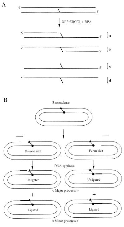
Figure 10 summarizes the findings of this study. Psoralen- and TBG-cross-linked linear DNA duplexes are specifically degraded by XPF-ERCC1 in the presence of RPA, producing two types of products. The first (Fig. 10A, products a and b) is the result of 3′-to-5′ exonucleolytic digestion, which is highly attenuated immediately past the cross-link site. The second types of products (products c and d) result from complete digestion of one strand by the 3′-to-5′ exonuclease activity by XPF-ERCC1. Given the exquisite sensitivity of rodent XPF and ERCC1 mutant cells to cross-linking agents, it is possible that the products in Fig. 10A are recombinogenic and potential substrates for XRCC2- and XRCC3-dependent steps of cross-link repair. Thus, it is conceivable that upon encountering a replication fork, the cross-link may give rise to a double-strand break (24) which creates an entry site for XPF-ERCC1 to process the DNA into a form which is then acted upon by XRCC2 and XRCC3 to generate a cross-link-free duplex. Further work is needed to test such a model.
FIG. 10.
Summary of the main findings of this study. (A) Cross-link-specific exonucleolytic degradation of a linear duplex by XPF-ERCC1 in the presence of RPA. Four types of products are generated. Products a and b result from the cross-link-attenuated progression of a 3′-to-5′ exonuclease activity of XPF-ERCC1. Products c and d represent terminal digestion. (B) Futile DNA synthesis induced by the cross-link. Nucleotide excision repair nuclease removes oligonucleotides of 22 to 28 nt from the immediate 5′ vicinity of the cross-link (1). The pyrone-side adducted strand is preferred over the furan-side adducted strand with the particular substrate used in this study. The gap is filled in by DNA polymerases. Following filling in, ligation to the unremoved cross-link is inefficient, leaving behind mostly unligated repair patch as the major product and a small fraction of molecules in which the repair patch is ligated to regenerate the original substrate.
Figure 10B summarizes the processing of psoralen cross-links by the mammalian nucleotide excision repair system which excises 22- to 28-nt oligomers from the 5′ side of the cross-link (1). In this reaction, the pyrone-side adducted strand is preferred over the furan-side adducted strand by a factor of about 10 to 1. The structural and mechanistic basis of this preference is unknown, but it is of interest that it is the opposite of that of the E. coli excinuclease, which prefers the furan-side adducted strand (32, 37) and which makes dual incisions bracketing the cross-linked bases. The resulting gap is filled by DNA polymerases in both cases. However, in mammalian cells the repair patch terminates at a nick adjacent to the cross-link in 90% of cases and is ligated to the parental DNA to regenerate the cross-linked substrate in the remaining 10% of molecules.
The reactions summarized in Fig. 10 are dependent on the excision repair system but independent of the XRCC2 and XRCC3 proteins which have been found by genetic studies to be required for the major pathway of cross-link repair (35, 36). These results raise two interrelated issues: are the dual incisions 5′ to the cross-link and the subsequent futile DNA synthesis relevant to cross-link repair, and what are the likely roles of XRCC2 and XRCC3 in cross-link repair? We do not have the answers to these questions. However, both the dual incisions and the futile DNA synthesis are such efficient reactions in vitro that we are inclined to believe that they occur with reasonably high frequency in vivo and may play some important roles in the cellular response to DNA cross-links. Thus, the excised oligomer may be a coactivator for an SOS-like response in mammalian cells and the gap filling by DNA polymerase ɛ may activate the DNA damage checkpoint response (31). Furthermore, it is possible that the repair synthesis which generates a nick immediately 5′ to the cross-link will produce a prerecombinogenic structure which eventually leads to cross-link removal by recombination. However, we failed to see further processing of the nicked intermediate in either the presence or absence of XRCC3 protein, which is believed to be required for cross-link repair. XRCC2 and XRCC3 encode RecA/HsRad51 homologs which are involved in homologous recombination (16, 29) and are thought to catalyze strand transfer and thus to participate in cross-link repair by promoting the recombination of the cross-linked duplex with a homologous duplex with no damage (35, 36). Hence, to provide a substrate for recombination, we included into our reaction mixtures homologous DNA duplex with no damage, single-stranded circular, or linear fragments complementary to the entire cross-linked plasmid or to the cross-linked region. Under no circumstances were we able to detect the elimination of cross-links from the substrate (data not shown).
In vivo data with cellular DNA (13) or transfected plasmid DNA (7) clearly show that mammalian cells are capable of removing interstrand cross-links. Indeed, previously, using randomly cross-linked DNA, we obtained data suggesting the removal of interstrand cross-links in vitro (30). In that study, however, the conclusion that repair synthesis was accompanied by the disappearance of cross-links was based on indirect evidence. In light of the results obtained in the present work with a substrate containing a single cross-link, we conclude that most of the cross-link-induced DNA synthesis observed in the previous study was not associated with cross-link removal. The data herein also constitute unambiguous evidence for damage-induced DNA synthesis that is not repair synthesis in the strict sense of the word. Finally, the “repair synthesis” results presented in this paper differ from those in a recent report suggesting that cross-link-induced DNA synthesis was independent of nucleotide excision repair but dependent on XPF-ERCC1, XRCC2, and XRCC3 and was greatly stimulated by homologous or nonhomologous DNA (18). As documented in Fig. 4, our repair synthesis (i) depends on a functional excision repair system, (ii) is independent of XRCC3, and (iii) is not affected by the presence of a second plasmid with or without homologous sequence in the reaction mixture (data not shown). We have no satisfactory explanations for the seemingly contradictory results of these two studies. However, in the previous study, cross-link-induced DNA synthesis was analyzed only in terms of radiolabel incorporated into the entire plasmid or large restriction enzyme fragments separated on nondenaturing agarose gels (18). Hence, it is not possible to ascertain whether the cross-link-induced DNA synthesis in that study was confined to the area of the cross-link, whether the newly synthesized DNA was ligated to the parental DNA, and whether there was preferential DNA synthesis in the region of homology when homologous nondamaged DNA was included in the reaction mixture. Clearly, more studies are needed with well-defined substrates and purified enzymes to gain better insight into the mechanism of cross-link repair in mammalian cells.
Finally, we would like to comment on the potential clinical significance of the cross-link-induced futile DNA synthesis. The concept of futile repair is currently being discussed with respect to both mismatch repair activity on damaged DNA (25) and transcription-coupled repair at transcription pause sites (11), but with little direct evidence. The work presented in this paper is the only direct demonstration so far of a potentially futile repair cycle in mammalian cells. It is plausible that this futile cycle and the potentially prpapoptotic signals resulting from this cycle, rather than the replication block per se, are the main causes of the lethality of cross-link-inducing anticancer drugs.
ACKNOWLEDGMENTS
David Mu and Tadayoshi Bessho contributed equally to this work.
This work was supported by grants GM32833 (A.S.) and EA74046 (D.J.C.) from the National Institutes of Health.
REFERENCES
- 1.Bessho T, Mu D, Sancar A. Initiation of DNA interstrand cross-link repair in humans: the nucleotide excision repair system makes dual incisions 5′ to the cross-linked base and removes a 22- to 28-nucleotide-long damage-free strand. Mol Cell Biol. 1997;17:6822–6830. doi: 10.1128/mcb.17.12.6822. [DOI] [PMC free article] [PubMed] [Google Scholar]
- 2.Bessho T, Sancar A, Thompson L H, Thelen M P. Reconstitution of human excision nuclease with recombinant XPF-ERCC1 complex. J Biol Chem. 1997;272:3833–3837. doi: 10.1074/jbc.272.6.3833. [DOI] [PubMed] [Google Scholar]
- 3.Cheng S A, Sancar A, Hearst J E. RecA-dependent incision of psoralen-DNA crosslinked DNA by (A)BC excinuclease. Nucleic Acids Res. 1991;19:657–663. doi: 10.1093/nar/19.3.657. [DOI] [PMC free article] [PubMed] [Google Scholar]
- 4.Cimino G D, Gamper H B, Isaacs S T, Hearst J E. Psoralens as photoactive probes of nucleic acid structure and function: organic chemistry, photochemistry, and biochemistry. Annu Rev Biochem. 1985;54:1151–1193. doi: 10.1146/annurev.bi.54.070185.005443. [DOI] [PubMed] [Google Scholar]
- 5.Cole R S. Repair of DNA containing interstrand cross-links in Escherichia coli: sequential excision and recombination. Proc Natl Acad Sci USA. 1973;70:1064–1068. doi: 10.1073/pnas.70.4.1064. [DOI] [PMC free article] [PubMed] [Google Scholar]
- 6.Collins A R. Mutant rodent cell lines sensitive to ultraviolet light, ionizing radiation and cross-linking agents: a comprehensive survey of genetic and biochemical characteristics. Mutat Res. 1993;293:99–118. doi: 10.1016/0921-8777(93)90062-l. [DOI] [PubMed] [Google Scholar]
- 7.Dooley P A, Tsarouhtsis D, Nechev L V, Kowaszyk A, Stone M P, Harris T M. Synthesis and structural studies of the saturated analog of malondialdehyde crosslink in a synthetic oligonucleotide. Proc Am Assoc Cancer Res. 1997;38:334–335. [Google Scholar]
- 8.Faruqi A F, Datta H J, Carroll D, Seidman M, Glazer P M. Triple-helix formation induces recombination in mammalian cells via a nucleotide excision repair-dependent pathway. Mol Cell Biol. 2000;20:990–1000. doi: 10.1128/mcb.20.3.990-1000.2000. [DOI] [PMC free article] [PubMed] [Google Scholar]
- 9.Fink S P, Reddy G R, Marnett L J. Mutagenicity in Escherichia coli of the major DNA adduct derived from the endogenous mutagen malondialdehyde. Proc Natl Acad Sci USA. 1997;94:8652–8657. doi: 10.1073/pnas.94.16.8652. [DOI] [PMC free article] [PubMed] [Google Scholar]
- 10.Fishman-Lobell J, Haber J E. Removal of nonhomologous DNA ends in double-strand break recombination: the role of the yeast ultraviolet repair gene RAD1. Science. 1992;258:480–484. doi: 10.1126/science.1411547. [DOI] [PubMed] [Google Scholar]
- 11.Hanawalt P C. DNA repair comes of age. Mutat Res. 1995;336:101–113. doi: 10.1016/0921-8777(94)00061-a. [DOI] [PubMed] [Google Scholar]
- 12.Henricksen L A, Umbricht G B, Wold M S. Recombinant replication protein A: expression, complex formation, and functional characterization. J Biol Chem. 1994;269:11121–11132. [PubMed] [Google Scholar]
- 13.Hoy C A, Thompson L H, Mooney C L, Salazar E P. Defective DNA cross-link removal in Chinese hamster cell mutants hypersensitive to bifunctional alkylating agents. Cancer Res. 1985;45:1737–1743. [PubMed] [Google Scholar]
- 14.Huang J C, Svoboda D L, Reardon J T, Sancar A. Human nucleotide excision nuclease removes thymine dimers from DNA by incising the 22nd phosphodiester bond 5′ and the 6th phosphodiester bond 3′ to the photodimer. Proc Natl Acad Sci USA. 1992;89:3664–3668. doi: 10.1073/pnas.89.8.3664. [DOI] [PMC free article] [PubMed] [Google Scholar]
- 15.Jachymczyk W J, von Borstel R C, Mowat M R, Hastings P J. Repair of interstrand cross-links in DNA of Saccharomyces cerevisiae requires two systems for DNA repair: the RAD3 system and the RAD51 system. Mol Gen Genet. 1981;182:196–205. doi: 10.1007/BF00269658. [DOI] [PubMed] [Google Scholar]
- 16.Johnson R D, Liu N, Jasin M. Mammalian XRCC2 promotes the repair of DNA double-strand breaks by homologous recombination. Nature. 1999;401:397–399. doi: 10.1038/43932. [DOI] [PubMed] [Google Scholar]
- 17.Kohn K W. Beyond DNA cross-linking: history and prospect of DNA-targeted cancer treatment. Cancer Res. 1996;56:5533–5546. [PubMed] [Google Scholar]
- 18.Li L, Peterson C A, Lu X, Wei P, Legerski R J. Interstrand cross-links induced DNA synthesis in damaged and undamaged plasmids in mammalian cell extract. Mol Cell Biol. 1999;19:5619–5630. doi: 10.1128/mcb.19.8.5619. [DOI] [PMC free article] [PubMed] [Google Scholar]
- 19.Liu N, Lamerdin J E, Tebbs R S, Schild D, Tucker J D, Shen M R, Brookman K W, Siciliano M J, Walter C A, Fan W, Narayana L S, Zhou Z Q, Adamson A W, Sorensen K J, Chen D J, Jones N J, Thompson L H. XRCC2 and XRCC3, new human Rad51-family members, promote chromosome stability and protect against DNA cross-links and other damages. Mol Cell. 1998;1:783–793. doi: 10.1016/s1097-2765(00)80078-7. [DOI] [PubMed] [Google Scholar]
- 20.Magana-Schwencke N, Henriques J A, Chanet R, Moustacchi E. The fate of 8-methoxypsoralen photoinduced crosslinks in nuclear and mitochondrial yeast DNA: comparison of wild-type and repair-deficient strains. Proc Natl Acad Sci USA. 1982;79:1722–1726. doi: 10.1073/pnas.79.6.1722. [DOI] [PMC free article] [PubMed] [Google Scholar]
- 21.Matsunaga T, Mu D, Park C-H, Reardon J T, Sancar A. Human DNA repair excision nuclease: analysis of the role of the subunits involved in dual incisions by using anti-XPG and anti-ERCC1 antibodies. J Biol Chem. 1995;270:20862–20869. doi: 10.1074/jbc.270.35.20862. [DOI] [PubMed] [Google Scholar]
- 22.Matsunaga T, Park C-H, Bessho T, Mu D, Sancar A. Replication protein A confers structure-specific endonuclease activities to the XPF-ERCC1 and XPG subunits of human DNA repair excision nuclease. J Biol Chem. 1996;271:11047–11050. doi: 10.1074/jbc.271.19.11047. [DOI] [PubMed] [Google Scholar]
- 23.McCutchen-Maloney S L, Giannecchini C A, Hwang M H, Thelen M P. Domain mapping of the DNA binding, endonuclease, and ERCC1 binding properties of the human DNA repair protein XPF. Biochemistry. 1999;38:9417–9425. doi: 10.1021/bi990591+. [DOI] [PubMed] [Google Scholar]
- 24.Michel B, Ehrlich S D, Uzest M. DNA double-strand breaks caused by replication arrest. EMBO J. 1997;16:430–438. doi: 10.1093/emboj/16.2.430. [DOI] [PMC free article] [PubMed] [Google Scholar]
- 25.Modrich P. Strand-specific mismatch repair in mammalian cells. J Biol Chem. 1997;272:24727–24730. doi: 10.1074/jbc.272.40.24727. [DOI] [PubMed] [Google Scholar]
- 26.Mu D, Hsu D S, Sancar A. Reaction mechanism of human DNA repair excision nuclease. J Biol Chem. 1996;271:8285–8294. doi: 10.1074/jbc.271.14.8285. [DOI] [PubMed] [Google Scholar]
- 27.Mu D, Tursun M, Duckett D R, Drummond J T, Modrich P, Sancar A. Recognition and repair of compound DNA lesions (base damage and mismatch) by human mismatch repair and excision repair systems. Mol Cell Biol. 1997;17:760–769. doi: 10.1128/mcb.17.2.760. [DOI] [PMC free article] [PubMed] [Google Scholar]
- 28.Mukai F H, Goldstein B D. Mutagenicity of malonaldehyde, a decomposition product of peroxidized polyunsaturated fatty acids. Science. 1976;191:868–869. doi: 10.1126/science.766187. [DOI] [PubMed] [Google Scholar]
- 29.Pierce A J, Johnson R D, Thompson L H, Jasin M. XRCC3 promotes homology-directed repair of DNA damage in mammalian cells. Genes Dev. 1999;13:2633–2638. doi: 10.1101/gad.13.20.2633. [DOI] [PMC free article] [PubMed] [Google Scholar]
- 30.Reardon J T, Spielmann H P, Huang J C, Sastry S, Hearst J E, Sancar A. Removal of psoralen monoadduct and crosslinks by human cell free extracts. Nucleic Acids Res. 1991;19:4623–4629. doi: 10.1093/nar/19.17.4623. [DOI] [PMC free article] [PubMed] [Google Scholar]
- 31.Russell P. Checkpoints on the road to mitosis. Trends Biochem Sci. 1998;24:399–402. doi: 10.1016/s0968-0004(98)01291-2. [DOI] [PubMed] [Google Scholar]
- 32.Sladek F M, Munn M M, Rupp W D, Howard-Flanders P. In vitro repair of psoralen-DNA cross-links by RecA, UvrABC, and the 5′-exonuclease of DNA polymerase I. J Biol Chem. 1989;264:6755–6765. [PubMed] [Google Scholar]
- 33.Spielmann H P, Sastry S S, Hearst J E. Methods for the large scale synthesis of psoralen furan-side monoadducted and diadducts. Proc Natl Acad Sci USA. 1992;89:4514–4518. doi: 10.1073/pnas.89.10.4514. [DOI] [PMC free article] [PubMed] [Google Scholar]
- 34.Svoboda D L, Taylor J S, Hearst J E, Sancar A. DNA repair by eukaryotic nucleotide excision nuclease: removal of thymine dimer and psoralen monoadduct by HeLa cell free extract and of thymine dimer by Xenopus oocytes. J Biol Chem. 1993;268:1931–1936. [PubMed] [Google Scholar]
- 35.Thompson L H. Evidence that mammalian cells possess homologous recombinational repair pathway. Mutat Res. 1996;363:77–88. doi: 10.1016/0921-8777(96)00008-0. [DOI] [PubMed] [Google Scholar]
- 36.Thompson L H, Schild D. The contribution of homologous recombination in preserving genome integrity in mammalian cells. Biochimie. 1999;81:87–105. doi: 10.1016/s0300-9084(99)80042-x. [DOI] [PubMed] [Google Scholar]
- 37.Van Houten B, Gamper H, Holbrook S R, Hearst J E, Sancar A. Action mechanism of ABC excision nuclease on a DNA substrate containing a psoralen crosslink at a defined position. Proc Natl Acad Sci USA. 1986;83:8077–8081. doi: 10.1073/pnas.83.21.8077. [DOI] [PMC free article] [PubMed] [Google Scholar]


