FIG 5.
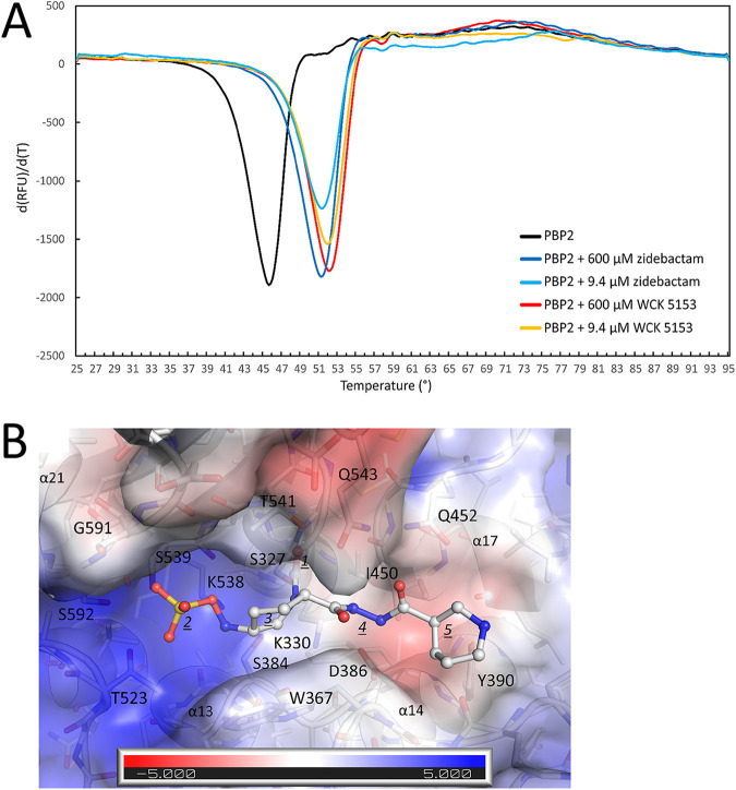
Differential scanning fluorimetry (DSF) measurement of WCK 5153 and zidebactam binding to PBP2 and modeling of zidebactam. (A) DSF thermal shift assay of WCK 5153 and zidebactam binding to PBP2. The derivative of the change in fluorescence is plotted versus temperature. Experiments were performed in duplicate (a representative curve is depicted). (B) Modeling of zidebactam in P. aeruginosa PBP2 active site. The coordinates of zidebactam were obtained by transplanting most of the atom coordinates from the similar WCK 5153 when bound to PBP2, yet with the pyrrolidine ring being replaced by the piperidine ring of zidebactam using the zidebactam piperidine conformation when complexed to KPC-2 (11). This modeling and superpositioning were done using COOT. The electrostatic potential map of the PBP2 active site is shown as generated using APBS in PyMOL. Zidebactam in shown in ball-and-stick representation, and the individual moieties are labeled as in Fig. 2B with the noted change that 5 now represents the (larger) piperidine ring of the R1-group.

