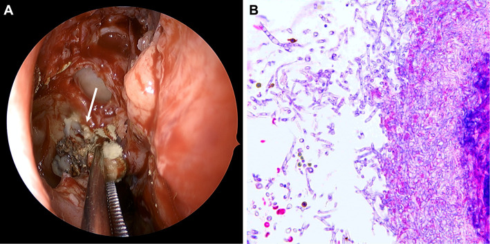
A 58-year-old woman presented to the otorhinolaryngology clinic with chronic unilateral purulent rhinorrhea. Her medical history included sarcoidosis, and she was being regularly treated with steroid therapy (prednisolone, 8 mg/day). Computed tomography revealed thickening of the nasal mucosa with calcification in the right maxillary sinus (Picture 1A). Magnetic resonance imaging demonstrated a high-intensity lesion on T1-weighted imaging (Picture 1B) and signal intensity loss on T2-weighted imaging (Picture 1C). Surgery was performed following the diagnosis of sinus mycosis based on imaging findings, and intraoperative imaging showed fungus mold in the maxillary sinus (Picture 2A). A biopsy specimen of the fungus was confirmed to be Aspergillus (Picture 2B). After surgery, the patient was completely relieved of her symptoms. Sinus mycetoma is frequently caused by a decrease in the immune function following steroid use, and the characteristic imaging findings of manganese and iron in the mycelium are useful for its diagnosis (1).
Picture 1.
Picture 2.
The authors state that they have no Conflict of Interest (COI).
Acknowledgement
We would like to thank Dr. Yoshio Suzuki of the Department of Pathology, Asahi General Hospital, Japan, for his supporting in pathological diagnosis.
References
- 1. Pierre G, Rainer W. Fungus balls of the paranasal sinuses: a review. Eur Arch Otorhinolaryngol 264: 461-470, 2007. [DOI] [PubMed] [Google Scholar]


