Abstract
Objectives
To analyze the biomechanical system of anterior retraction with clear aligner therapy (CAT) with and without an anterior mini-screw and elastics.
Materials and Methods
Models including a maxillary dentition (without first premolars), maxilla, periodontal ligaments (PDLs), attachments, and aligners were constructed and imported to finite element software. Three model groups were created: (1) control (CAT alone), (2) labial elastics (CAT with elastics between the anterior mini-screw and buttons on central incisors), and (3) linguoincisal elastics (CAT with elastics between the anterior mini-screw and precision cuts on the lingual sides of the aligner). Elastic forces (0–300 g, in 50 g increments) were applied.
Results
CAT alone caused lingual tipping and extrusion of the incisors. Labial elastics caused palatal root torquing and intrusion and mesial tipping of the central incisors, while linguoincisal elastics produced palatal root torquing and intrusion of both central and lateral incisors. Second premolars were intruded in all three groups, with less intrusion in the linguoincisal elastics group. For the control group, stress was concentrated on both labial and lingual root surfaces, alveolar ridge, and cervical and apical PDLs. Stress was more concentrated in the labial elastics group and less concentrated in the linguoincisal elastics group.
Conclusions
CAT produced lingual tipping and extrusion of incisors during anterior retraction. Anterior mini-screws and elastics can achieve incisor intrusion and palatal root torquing. Linguoincisal elastics are superior to labial elastics with a lower likelihood of buccal open bite. Root resorption and alveolar defects may occur in CAT, more likely for labial elastics and less likely for linguoincisal elastics.
Keywords: Clear aligner, Finite element, Mini-screw, Elastics, Anterior retraction
INTRODUCTION
With recent advances in orthodontic materials and innovations in orthodontic biomechanics, clear aligner therapy (CAT) is gaining popularity among orthodontists and patients for its esthetics and comfort. However, it has been suggested that complex tooth movement cannot be achieved with CAT, especially for bodily movement and root torquing,1,2 which are important for anterior retraction in extraction cases. It was shown that CAT was not effective for achieving anterior bodily retraction and often causes lingual tipping and extrusion of anterior teeth.3 Thus, sophisticated force systems that apply anticlockwise moments and intrusion to anterior teeth are required for effective anterior retraction in extraction CAT cases.
Several studies have demonstrated that the combination of mini-screws and CAT could provide an exciting solution to the biomechanical disadvantages of CAT.4,5 With the aid of elastics, additional force systems could be built on those of clear aligners,6 thereby improving the biomechanical systems of clear aligners. The combination of an anterior mini-screw and elastics may apply additional anticlockwise moments and an intrusive force to anterior teeth, facilitating anterior bodily retraction. However, this idea has never been investigated.
Three-dimensional finite element analysis (FEA), an effective computer simulation technique, is widely used to calculate stress and deformation developed on a geometric solid subjected to external forces.7 In the field of orthodontics, it provides a noninvasive, accurate method that permits the estimation of the responses generated within different tissues (eg, the periodontal ligament [PDL], teeth, and alveolar bone). Therefore, FEA has been suggested as a solution for complex biomechanical analyses in orthodontics.8
The aim of this study was to analyze the displacement tendency of teeth and aligners and stress distribution of teeth, PDL, and alveolar bone in an extraction case treated with CAT and to compare the biomechanical systems with and without an anterior mini-screw and elastics.
MATERIALS AND METHODS
An orthodontic patient with permanent dentition (U1-SN = 118°) was selected as the subject for this study. Cone-beam computed tomography (CBCT) and three-dimensional intraoral scanning were used to obtain digital models of the maxillary bone and maxillary dentition. The study was approved by the Ethical Committee of West China Hospital of Stomatology, Sichuan University (WCHSIRB-OT-2020-160).
The digital data of the maxilla and maxillary dentition were used to construct three-dimensional geometric surface models of the maxilla and maxillary teeth using Mimics Research 17.0 and Geomagic Studio 2014 (Materialise, Leuven, Belgium). The PDL was modeled on the root shape with an average uniform thickness of 0.27 mm.9 The alveolar sockets of the maxilla were obtained after subtracting the teeth and the PDL from the maxilla by Boolean operation.
First premolars and their PDL were removed to obtain the extraction dentition model (D1). Horizontal rectangular attachments (3 × 2 × 1 mm) were designed on the buccal surfaces of the posterior teeth of D1. Then, a new extraction dentition model (D2) was created through retracting the anterior teeth by 0.25 mm on D1. The aligner was developed by making an external offset on D2 with the thickness of 0.5 mm.7
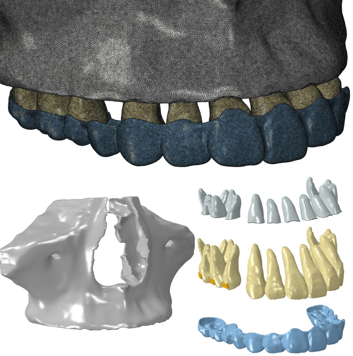
All components (dentition model D1, maxilla, PDL, attachments, and aligner) were assembled and converted into three-dimensional FEA solid models with Hypermesh 14.0 software (Altair, Troy, Mich). The models were meshed to unstructured four-noded tetrahedral elements. Mesh sizes were 0.20 mm for the dentition, 0.15 mm for the PDL, 0.25 mm for the maxilla, 0.20 mm for attachments, and 0.20 mm for aligners. The mesh sizes were defined through a convergence study on the control group, and the results tended to be stable if the mesh sizes became smaller. The process of discretization resulted in a total of 1,910,491 nodes and 8,367,215 elements. The numbers of nodes and elements for all components are summarized in Table 1.
Table 1.
Number of Nodes and Elements of the Components of the Finite Element Model
| Component |
Number of Elements |
Number of Nodes |
| Teeth | 1,485,245 | 315,650 |
| Periodontal ligament | 1,544,678 | 417,080 |
| Bone | 4,364,816 | 921,615 |
| Attachment | 18,608 | 5227 |
| Clear aligner | 883,404 | 215,228 |
The models were then imported into Abaqus/CAE software (SIMULIA, Providence, RI). The finite element models of all components are displayed in Figure 1. The teeth, maxilla, attachments, and aligners were considered as linear elastic. The teeth and maxilla were regarded as isotropic and homogeneous materials without discriminating internal tissues.6 No materials filled in the extraction space. The PDL was set as a linear elastic material for the best accuracy-computational ratio.10 The material properties of the components are presented in Table 2.1,7,11
Figure 1.
Computer-aided design model.
Table 2.
Material Properties
| Component |
Young's Modulus (MPa) |
Poisson's Ratio |
| Teeth | 1.96*104 | .30 |
| Periodontal ligament | 0.67 | .45 |
| Bone | 1.37*104 | .30 |
| Attachment | 12.5*103 | .36 |
| Clear aligner | 528 | .36 |
The upper part of the maxilla was set as fixed support when the force was loaded. Bonded contacts were set between the internal surface of the PDL and teeth and between the external surface of the PDL and alveolar bone. Surface-to-surface contact was used between the aligner and the surfaces of the teeth and attachments with a Coulomb friction coefficient of 0.2.1,2 To simulate the clinical situation replicating the order of putting on aligners (the aligner was first fitted onto the anterior teeth and then onto the posterior teeth), the anterior part of the aligner and teeth were in close contact, and the posterior part was set up with an interference fit. With the above settings, the contact calculation convergence of the models was realized.
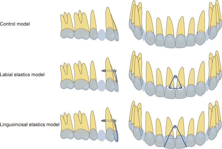
Three model groups were developed. The control group was obtained with the aforementioned components. In addition, as displayed in Figure 2, two experimental groups were developed by adding a simplified mini-screw (fixed support) in the interradicular region between the roots (middle third) of the central incisors. For the labial elastics group, an elastic band was worn from two buttons on the labial surfaces of the central incisors to the mini-screw. For the linguoincisal elastic group, an elastic band was worn from the precision cuts on the lingual side of the aligner and went beneath the incisal edge of the aligner between the central incisors and lateral incisors to the mini-screw. In these two groups, the elastic band was simulated by spring elements. Different elastic force values were designed: 0, 50, 100, 150, 200, 250, and 300 g.
Figure 2.
Three model groups.
Tooth and aligner displacement tendencies and Von Mises12 equivalent stress and compressive and tensile stress of teeth, PDL, and alveolar bone were analyzed. The incisal and apical center of the anterior teeth and the occlusal center and apical center of the mesial buccal root of the posterior teeth were taken as the measuring points.
RESULTS
Displacement Tendency of Teeth and Aligner (Unit: m)
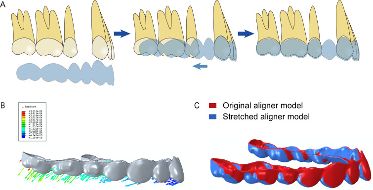
The aligner was displaced backward and downward, with this effect more significant in the posterior part of the aligner (Figure 3).
Figure 3.
Schematic of wearing (A), displacement tendency (B), and overlay (C) of aligner.
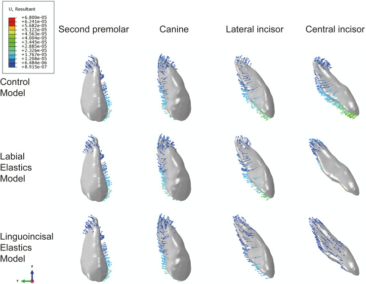
The displacement tendencies of the central incisor, lateral incisor, canine, and second premolar in the sagittal dimension are displayed in Figure 4. For the control group, the incisors were lingually tipped and extruded. The canine exhibited distal tipping and extrusion, whereas the second premolar was mesially tipped and intruded. For the labial elastics group and the linguoincisal elastics group, the displacement tendencies of canines and second premolars were similar to those of the control group, while those of the central and lateral incisors differed among the three groups. For central incisors, they were intruded in the labial elastics group whereas they were intruded and retracted in the linguoincisal elastics group. For lateral incisors, the displacement tendencies were similar between the control group and the labial elastics group, while the lateral incisor was intruded and retracted in the linguoincisal elastics group.
Figure 4.
Displacement tendencies of central incisor, lateral incisor, canine, and second premolar in sagittal dimension.
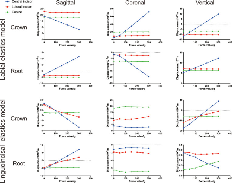
The displacement tendencies of the anterior teeth with different elastic forces are shown in Figure 5. For the labial elastics group, the displacement tendencies of the lateral incisors and canines in all three dimensions (sagittal, coronal, and vertical) were similar among different force magnitudes. For the central incisor, sagittally, the crown was displaced labially while the root moved palatally with increased elastic forces, producing an anticlockwise moment. Coronally, the crown was displaced mesially while the root moved distally, resulting in mesial tipping. Vertically, both the crown and root were intruded. For the linguoincisal elastics group, the displacement tendency of the canine in all three dimensions was similar with different force magnitudes, except that the canine root was intruded slightly with increased force magnitudes. Sagittally, the crowns of both the central and lateral incisors were displaced labially and their roots displaced palatally, producing an anticlockwise moment. Coronally, no significant displacements were observed for the incisors. Vertically, the crowns of the central and lateral incisors were intruded while their roots were extruded, consistent with the anticlockwise moment. Bodily retraction of central incisors was achieved with the elastic force being 250 g for both the labial and linguoincisal groups.
Figure 5.
Displacement tendencies of anterior teeth with different elastic forces.
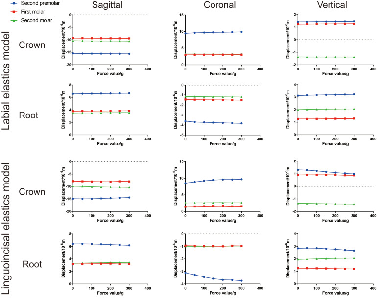
As displayed in Figure 6, the displacement tendencies of all three posterior teeth were similar in all three dimensions with different elastic forces for the two elastic groups, except that the second premolars were less intruded in the linguoincisal group with increased elastic force.
Figure 6.
Displacement tendencies of posterior teeth with different elastic forces.
The displacements of all upper teeth for the three groups are summarized in Figure 7.
Figure 7.
Schematic displacement tendencies of all upper teeth for the three groups.
Stress Distribution of Roots, PDL, and Alveolar (Unit: Pa)
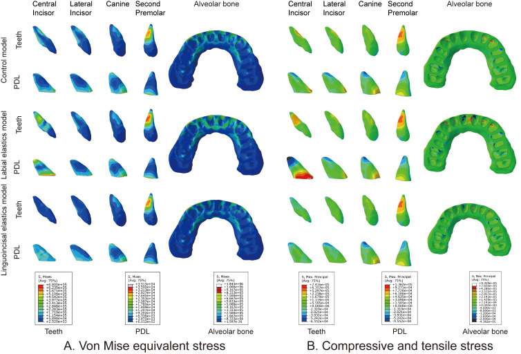
As displayed in Figure 8, stress was concentrated on the labial and lingual surfaces (apical and cervical third) of the incisor roots in the control group. For the labial elastics group, the stress was more concentrated on the mesial and distal surface of the central incisor roots. In contrast, the stress was well distributed and less concentrated on the incisor root surfaces in the linguoincisal elastics group.
Figure 8.
Equivalent stress (A) and compressive and tensile stress (B) of roots, periodontal ligament, and alveolar bone.
For the stress on the PDL, similar results were found for those of dental roots.
For the stress on alveolar bone, the stress was concentrated at the labial and lingual ridges of the alveolus in the control group. The stress was more concentrated at the labial, lingual, and apical ridges of the alveolus in the labial elastics group, while the stress was more evenly distributed in the linguoincisal elastics group.
DISCUSSION
CAT offers advantages such as esthetics and comfort, but the predictability of tooth movement is much less certain, especially for extraction cases.13 A recent study revealed that upper incisor retraction and posterior anchorage control were not fully achieved with CAT, resulting in lingual tipping and extrusion of central incisors and mesial tipping of posterior teeth.3 This was consistent with the present study, which shows that the incisors were lingually tipped and extruded and posterior teeth were mesially tipped and intruded in the control group. From a biomechanical perspective, the retraction force generated by clear aligners was applied on the crowns and passed through the occlusal side of the center of resistance, resulting in lingual tipping of the incisors and mesial tipping of the posterior teeth. This would deepen the bite and cause incisor occlusal interferences and buccal open bite, preventing the upper incisors from being retracted.
To avoid these side effects, several innovations are built into clear aligners for incisor intrusion and palatal root torquing. In conjunction, mini-screws and elastics offer additional force systems for CAT. In this study, the effectiveness of two modes of elastics for incisor intrusion and root torquing (ie, labial elastics and linguoincisal elastics) were examined. As summarized in Figure 7, for the labial elastics group, the elastic force was applied only on the central incisors. The central incisors were intruded, and their roots were torqued palatally. Because of Newton's third law, palatal torquing of the central incisors resulted in elastic changes of the aligners that should have subsequently generated palatal torque on the lateral incisors. Interestingly, it was found that the palatal root torquing of the lateral incisors was ineffective, which could be attributed to the inadequacy of force on the lateral incisors. In addition, in the coronal dimension, the elastic force had a mesial component, causing mesial tipping of the central incisors. For the linguoincisal elastics group, the elastic force was applied on the aligner so that intrusion and palatal root torquing of both the central and lateral incisors were well achieved. In addition, no mesial tipping of the central incisors was observed. Thus, linguoincisal elastics, not labial elastics, were able to achieve incisor intrusion and root palatal torquing effectively, producing an anticlockwise moment that promotes bodily movement for anterior retraction.
Posterior teeth were mesially tipped and intruded, resulting in a buccal open bite. In the current study, the displacement tendencies of posterior teeth in the labial elastics group did not change significantly in response to increased anterior elastic force. This could be attributed to the fact that only central incisors were intruded and root torqued. In contrast, in the linguoincisal elastics group, second premolars were less intruded in response to increased anterior elastic force, rendering buccal open bite less likely. Thus, this result suggested that linguoincisal elastics were superior to labial elastics in avoiding buccal open bite.
Ironically, the magnitudes of tooth displacement in this study were on the order of micrometers (eg, 0.02 mm), which was not close to 0.25 mm (the amount of anterior retraction). However, the results were in agreement with Jones et al.14 (tooth displacements ranged from 0.012 to 0.133 mm) and with a recent finite element study on clear aligners by Jiang et al.15 This inconsistency (0.02 vs 0.25 mm) may be attributed to the fact that initial displacement tendency, and not biological displacement, was determined through FEA. For biological displacement, it would take about 2–4 weeks for alveolar remodeling to achieve the actual tooth movement (0.25 mm).
In the sagittal dimension, the aligner was shorter than the dentition, and the aligner should be stretched to fit onto the dentition. In this way, the aligner exerted a retraction force on anterior teeth and a protraction force on posterior teeth. Patients are often instructed to push aligners onto anterior teeth first and then onto posterior teeth. Thus, to simulate the actual situation, the central incisors were set first and their corresponding parts of the aligner at the same location in the sagittal dimension. Upon stretching the aligner and subsequently fitting it onto the dentition, the whole aligner was displaced posteriorly, with the displacement more significant at the posterior part of the aligner (Figure 3).
Root resorption, an inflammation-mediated resorption, is commonly encountered in orthodontic practice. Maxillary incisors are susceptible to root resorption.16 Although the incidence of root resorption is lower with CAT than with fixed appliances, root resorption cannot be avoided.17 Because root resorption is caused by force stresses concentrated on the root surfaces,18 the stress on the roots and PDL were analyzed. For the control group, the stress was concentrated at the apical third of the labial and lingual surfaces of the incisor roots and apical regions of the PDL, rendering the incisor roots susceptible to root resorption. This was consistent with previous studies showing that premolar extraction followed by incisor retraction was a predisposing factor for incisor root resorption.19 Conceivably, since the central incisors were intruded in the labial elastics group, the stress value was higher at the apical third of central incisor roots and apical regions of the PDL, making central incisors more susceptible to root resorption. Interestingly, the stress was evenly distributed and less concentrated in the linguoincisal elastics group, reducing the likelihood for incisor root resorption.
In addition to root resorption, alveolar defects (fenestration and dehiscence) are closely monitored in orthodontic practice. Unlike the palatal alveolar bone, the labial alveolar bone is thinner and more susceptible to bone defects. As displayed in Figure 8, stress was concentrated on the labial sides of alveolar bone in the control group and even more concentrated in the labial elastics group. In addition, minimal stress was detected on the labial alveolar bone in the linguoincisal elastics group. These findings suggest that labial alveolar bone was susceptible to bone defects during anterior retraction, with a higher likelihood with labial elastics and lower likelihood with linguoincisal elastics.
Limitation
FEA represents one of the best ways to analyze force systems delivered by orthodontic appliances. However, in vitro study and clinical study results may differ, and the findings must be clinically validated to support the effect on clear aligner treatment with elastics and a mini-screw.
CONCLUSIONS
For anterior retraction, CAT produced lingual tipping and extrusion of incisors and mesial tipping and intrusion of posterior teeth, resulting in a buccal open bite tendency.
An anterior mini-screw with intrusive elastics was able to achieve incisor intrusion and palatal root torquing effectively, with linguoincisal elastics being more advantageous over labial elastics.
Buccal open bite may occur due to the intrusion of second premolars, which was less likely for linguoincisal elastics.
Incisor root resorption and alveolar defects may occur with CAT, and they were more likely to occur for labial elastics and less likely for linguoincisal elastics.
ACKNOWLEDGMENTS
This work was supported by National Natural Science Foundation of China (NSFC, No. 82071147), CSA Clinical Research Fund (CSA-02020-02), and Sichuan Science and Technology Program (Nos. 2018JY0558, 2021YJ0428 & 2018SZ0232).
REFERENCES
- 1.Pablo Gomez J, Marcelo Pena F, Martinez V, Giraldo DC, Ivan Cardona C. Initial force systems during bodily tooth movement with plastic aligners and composite attachments: a three-dimensional finite element analysis. Angle Orthod. 2015;85:454–460. doi: 10.2319/050714-330.1. [DOI] [PMC free article] [PubMed] [Google Scholar]
- 2.Hahn W, Zapf A, Dathe H, et al. Torquing an upper central incisor with aligners-acting forces and biomechanical principles. Eur J Orthod. 2010;32:607–613. doi: 10.1093/ejo/cjq007. [DOI] [PubMed] [Google Scholar]
- 3.Dai FF, Xu TM, Shu G. Comparison of achieved and predicted tooth movement of maxillary first molars and central incisors: first premolar extraction treatment with Invisalign. Angle Orthod. 2019;89:679–687. doi: 10.2319/090418-646.1. [DOI] [PMC free article] [PubMed] [Google Scholar]
- 4.Celenza F. Implant interactions with orthodontics. J Evid Based Dent Pract. 2012;12(3 suppl):192–201. doi: 10.1016/S1532-3382(12)70036-2. [DOI] [PubMed] [Google Scholar]
- 5.Lin JC, Tsai SJ, Liou EJ, Bowman SJ. Treatment of challenging malocclusions with Invisalign and miniscrew anchorage. J Clin Orthod. 2014;48:23–36. [PubMed] [Google Scholar]
- 6.Barone S, Paoli A, Razionale AV, Savignano R. Computational design and engineering of polymeric orthodontic aligners. Int J Numer Method Biomed Eng. 2017;33:e2839. doi: 10.1002/cnm.2839. [DOI] [PubMed] [Google Scholar]
- 7.Cortona A, Rossini G, Parrini S, Deregibus A, Castroflorio T. Clear aligner orthodontic therapy of rotated mandibular round-shaped teeth: a finite element study. Angle Orthod. 2020;90:247–254. doi: 10.2319/020719-86.1. [DOI] [PMC free article] [PubMed] [Google Scholar]
- 8.Ammar HH, Ngan P, Crout RJ, Mucino VH, Mukdadi OM. Three-dimensional modeling and finite element analysis in treatment planning for orthodontic tooth movement. Am J Orthod Dentofacial Orthop. 2011;139:e59–e71. doi: 10.1016/j.ajodo.2010.09.020. [DOI] [PubMed] [Google Scholar]
- 9.Schmidt F, Lapatki BG. Effect of variable periodontal ligament thickness and its non-linear material properties on the location of a tooth's centre of resistance. J Biomech. 2019;94:211–218. doi: 10.1016/j.jbiomech.2019.07.043. [DOI] [PubMed] [Google Scholar]
- 10.Kawamura J, Tamaya N. A finite element analysis of the effects of archwire size on orthodontic tooth movement in extraction space closure with miniscrew sliding mechanics. Prog Orthod. 2019;20:3. doi: 10.1186/s40510-018-0255-8. [DOI] [PMC free article] [PubMed] [Google Scholar]
- 11.Liang W, Rong Q, Lin J, Xu B. Torque control of the maxillary incisors in lingual and labial orthodontics: a 3-dimensional finite element analysis. Am J Orthod Dentofacial Orthop. 2009;135:316–322. doi: 10.1016/j.ajodo.2007.03.039. [DOI] [PubMed] [Google Scholar]
- 12.Jindal P, Worcester F, Siena FL, et al. Mechanical behaviour of 3D printed vs thermoformed clear dental aligner materials under non-linear compressive loading using FEM. J Mech Behav Biomed Mater. 2020;112:104045. doi: 10.1016/j.jmbbm.2020.104045. [DOI] [PubMed] [Google Scholar]
- 13.Rossini G, Parrini S, Castroflorio T, Deregibus A, Debernardi CL. Efficacy of clear aligners in controlling orthodontic tooth movement: a systematic review. Angle Orthod. 2015;85:881–889. doi: 10.2319/061614-436.1. [DOI] [PMC free article] [PubMed] [Google Scholar]
- 14.Jones ML, Hickman J, Middleton J, Knox J, Volp C. A validated finite element method study of orthodontic tooth movement in the human subject. J Orthod. 2001;28:29–38. doi: 10.1093/ortho/28.1.29. [DOI] [PubMed] [Google Scholar]
- 15.Jiang T, Wu RY, Wang JK, Wang HH, Tang GH. Clear aligners for maxillary anterior en masse retraction: a 3D finite element study. Sci Rep. 2020;10:10156. doi: 10.1038/s41598-020-67273-2. [DOI] [PMC free article] [PubMed] [Google Scholar]
- 16.Linkous ER, Trojan TM, Harris EF. External apical root resorption and vectors of orthodontic tooth movement. Am J Orthod Dentofacial Orthop. 2020;158:700–709. doi: 10.1016/j.ajodo.2019.10.017. [DOI] [PubMed] [Google Scholar]
- 17.Fang X, Qi R, Liu C. Root resorption in orthodontic treatment with clear aligners: a systematic review and meta-analysis. Orthod Craniofac Res. 2019;22:259–269. doi: 10.1111/ocr.12337. [DOI] [PubMed] [Google Scholar]
- 18.Dindaroglu F, Dogan S. Root resorption in orthodontics. Turk J Orthod. 2016;29:103–108. doi: 10.5152/TurkJOrthod.2016.16021. [DOI] [PMC free article] [PubMed] [Google Scholar]
- 19.Nanekrungsan K, Patanaporn V, Janhom A, Korwanich N. External apical root resorption in maxillary incisors in orthodontic patients: associated factors and radiographic evaluation. Imaging Sci Dent. 2012;42:147–154. doi: 10.5624/isd.2012.42.3.147. [DOI] [PMC free article] [PubMed] [Google Scholar]


