Abstract
Schmallenberg virus (SBV—Orthobunyavirus serogroup Simbu) is an emerging RNA vector-borne virus which has an important impact in animal health within Europe, and some Asian and African countries. It is mainly reported in ruminants, causing congenital malformations and stillbirths. However, there are no studies regarding the occurrence, diagnosis, or surveillance of SBV in Brazil, due to the lack of diagnostic techniques available so far. This study aimed to implement a reliable diagnostic technique able to detect the SBV in Brazil and also to investigate occurrence of the virus in this country. A molecular technique, quantitative reverse transcription polymerase chain reaction (RT-qPCR), was used to analyze 1665 bovine blood samples and 313 aborted fetuses, as well as 596 serum samples were analyzed by serological analysis. None of the blood and fetus samples analyzed was positive for SBV, and neither serum samples were reactive for antibodies anti-SBV. Thus, although Brazil presents suitable conditions for the dissemination of the SBV, results of the present study suggest that SBV did not propagate in the analyzed bovine population.
Supplementary Information
The online version contains supplementary material available at 10.1007/s42770-021-00637-6.
Keywords: Bunyavirales, Arbovirus, Congenital malformations in ruminant
Introduction
Schmallenberg virus (SBV) is an RNA virus member of the order Bunyavirales, family Peribunyaviridae, genus Orthobunyavirus, species Schmallenberg orthobunyavirus, and Simbu serogroup. It was first reported in North Rhine-Westphalia (Germany) in November 2011 and rapidly disseminated through all European continent [1]. Since then, the virus has emerged and re-emerged in several European countries, and has also been detected in other regions, including Asian and African countries [2–10].
The SBV is an enveloped virus and the genome includes three segments of negative-sense single-stranded RNA. Designated as small (S), medium (M), and large (L), they encode four structural proteins: nucleocapsid (N), two glycoproteins (Gn and Gc), and viral polymerase. They also encode two nonstructural proteins: NSs and NSm [11].
This virus infects predominantly ruminants, wild and domestics, causing a short period of viremia (2–6 days), seroconverting within approximately 1–3 weeks after infection [3, 12–15]. In adult animals, it is associated with a mild and transient disease characterized by fever, diarrhea, and decrease in milk production [16–18]. When a cow or a sheep gets infected during the period of gestation, SBV may cross the placental barrier and infect the fetus, leading to abortions, stillbirths, and severe congenital malformations (arthrogryposis-hydranencephaly syndrome) [3, 19]. SBV is mainly transmitted by Culicoides sp. biting midges [20, 21], but can also be transmitted vertically through the placenta of infected pregnant animals. Moreover, viral RNA was already detected in the semen of naturally infected bulls [22].
Knowing how the virus can disseminate between the population, Brazil has promising conditions for dissemination of SBV (climatic conditions, presence of the potential competent vector, and high density of susceptible animals). However, in Brazil, there is still a lack of diagnostic resources available so far. There are no studies regarding the occurrence, diagnosis, research, and neither surveillance of SBV in this territory. The present study aimed to implement a satisfactory laboratory diagnostic method able to detect SBV in Brazil and to investigate whether there is a circulation of the virus in this country.
Materials and methods
Sample selection
Whole blood
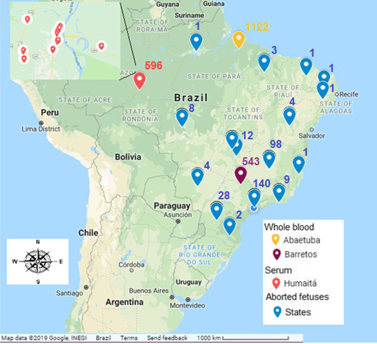
A total 1665 blood samples from bovine, aged 0–12 months, were analyzed. Within these samples, 543 were collected in November 2016, from the county of Barretos, state of São Paulo; 162 were collected between April and May 2017 and 960 collected in June 2017, from the county of Abaetetuba, state of Pará. The samples were previously sent to the Biological Institute for detection of Bluetongue virus (BTV) and stored at − 20 °C.
Aborted fetuses
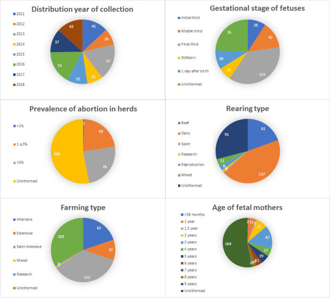
A total of 313 samples of aborted fetuses were retrospectively analyzed, including 283 bovine, 5 caprine, 23 ovine, and 2 buffalo from 255 farms in different Brazilians states. All of those samples were collected during SBV emergencies and re-emergencies in Europe and Asia (from January 2011 to December 2018, Fig. 1). With the fetus’s samples, a pool of body organs was made and analyzed by RT-qPCR. All the pools were composed of the central nervous system, heart, liver, spleen, and kidney. Blood and serum samples of the fetuses were not available for antibody anti-SBV detection and research RNA SBV.
Fig. 1.
Data on samples, properties, and herds,
taken from the referral forms for samples of aborted fetuses
According to the official sample’s form data, the fetuses received in the lab were in different gestational stages, and the age of the pregnant cows ranged from < 36 months to 9 years. Most of the studied properties that had pregnancy loss had an abortion prevalence above 3% (52%—76/147) and reports of increased abortions over the past 3 months (Fig. 1). It is important to notice that most of these animals were vaccinated against the most common causes of abortion in bovine, including brucellosis, rabies, leptospirosis, foot-and-mouth disease, carbuncle, bovine viral diarrhea (BVD), botulism, clostridiums, and infectious bovine rhinotracheitis (IBR). These samples were previously sent to the Biological Institute for differential diagnosis of abortion and stored at − 20 °C.
Serum
The serum bank, provided by the Agency for Agriculture and Livestock and Forestry Defense of the State of Amazonas (ADAF), was used to evaluate the presence of antibodies against SBV.
A total of 596 bovine serum samples from Humaitá, state of Amazonas/Brazil were analyzed. Nellore and Girolando dairy cows are between 13 and 36 months, from 26 different farms which cattle, goats, sheep, pigs, horses, and mules were grown together.
Samples were collected from October 2017 to March 2018 according to a previously study designed for other viral diseases in different parts of the state of Amazonas, sent to the Biological Institute, and stored at − 20 °C. The studied properties had mixed cattle production systems, dairy, or beef and were defined as family or commercial agriculture (online Supplementary Information) and were selected by drawing lots from the ADAF register.
Molecular diagnosis of SBV in Brazil
Standard curve
For biosafety reasons, a standard positive control in the molecular diagnosis of SBV, a gBlocks® Gene Fragments (Integrated DNA Technologies-IDT), was built. Highly conserved, the S segment of the SBV genome (strain BH80/11–4, GenBank accession number HE649914.1) was used as a reference. The gBlocks® sequence was composed by the entire S segment that include the primer and probe hybridization region, coupled with EcoRI and BamHI restriction site at 5′ and 3′ end, respectively (Table 1). After the restriction enzyme sequences, 5 thymines were added at each end to avoid nonspecificities.
Table 1.
Primers and probes used in this study [19]
| Name | Sequence | Amplification (nt) |
|---|---|---|
| SBV-S-382F | 5′ TCA GAT TGT CAT GCC CCT TGC 3′ | 88 |
| SBV-S-469R | 5′ TTC GGC CCC AGG TGC AAA TC 3′ | |
| SBV-S-408FAM | 5′ FAM-TTA AGG GAT GCA CCT GGG CCG ATG GT (ZEN-Iowa BQ) 3′ | |
| ACT-1005F | 5′ CAG CAC AAT GAA GAT CAA GAT CAT C 3′ | 130 |
| ACT-1135R | 5′ CGG ACT CAT CGT ACT CCT GCT T 3′ | |
| ACT-1081HEX | 5′ HEX-TCG CTG TCC ACC TTC CAG CAG ATG T (ZEN-Iowa BQ) 3′ |
The gBlocks® was cloned into plasmid pTZ57R/T control included in InsTAclone PCR Cloning Kit (Thermo Fisher Scientific) after double digestion with EcoRI and BamHI enzymes, according to the manufacturer’s instructions. The plasmid was propagated in Escherichia coli (JM109), purified with kit GeneJET Plasmid Miniprep (Thermo Fisher Scientific) also according to the manufacturer’s instructions, and linearized by EcoRI digestion. The presence and identity of the insert were confirmed by genetic sequencing. The plasmid was subsequently used as the template for the synthesis of the SBV S segment RNA standard using the MEGAscript® Transcription Kit (Thermo Fisher Scientific) according to the manufacturer’s instructions. The RNA was quantified by NanoDrop (Thermo Fisher Scientific) and QuantiFluor® RNA System (Promega) according to the manufacturer’s instructions. The number of RNA standard copies was calculated, and standard curve was constructed using a log10 dilution series ranging from 106 to 100 RNA copies/μL, to determine the analytical sensitivity, limit of detection (LOD), and RT-qPCR assay performance (efficiency [E = 10−1/slope – 1] and r2 value) [23].
The intra-assay variation was assessed with each viral dilution of the standard curve tested in triplicate. The inter-run variation was determined by three runs independent of the standard curve, in triplicate.
Specificity of primes and probes was tested against samples which showed positive results for other agents that cause abortion and stillbirth (BVDV, BTV, and Neopora caninum).
RNA extraction and real-time RT-PCR assays
Genetic material was extracted with the QIAcube HT (Qiagen) using the cador® Pathogen 96 QIAcube® HT Kit (Qiagen) according to the manufacturer’s recommendations.
The genetic material from the whole blood samples and aborted fetuses were extracted and amplified. The total reaction volume was 20 μL, containing 10 µL of GoTaq® Probe qPCR Master Mix (2x) (Promega), 0.4 µL of GoScript™ RT Mix for 1-Step RT-qPCR (50x) (Promega), 700 nM of SBV-S-382F primer, 800 nM of SBV-S-469R primer, 90 nM of SBV-S-408 probe, 200 nM of each actin-β primers and 100 nM of beta-actin probe, 2 µL of extracted RNA, and nuclease free water to complete the volume. The following thermal program was applied: 1 cycle of 45 °C for 15 min and 95 °C for 2 min, followed by 40 cycles of 95 °C for 15 s, 60 °C for 1 min, and 40 °C for 10 s.
All negative samples for SBV, in addition to the use of endogenous control (actin-β), were contaminated with the standard control in such a way that the sample had a final concentration of 103 copies of RNA/µL and submitted again to RT-qPCR, for evaluation of PCR inhibitors. Then, the Cq of the standard curve of 103 copies of RNA/µL was compared with the Cq of the contaminated samples and the variation of the Cq obtained, as well as the non-amplification, was evaluated.
Serological analysis
All serum samples were analyzed to detect antibody anti-SBV IgG, using ELISA Kit ID Screen® Schmallenberg virus Indirect Multi-species (IDvet), according to the manufacturer’s instructions.
From the total serum samples analyzed, one sample from each property was randomly selected, totaling 26 samples, for detection of antibodies against BTV. The competitive ELISA was performed using the commercial Bluetongue Virus Antibody Test Kit, cELISA v2 (VMRD) according to the manufacturer’s manual.
Results and discussion
Based on current literature, there are no studies related to the occurrence of SBV in Brazil, as well as there is no reliable laboratory diagnosis for the virus in the country. Thus, so far no study exists regarding the diagnosis and surveillance of SBV in Brazil, and at our knowledge, this is the first study that was performed in Brazilian animals on the behalf.
The design and cloning of the synthetic positive control of the S segment SBV genome, non-infective and without a biological risk, were successfully performed. RT-qPCR was used for direct detection of SBV.
The standard curve with 106 to 100 copies of RNA/µL showed Cq values ranging from 15 to 36. The efficiency of SBV RT-qPCR was 1.98, with slope of − 3.365 and error of 0.01.
The analytical sensitivity of the reaction was 1 copy of RNA per microliter (LOD) and mean of Cq 36.03 ± 0.27 (standard deviation 0.41) that is, the last dilution in triplicate in which a positive result was obtained.
Thus, the values obtained in this study were within the expected standard (slope of approximately − 3.3, efficiency of approximately 2.0, and error > 0.02).
For the inter-assay and intra-assay precision, the coefficient of variation of the Cq obtained for each standard dilution was evaluated, and tested in three independent reactions and in triplicate. As there was no noteworthy variation of the Cq, it is concluded that these are precise reactions, as well as the reactions showed repeatability and reproducibility, as there was no variation in the standard curve in the three independent runs, in triplicate.
All samples were positive for β-actin gene. Those manually contaminated with the standard control 103 copies of RNA/µL were positive for SBV and without significant Cq variation (Cq 24.8 ± 0.7), indicating that there are no PCR inhibitors, as well as the quality of sample extraction and RNA integrity.
Specificity of primes and probes was tested against samples which showed positive results for other agents that cause abortion and stillbirth (BVDV, BTV, and Neopora caninum) and no sample was positive.
SBV was first reported in the summer and fall of 2011 in North Rhine-Westphalia (Germany) and the Netherlands. After the first reports in 2011 and 2012, there was a period of absence of clinical disease and a decline in seropositivity in the population of ruminants residing in the affected countries. But despite circulating at a low level and with little occurrence in Europe in 2013, the SBV continued its expansion to other countries. There was evidence of increased circulation in the UK in 2016. In 2017 and 2018 in Germany, the level of circulation of the SBV remained low, but in 2019 again cases were reported to a greater extent, indicating that the SBV circulated again in German territory, genetically stable over 9 years [24].
This alternation between low-level circulation and resurgence to a greater extent in endemic regions occurs from time to time due to a combination of the number of susceptible animals reaching a critical level (the herd replacement rate strongly influences the number of susceptible animals), favorable conditions for the vectors, and presence of wild and/or domestic reservoir host. Therefore, it should be anticipated that the SBV has established an enzootic status in Europe and will reappear on a larger scale and at regular intervals in the future with emergencies and re-emergencies (every 3–5 years in European countries) [25, 26].
SBV continues to spread and is willing to cross borders. If susceptible vectors and susceptible hosts are available, it will continue to spread to new territories and could become endemic [24, 25].
Assuming that Brazilian animals are immunologically naïve to the SBV and Brazil has a several species of Culicoides sp. in the territory, a potential vector, if SBV were introduced in Brazil, it would rapidly disseminate through the country and would be difficult to control the disease, reinforcing the importance of the present study.
The Oropouche virus, which also belongs to the Orthobunyavirus of the Simbu serogroup as well as SBV, causes outbreaks of acute febrile illnesses similar to dengue, including some cases of meningitis in Brazil. The first urban outbreak of OROV fever was reported in Belém (state of Pará/Brazil) in 1961. It causes large and explosive outbreaks in cities and towns in the Amazon and Central Plateau regions (where the serum and blood samples for this study come from). OROV became the second most frequent arbovirus in the number of reported cases in Brazil, being surpassed only by dengue [27–29].
Bluetongue virus (BTV) and SBV are different viruses that present similar epidemiological characteristics and clinical manifestations. Both arboviruses are transmitted by midges Culicoides sp. and can cause congenital malformations, abortion, and stillbirths. After the unexpected appearance of bluetongue serotype 8 (BTV-8) in northern Europe in 2006 and then different new serotypes, BTV-6, and BTV-11 in the same region, the SBV virus emerged in 2011 causing a new economically important disease in ruminants [30].
The blood samples analyzed in this study were collected for certification of animals free from BTV, and some animal samples were positive for BTV in RT-qPCR (about 2%). The presence of RNA of SBV was investigated in these samples, due to the epidemiological similarity between BTV and SBV in Europe (same vector and occurrence regions). BTV positivity in blood samples indicates recent circulation and activity of the vector in the region (age of animals 0–12 months), in addition to the regions of origin of the samples (Abaetetuba/PA and Barretos/SP), being favorable for spread of the SBV (climatic conditions, presence of the potential competent vector, and high density of susceptible animals). However, there were no SBV positive blood samples (0/1665).
The pool of organs from aborted and stillborn were not positive for SBV by RT-qPCR. Most of the aborted fetuses were obtained from southeastern Brazil (states of São Paulo and Minas Gerais) for logistical and non-epidemiological reasons. The samples belonged to the convenience bank of the Biological Institute of São Paulo. This does not exclude the importance and reliability of investigating the occurrence of SBV in samples from this bank, because samples from all Brazilian regions were analyzed (Fig. 2).
Fig. 2.
Spatial distribution of whole blood samples, aborted fetuses, and serum analyzed. The inclusion shows details of the area where serum samples were collected. Map created by Google Maps
The fetal samples analyzed were collected from January 2011—when the first SBV occurrence was reported in Europe—until December 2018, because of the possible risk of SBV introduction in Brazil at any time. These samples represented various gestational stages, but the vast majority were collected in the 3rd trimester of pregnancy (37%—115/313), as well as stillbirths or 1 day after birth, and were collected from cows of various ages (under 36 months to 9 years). Previous studies showed no correlation between maternal age and the gestational period of the fetus to the occurrence of abortion due to SBV. However, it is known that teratogenic defects and their severity are associated with the gestational stage when the infection occurred [5, 31].
The serum samples analyzed in this study corresponded to 20% of the bovine herd in the county of Humaitá (second largest herd in the Brazil), registered with the ADAF in 2017. Antibodies against SBV were not detected by ELISA (0/596).
One sample from each property was randomly selected for detection of antibodies anti-BTV. All of them were reactive and with high antibody titers by ELISA (26/26), indicating vector circulation in the region. This is an important result, since BTV and SBV are transmitted by the same vector and present similar epidemiological characteristics in Europe.
The clinical manifestations of SBV infection are nonspecific and can be confused with BVD, IBR, BTV, and Cache Valley (in sheep), among others.
Serology for IBR and BVD was performed on the sera analyzed and shows positive samples in 100% of the properties analyzed for IBR and 42% for BVD (the animals were not vaccinated). Molecular diagnosis has also been carried out previously in some of the aborted and stillborn, for differential abortion diagnosis. The main infectious agent associated with the abortion was Neospora caninum (11%—27/256), and followed by BVD (2%—6/290). No positive fetuses were found for IBR (0/286) or for BTV (0/309). None of the samples was positive for Leptospira sp. (0/146). All of these results were kindly provided by the Biological Institute.
In early 2012, regulations regarding the importation of live animals and germplasm from the European Union, and other countries that have notified the disease, were made to mitigate the introduction of SBV in Brazil. According to the results found in this study, these measures were efficient, since no molecular or serological evidence of the presence of SBV was found in the samples analyzed.
This approach resulted in important knowledge about SBV for the scientific community, veterinary authorities, and decision makers. It also should be considered to continue the surveillance studies with SBV in Brazil in the future and in the countries that had already reported the virus circulation.
Conclusion
A standard positive control, innocuous and safe, was developed for molecular diagnosis of SBV in Brazil. SBV was not detected in the analyzed samples of whole blood, aborted fetuses, and stillbirths of Brazilian ruminants. Therefore, viremia and transplacental transmission of SBV was not evidenced in the analyzed samples. No anti-SBV antibodies were detected in the bovine serum samples analyzed; thus, SBV infection and subsequent seroconversion of the analyzed animals were not evidenced. Although the samples belonged to a convenience bank, the data obtained suggest that the SBV did not circulate in the bovine population studied in the Brazilian territory.
Supplementary Information
Below is the link to the electronic supplementary material.
Supplementary file 1 Classification, species raised, number of animals, type, size, age and number of animals collected from each property. ha: hectares. (DOCX 19 kb)
Author contribution
M. S. N. M. conceived, performed the experiment, analyzed the results, and wrote the manuscript, E. M. P. and L. J. R. conducted the experiment, and S. A. T. and L. H. O. contributed with the practical part. All authors have reviewed the manuscript.
Funding
This study received support from the CAPES, Coordination for the Improvement of Higher Education Personnel (process number: 88882.327996/2019–01).
Availability of data and material
The data that support the conclusions of this study are available from the corresponding author upon reasonable request.
Code availability
Not applicable.
Declarations
Ethics approval
This research is in accordance with Law 11.794 of October 8, 2008, Decree 6899 of July 15, 2009, as well as with the rules issued by the National Council for Control of Animal Experimentation (CONCEA), and was approved by the Ethic Committee on Animal Use of the School of Veterinary Medicine and Animal Science (University of São Paulo) (CEUA/FMVZ) protocol number CEUA 2892260716.
Consent to participate
Not applicable.
Consent for publication
Not applicable.
Competing interests
The authors declare no competing interests.
Footnotes
Publisher's note
Springer Nature remains neutral with regard to jurisdictional claims in published maps and institutional affiliations.
References
- 1.Hoffmann B, Scheuch M, Höper D, et al. Novel orthobunyavirus in cattle, Europe, 2011. Emerg Infect Dis. 2012;18:469–472. doi: 10.3201/eid1803.111905. [DOI] [PMC free article] [PubMed] [Google Scholar]
- 2.Collins B, Barrett DJ, Doherty ML, et al. Significant re-emergence and recirculation of Schmallenberg virus in previously exposed dairy herds in Ireland in 2016. Transbound Emerg Dis. 2017;64:1359–1363. doi: 10.1111/tbed.12685. [DOI] [PubMed] [Google Scholar]
- 3.Doceul V, Lara E, Sailleau C, et al. Epidemiology, molecular virology and diagnostics of Schmallenberg virus, an emerging orthobunyavirus in Europe. Vet Res. 2013;44:1–13. doi: 10.1186/1297-9716-44-31. [DOI] [PMC free article] [PubMed] [Google Scholar]
- 4.Dominguez M, Gache K, Touratier A, et al. Spread and impact of the Schmallenberg virus epidemic in France in 2012–2013. BMC Vet Res. 2014;10:1–10. doi: 10.1186/s12917-014-0248-x. [DOI] [PMC free article] [PubMed] [Google Scholar]
- 5.Esteves F, Mesquita JR, Esteves F, et al (2016) Epidemiology and emergence of Schmallenberg virus part 2: pathogenesis and risk of viral spread. In: Epidemiology of communicable and non-communicable diseases - attributes of lifestyle and nature on humankind. pp 58–71
- 6.Mcgowan SL, La RSA, Grierson SS, et al. Incursion of Schmallenberg virus into Great Britain in 2011 and emergence of variant sequences in 2016. Vet J. 2018;234:77–84. doi: 10.1016/j.tvjl.2018.02.001. [DOI] [PubMed] [Google Scholar]
- 7.Tarlinton R, Daly J, Dunham S, Kydd J. The challenge of Schmallenberg virus emergence in Europe. Vet J. 2012;194:10–18. doi: 10.1016/j.tvjl.2012.08.017. [DOI] [PubMed] [Google Scholar]
- 8.Zhai SL, Lv DH, Wen XH et al (2017) Preliminary serological evidence for Schmallenberg virus infection in China. Trop Anim Health Prod 1–5.10.1007/s11250-017-1433-2 [DOI] [PubMed]
- 9.Sibhat B, Ayelet G, Gebremedhin EZ, et al. Seroprevalence of Schmallenberg virus in dairy cattle in Ethiopia. Acta Trop. 2018;178:61–67. doi: 10.1016/j.actatropica.2017.10.024. [DOI] [PubMed] [Google Scholar]
- 10.Molini U, Dondona AC, Hilbert R, Monaco F. Antibodies against Schmallenberg virus detected in cattle in the Otjozondjupa region, Namibia. J S Afr Vet Assoc. 2018;89:1–2. doi: 10.4102/jsava.v89i0.1666. [DOI] [PMC free article] [PubMed] [Google Scholar]
- 11.Varela M, Schnettler E, Caporale M, et al. Schmallenberg virus pathogenesis, tropism and interaction with the innate immune system of the host. PLoS Pathog. 2013;9:1–13. doi: 10.1371/journal.ppat.1003133. [DOI] [PMC free article] [PubMed] [Google Scholar]
- 12.García-Bocanegra I, Cano-Terriza D, Vidal G, et al. Monitoring of Schmallenberg virus in Spanish wild artiodactyls, 2006–2015. PLoS ONE. 2017;12:1–12. doi: 10.1371/journal.pone.0182212. [DOI] [PMC free article] [PubMed] [Google Scholar]
- 13.Malmsten A, Malmsten J, Blomqvist G, et al. Serological testing of Schmallenberg virus in Swedish wild cervids from 2012 to 2016. BMC Vet Res. 2017;13:1–5. doi: 10.1186/s12917-017-1005-8. [DOI] [PMC free article] [PubMed] [Google Scholar]
- 14.Poskin A, Martinelle L, Van Der SY, et al. Genetically stable infectious Schmallenberg virus persists in foetal envelopes of pregnant ewes. J Gen Virol. 2017;98:1630–1635. doi: 10.1099/jgv.0.000841. [DOI] [PubMed] [Google Scholar]
- 15.Schulz C, van der Poel WHM, Ponsart C, et al. European interlaboratory comparison of Schmallenberg virus (SBV) real-time RT-PCR detection in experimental and field samples: the method of extraction is critical for SBV RNA detection in semen. J Vet Diagnostic Investig. 2015;27:422–430. doi: 10.1177/1040638715593798. [DOI] [PubMed] [Google Scholar]
- 16.Garigliany MM, Hoffmann B, Dive M, et al. Schmallenberg virus in calf born at term with porencephaly, Belgium. Emerg Infect Dis. 2012;18:1005–1006. doi: 10.3201/eid1806.120104. [DOI] [PMC free article] [PubMed] [Google Scholar]
- 17.Lievaart-peterson K, Luttikholt S, Peperkamp K, et al. Schmallenberg disease in sheep or goat: past, present and future. Vet Microbiol. 2015;181:147–153. doi: 10.1016/j.vetmic.2015.08.005. [DOI] [PubMed] [Google Scholar]
- 18.Pawaiya RVS, Gupta VK. A review on Schmallenberg virus infection: a newly emerging disease of cattle, sheep and goats. Vet Med (Praha) 2013;58:516–526. doi: 10.17221/7083-VETMED. [DOI] [Google Scholar]
- 19.Bilk S, Schulze C, Fischer M, et al. Organ distribution of Schmallenberg virus RNA in malformed newborns. Vet Microbiol. 2012;159:236–238. doi: 10.1016/j.vetmic.2012.03.035. [DOI] [PubMed] [Google Scholar]
- 20.Rasmussen LD, Kristensen B, Kirkeby C, et al. Culicoids as vectors of Schmallenberg virus. Emerg Infect Dis. 2012;18:1204–1206. doi: 10.3201/eid1807.120385. [DOI] [PMC free article] [PubMed] [Google Scholar]
- 21.De RN, Deblauwe I, De DR, et al. Detection of Schmallenberg virus in different Culicoides spp. by real-time RT-PCR. Transbound Emerg Dis. 2012;59:471–475. doi: 10.1111/tbed.12000. [DOI] [PubMed] [Google Scholar]
- 22.Hoffmann B, Schulz C, Beer M. First detection of Schmallenberg virus RNA in bovine semen, Germany, 2012. Vet Microbiol. 2013;167:289–295. doi: 10.1016/j.vetmic.2013.09.002. [DOI] [PubMed] [Google Scholar]
- 23.Bustin SA, Benes V, Garson JA, et al. The MIQE guidelines: minimum information for publication of quantitative real-time PCR experiments SUMMARY. 2009;622:611–622. doi: 10.1373/clinchem.2008.112797. [DOI] [PubMed] [Google Scholar]
- 24.Wernike K, Beer M (2020) Re-circulation of Schmallenberg virus, Germany, 2019. Transbound Emerg Dis 1–6. 10.1111/tbed.13592 [DOI] [PubMed]
- 25.Stavrou A, Daly JM, Maddison B, et al. How is Europe positioned for a re-emergence of Schmallenberg virus? Vet J. 2017;230:45–51. doi: 10.1016/j.tvjl.2017.04.009. [DOI] [PubMed] [Google Scholar]
- 26.Wernike K, Beer M. International proficiency trial demonstrates reliable Schmallenberg virus infection diagnosis in endemic and non-affected countries. PLoS ONE. 2019;14:1–11. doi: 10.1371/journal.pone.0219054. [DOI] [PMC free article] [PubMed] [Google Scholar]
- 27.Bastos MDS, Tadeu L, Figueiredo M, et al. Short report: identification of Oropouche Orthobunyavirus in the cerebrospinal fluid of three patients in the Amazonas. Brazil. 2012;86:732–735. doi: 10.4269/ajtmh.2012.11-0485. [DOI] [PMC free article] [PubMed] [Google Scholar]
- 28.Tadeu L, Figueiredo M. Emergent arboviruses in Brazil Arboviroses emergentes no Brasil. 2007;40:224–229. doi: 10.1590/s0037-86822007000200016. [DOI] [PubMed] [Google Scholar]
- 29.Cardoso BF, Serra OP, Borges L, et al. Detection of Oropouche virus segment S in patients and in Culex quinquefasciatus in the state of Mato Grosso. Brazil. 2015;110:745–754. doi: 10.1590/0074-02760150123. [DOI] [PMC free article] [PubMed] [Google Scholar]
- 30.Sedda L, Rogers DJ. The influence of the wind in the Schmallenberg virus outbreak in Europe. Sci Rep. 2013;3:1–8. doi: 10.1038/srep03361. [DOI] [PMC free article] [PubMed] [Google Scholar]
- 31.Martinelle L, Poskin A, Pozzo FD, et al. Three different routes of inoculation for experimental infection with Schmallenberg virus in sheep. Transbound Emerg Dis. 2017;64:305–308. doi: 10.1111/tbed.12356. [DOI] [PubMed] [Google Scholar]
Associated Data
This section collects any data citations, data availability statements, or supplementary materials included in this article.
Supplementary Materials
Supplementary file 1 Classification, species raised, number of animals, type, size, age and number of animals collected from each property. ha: hectares. (DOCX 19 kb)
Data Availability Statement
The data that support the conclusions of this study are available from the corresponding author upon reasonable request.
Not applicable.


