PLOS ONE logoLink to PLOS ONE
. 2021 Oct 28;16(10):e0258688. doi: 10.1371/journal.pone.0258688

Characteristics of astigmatism before and 1 month after blepharoptosis surgery in patients with acquired ptosis

Kazuhiko Dannoue 1,*, Seiji Takagi 1,2, Keiko Uemura 1, Anna Takei 1, Tomohiko Usui 1,3
Editor: Ahmed Awadein4
PMCID: PMC8553058  PMID: 34710107

Abstract

In this study, we aimed to evaluate the characteristics of astigmatism preoperatively and 1 month postoperatively in patients with age-related ptosis (AP) and contact lens-related ptosis (CLP), and investigate surgery-induced astigmatism (SIA) using the Jaffe vector analysis and the Cravy method. Consecutive patients who underwent blepharoptosis surgery between January 2019 and December 2019 were included. The patients were divided into AP and CLP groups. Computerized corneal topography was used to assess the magnitude and axis of corneal astigmatism. Astigmatism was classified as with-the-rule (WTR), against-the-rule (ATR), or oblique astigmatism (OA) pre- and postoperatively. SIA was calculated by vector analysis using the Cravy and Jaffe methods. The correlation between SIA and margin reflex distance (MRD) was calculated. One hundred and eight eyes from 58 patients (AP group: 85 eyes from 45 patients, CLP group: 23 eyes from 13 patients) were included. The AP group (73.8±7.6 years) was significantly older than the CLP group (47.7±6.6 years). The MRD increased significantly after treatment in both groups. The proportions of WTR, ATR, and OA were 52%, 22%, and 25%, and 86%, 9%, and 4% in the AP and CLP groups, respectively. A shift in astigmatism type was observed in 41% and 13% of patients in the AP and CLP groups, respectively. The average SIA measured using the Cravy method was 0.11±1.22 D in the AP group and −0.28±1.07 D in the CLP group (WTR astigmatism). The SIA calculated using the Jaffe method was 0.78±0.70 D in the AP group and 0.82±0.88 D in the CLP group. There was no significant correlation between SIA calculated using the Cravy and Jaffe methods and MRD. ATR was most common in age-related ptosis and WTR was most common in contact lens-related ptosis. Upper eyelid re-positioning may affect visual functions due to astigmatic changes in the short term postoperatively.

Introduction

Acquired ptosis is a common ophthalmological condition and is associated with aging [1], prolonged use of hard contact lens [2], and anterior segment surgery [3]. Acquired ptosis does not just cause esthetic problems but also impairment of visual functions. Altitudinal visual field defects that impair the upward gaze and are measured using manual kinetic perimetry by the Goldmann perimeter [4] or static automated perimetry [5] have been well documented. Limiting of light influx due to palpebral narrowing could cause impairment of contrast sensitivity [6]. Moreover, a retracted eyelid affects the shape of the cornea as the upper eyelids are directly adhered to the cornea [7]. Previous studies have reported on the influence of eyelids on the corneal surface in various eyelid conditions, including chalazion [8], hemangiomas [9], downward gaze [10], and ptosis [11], using localized topographical analysis.

The corneal surface plays an important role in refraction. Hence, increased pressure on the upper eyelid could result in corneal refractive changes, which could cause amblyopia [12] in congenital ptosis and persistent blurred vision in acquired ptosis [13]. Blepharoplasty is performed to correct the upper eyelid position. Various studies have reported astigmatic recovery after upper eyelid surgery [1319]; however, patients who underwent blepharoptosis surgery occasionally complained of blurred vision in the operated eye [20]. The other astigmatic change observed after blepharoptosis surgery is change in the axis of the cylinder. Classically, the mechanical effect of ptosis may result in a steep corneal curvature in the vertical meridian, i.e., with-the-rule (WTR) corneal astigmatism [13,16]. However, a limited number of studies have reported on the astigmatic axis pre- and postoperatively for acquired ptosis [16]. Surgery-induced astigmatism should be assessed in both direction and magnitude, calculation methods for which have been discussed and reported, especially following cataract surgery [2128]. In this study, we aimed to evaluate the characteristics of astigmatism in patients with two representative acquired ptosis (age-related and contact lens-related ptosis), preoperatively and 1 month postoperatively, and investigate blepharoptosis surgery-induced astigmatism (SIA) using the Jaffe vector analysis [21] and the Cravy method [22]. Moreover, the correlation among SIA assessed using the Jaffe method, that using the Cravy method, and the patient background was analyzed.

Patients and methods

Ethics

The Dannoeu Eye Clinic Ethics Committee reviewed and approved the protocol of this retrospective observational study and waived the requirement for informed consent with an opt-out display in the hospital. This study conformed to the tenets of the Declaration of Helsinki.

Patients

Consecutive patients with acquired ptosis (AP) (age, >60 years; no history of any intraocular surgery and prolonged contact lens wear) and contact lens-related ptosis (CLP) (age, <59 years; long history of hard contact lens wear) who underwent blepharoptosis surgery between January 2019 and December 2019 were included. On the last visit before surgery, all patients underwent complete ocular examinations, including best-corrected visual acuity (BCVA), slit-lamp biomicroscopy, dilated fundoscopy, and optical coherence tomography. BCVA was obtained using Landolt C charts. These values were then converted to the logarithm of the minimum angle of resolution equivalent for statistical comparisons. Other potentially confounding retinal pathologies were excluded, and patients with a levator function of <4 mm were excluded. Ptosis severity was evaluated using the margin reflex distance (MRD), a measurement of the distance from the middle upper eyelid to the corneal light reflex [29]. Levator function was evaluated as eyelid excursion; upper lid was measured from the maximum downward gaze to the maximum upward gaze, with the frontalis muscle blocked by the examiner.

Blepharoptosis surgery

Blepharoptosis surgery was conducted by one surgeon (KD) mainly using a CO2 laser using the Müller’s muscle tracking method. All surgeries were performed under local anesthesia using 2% xylocaine. In blepharoplasty, the excess skin is excised after tracing the skin that needs to be removed. After the tarsal plate was exposed, the levator brevis aponeurosis and the Müller’s muscle were identified and detached carefully. Two or three places were tacked and crimped using 6–0 nylon, and the skin was sutured with a 7–0 nylon thread.

Astigmatism

Corneal topography was measured using a computed topography system with a Scheimpflug camera (TMS5 Tomey, Japan) to measure the corneal elevation and the surface curvature. The patients were asked to blink twice to obtain reproducible measurements. To avoid any pressure from the eyelids on the corneal surface, the examination was performed with the patients’ eyes open. Measurements were taken pre- and 1 month postoperatively when wound healing was observed. The refractive power was calculated from the radius of curvature, where the center of curvature was defined on the optical axis. The color map was constructed from one of the four scans with the least data variation.

Astigmatism was classified as follows: WTR astigmatism: steepest meridian close to the vertical meridian (30° on either side of the 90° meridian); “against the rule” (ATR) astigmatism: steepest meridian close to the horizontal meridian (30° on either side of the 180° meridian); and “oblique astigmatism” (OA): steepest meridians not close to either side of the vertical or horizontal meridians, within the aforementioned range but perpendicular to each other.

The degree of blepharoptosis SIA was calculated using Jaffe’s vector analysis [21] and Cravy’s vector analysis [22]. The following equations were used for each vector analysis:

Jaffe method

SIA(D)=|C12+C222C1C2cos2(A1A2)|
SIA(Ax)=0.5tan1C2sin2A2C1sin2A1C2cos2A2C1cos2A1

C1: preoperative cylinder, C2: postoperative cylinder, A1: axis of the cylinder of C1, A2: axis of the cylinder of C2.

Cravy method:

CravyΔK=(Δx)1(Δy)
Δx=|K2cosA2K1cosA1|
Δy=|K2sinA2K1sinA1|

K1: preoperative cylinder, K2: postoperative cylinder.

A1: strong meridian of K1, A2: strong meridian of K2.

Statistical analyses

All statistical analyses were performed using SPSS®, version 21 (SPSS Science, Chicago, IL, USA). The results of the descriptive analyses are reported as mean ± standard deviation. Astigmatism was compared using the two-tailed Mann–Whitney U test. The preoperative and postoperative topographic parameters and BCVA were compared using Student’s t-tests. The association among the individual SIA was calculated using the Cravy method, the Jaffe method, and the preoperative MRD. Statistical significance was set at P <0.05.

Results

The demographic data of the patients are presented in Table 1. One hundred and eight eyes from 58 patients (AP group: 85 eyes from 45 patients; females, 32; age, 73.8±7.6 years; CLP group: 23 eyes from 13 patients; females, 10; age, 47.7±6.6 years) were included. There was a significant difference in age, though not in sex distribution, between the two groups. All participants were Asians. The MRD increased significantly after treatment in both the AP group (0.78±0.9 mm to 3.22±1.2 mm; P<0.001) and CLP group (0.70±0.5 mm to 3.85±0.5 mm; P<0.001). No significant change was observed in the visual acuity postoperatively in both groups.

Table 1. Demographic data of the age-related ptosis (AP) and contact lens-related ptosis (CLP) groups.

AP CLP P-value
Number of eyes 85 23 -
Age (years) 73.8±7.6 47.7±6.6 <0.01*
Sex (male:female) 13:32 3:10 >0.05**
Pre-operative value Post-operative value P-value *** Pre-operative value Post-operative value P-value *** -
MRD (mm) 0.78±0.92 3.22±1.15 <0.001 0.70±0.51 3.85±0.53 <0.001 -
Visual acuity (logMar) 0.05±0.92 0.04±0.89 >0.05 −0.08±1.00 −0.08+1.15 >0.05 -

*Mann–Whitney U test,

**chi-square test, and

***student’s t-test.

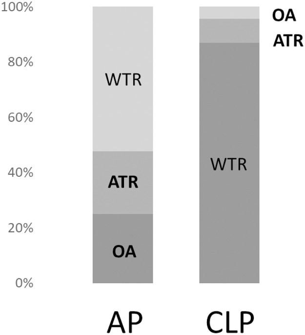
The proportions of WTR, ATR, and OA were 52%, 22%, and 25% and 86%, 9%, and 4% in the AP and CLP groups, respectively (Fig 1).

Fig 1. Proportions of the preoperative astigmatism type in the acquired ptosis (AP) and contact lens-related ptosis (CLP) groups.

Fig 1

WTR: With-the-rule; ATR: Against the rule; OA: Oblique astigmatism.

The shift in the type of astigmatism postoperatively is shown in Table 2. In the AP group, 41% of patients showed a shift in the type of astigmatism. WTR changed to OA in 23.8% and ATR in 4.8% of the patients, OA changed to WTR in 26.4% and ATR in 26.3% of the patients, and ATR changed to WTR in 11.8% and OA in 31.8% of the patients. In the CLP group, 13% of patients showed a shift in the type of astigmatism. WTR changed to ATR in 4.8%, OA changed to WTR in 100%, and ATR changed to OA in 100% of the patients.

Table 2. Shift in the type of astigmatism postoperatively in the acquired ptosis (AP) and contact lens-related ptosis (CLP) groups.

AP CLP
Preoperative (cases) Postoperative (cases/%) Preoperative (cases) Postoperative (cases/%)
WTR (21) WTR (15/71.4) WTR (20) WTR (1/95.0)
OA (5/23.8) OA (0/0)
ATR (1/4.8) ATR (1/5.0)
OA (19) OA (9/47.3) OA (2) OA (0/0)
WTR (5/26.3) WTR (2/100)
ATR (5/26.3) ATR (0)
ATR (44) ATR (25/56.8) ATR (1) ATR (0)
OA (14/31.8) OA (100)
WTR (5/11.3) WTR (0)

The shaded cells represent a change in the type of astigmatism postoperatively.

WTR: With-the-rule; ATR: Against the rule; OA: Oblique astigmatism.

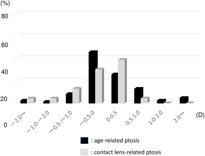
The distribution of blepharoptosis SIA in the AP and CLP groups measured using the Cravy method is shown in Fig 2. The average SIA measured using the Cravy method was a mean WTR astigmatism of 0.11±1.22 D in the AP group and −0.28±1.07 D in the CLP group. In the AP group, 58% of patients showed a WTR shift and 42% showed an ATR shift. In the CLP group, 52% patients showed a WTR shift and 48% showed an ATR shift. We observed that the astigmatic axis changed significantly (>1.00 D) in some patients in both groups.

Fig 2. Distribution of blepharoptosis surgery-induced astigmatism as measured using the Cravy method in the age-related ptosis group (black bar) and the contact lens-related ptosis groups (gray bar).

Fig 2

D: Diopter.

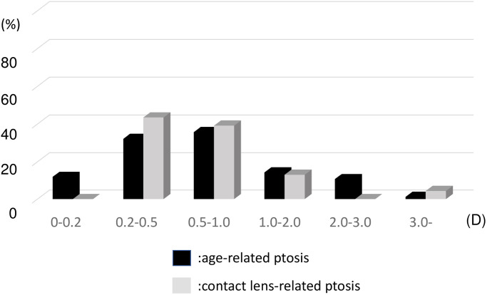
The SIA measured using the Jaffe method was 0.78±0.70 D in the AP group and 0.82±0.88 D in the CLP group (P>0.05) (Fig 3). In 25% of patients, the SIA was >1.00 D as measured using the Jaffe method. No significant difference was observed between patients with mild and severe ptosis in the AP group (0.77±0.71 D vs. 0.8±0.75 D, P>0.05).

Fig 3. Distribution of blepharoptosis surgery-induced astigmatism measured using the Jaffe method in the age-related ptosis and contact lens-related ptosis groups.

Fig 3

D: Diopter.

The correlation between SIA measured using the Cravy and Jaffe methods method and MRD did not show a significant relationship in the AP and CLP groups.

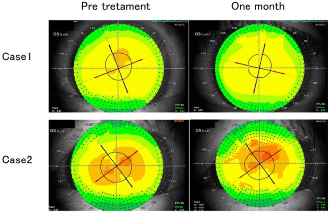
The representative cases are shown in Fig 4.

Fig 4.

Fig 4

Case 1: Postoperative topography showing change in axis with improvement in focal steepening in the superior corneal hemisphere. Case 2: Postoperative topography showing increased magnitude of astigmatism and decreased uncorrected visual acuity 1 month postoperatively.

In case 1, steep asymmetric deformation of the cornea was observed preoperatively in the upper part of the cornea, with improvement in corneal astigmatism after the surgical correction of ptosis; however, the astigmatic axis showed a change of about 30°.

In case 2, oblique astigmatism was observed preoperatively and postoperatively, with increased degree of astigmatism 1 month postoperatively. In this case, the preoperative visual acuity was VS = 0.9 (1.2 × +0.75cly-0.75A120), and the postoperative visual acuity was VS = 0.5 (1.2 × +1.00 × -1.75A130), indicating a decrease in uncorrected visual acuity.

Discussion

A retracted eyelid could affect the corneal morphology pre- and postoperatively, and this deformation could affect visual functions as a result of the astigmatic change. We focused on the change in astigmatic axis and calculated SIA using the Cravy and Jaffe methods. We observed that the type of astigmatism was different between the AP and CLP groups. The proportion of ATR was increased in both groups; in the AP group, the proportions of WTR and OA had increased, while in the CLP group, the proportion of WTR was high. Previously, it has been assumed that the mechanical pressure from ptosis of the upper eyelid causes the steepest corneal curvature in the vertical meridian, which leads to WTR drift. Gullstrand has reported that flattening of the peripheral cornea by eyelid pressure causes corneal astigmatism in the WTR direction [30]. We have also come across several cases in which the topography showed a strong asymmetric corneal astigmatism on the superior corneal hemifield preoperatively. WTR was most frequent in the CLP group, while the proportions of WTR and OA were 25% and 53%, respectively, in the AP group. A possible reason for this difference might have been related to the age difference between the two groups. A previous study that investigated corneal astigmatism using videokeratometry in two different age groups reported that the preoperative proportion of WTR was 70% in the younger ptosis surgery group and 22% in the older blepharoptosis group [16], which is consistent with our results. Age-related changes in astigmatism in the normal population have been reported. Namba et al. reported that age-related variations in the corneal geometry showed horizontal steepening and vertical flattening of the corneal surface with increasing age in a large Japanese cohort study [31]. This age-related mechanism could significantly affect a large WTR proportion in the young CLP group and ATR proportion in the older AP group pre- and postoperatively.

Changes in the degree of astigmatism after blepharoptosis surgery have been reported previously [1419]; however, changes in the astigmatic axis after blepharoptosis surgery have been reported in only a few studies [13,16]. This is the first study to report on the astigmatic shift after blepharoptosis using the Cravy and Jaffe method.

The method of calculating the astigmatic change, especially following cataract surgery, has been reported by Jaffe and Clayman [21], Cravy [22], Holladay [23], Olsen [24], Naeser [25,26] and others [27,28]. The Jaffe analysis uses the polar value method that is conceptually based on the surgically induced flattening and torque of the preoperative cylinder. In contrast, the Cravy method is a more mathematical method that focuses on the axial variation rather than the cylindrical; a positive value represents a WTR shift, while a negative value indicates an ATR shift [22]. The AP group showed a slight WTR shift, and the CLP group showed a slight ATR shift. This result might have been normal since the AP group had more patients with ATR and the CLP group had more patients with WTR preoperatively. The average change in diopter was −0.28 D and +0.11 D in the AP and CLP groups, respectively, which was minimal and not clinically significant. However, we also observed nine patients (11%) in the AP group and two patients (8%) in the CLP group who showed a significant astigmatic change of >1.00 D. We observed similar results in terms of the type of astigmatism: 40% of patients in the AP group and 17% of patients in the CLP group showed a change in the type of astigmatism postoperatively (Table 2). These results indicate that a large change in the astigmatic axis develops at a constant rate at 1 month postoperatively.

Brown et al. reported that persistent astigmatic changes were observed in ~10% of patients with ptosis and blepharoptosis [16]. This result is consistent with that of ours. The significant change in the astigmatic axis might have been due to the release of downward pressure on the cornea from the retracted contacted eyelid. However, we could not find a correlation between MRD and SIA measured using the Cravy and Jaffe methods. This result indicates that the relationship between astigmatism and the ptosis severity in the short term may not be linear but multifactorial. We speculated that one of the factors affecting this may be postoperative eyelid swelling. The eyelids are histologically prone to swelling and, occasionally, significant swelling after ptosis surgery may put pressure on the corneal surface. In this regard, our results should be treated with caution. That is, postoperative topographic measurements performed after complete healing of the eyelid might have shown altered results.

Axis rotation is an important factor for the implantation of toric intraocular lens, especially bifocal or trifocal lens. Patients in their 70s are likely to undergo cataract surgery, which is consistent with the average age of the AP group in this study. Numerous patients undergoing cataract surgery have corneal astigmatism and are eligible for toric lens implantation to restore the vision quality [32,33]. When planning cataract surgery, it is ideal to use corneal topography to identify whether the eyelids affect the cornea. The astigmatic axis after blepharoptosis surgery could interfere with the clinical improvement. Therefore, it is also important to consider the sequence of cataract surgery and blepharoptosis surgery [13].

We observed that the SIA measured using the Jaffe method was 0.78±0.70 D in the AP group and 0.82±0.88 D in the CLP group, and 25% of patients showed an SIA of >1.00 D. Several studies have reported on topographic changes after ptosis or blepharoptosis surgery. Brown et al. investigated astigmatic changes after ptosis and blepharoptosis surgery in a case series of 82 eyes and reported that 30% of patients undergoing ptosis surgery and 11% of patients undergoing blepharoptosis surgery had a transient astigmatic change of >1.00 D [16]. This result was consistent with ours. Savino et al. reported that corneal topography demonstrated a reduction in the average keratometry of 0.15±0.47° in 20 eyes of 17 patients with acquired ptosis using computerized topography. This study also reported that postoperative topographic maps showed a reduction of asymmetry [18]. Zinkernagel et al. reported the effects of different eyelid procedures with various degrees of dermatochalasis or ptosis on corneal topography, comparing the mean change in simulated keratometry after blepharoplasty (0.21 ± 0.20 D) and ptosis surgery (0.25 ± 0.25 D) [17]. Our results showed larger values than those previous reported, which implied that the results obtained with the use of simple subtraction is not consistent with those obtained using algebraic methods. [26] We believe that blepharoptosis SIA, much like astigmatism induced by any other surgery, should be assessed using both direction and magnitude. To the best of our knowledge, this is the first study to report on SIA after blepharoptosis surgery using the “Jaffe vector analysis,” which is based on the formula for calculating the resultant cylinder when the optical cylinder is crossed.

There are some limitations to this study. We did not perform a long-term follow-up. Holck et al. reported that the increased WTR at 6 weeks decreased by 12 months [14]. Gingold et al. reported no statistically significant refractive change 6 months after surgery in patients with acquired ptosis [15]. Moreover, as mentioned above, the eyelid swelling might have been one of the factors affecting this postoperative result, and the result may change after complete healing of the eyelid. These results findings indicate that a long-term follow-up is required to investigate the axis change after blepharoptosis surgery.

To conclude, we observed that ATR was most common in age-related ptosis, and WTR was most common in contact lens-related ptosis. Re-positioning of the upper eyelid may affect visual functions due to astigmatic changes. Therefore, it is important to consider the eyelid position prior to cataract surgery for the best refractive outcome. Further, the cornea was dynamically affected by ptosis both pre- and postoperatively, and the ways in which early postoperative changes and long-term astigmatic changes affect the quality of vision should be taken into consideration.

Supporting information

S1 STROBE checklist. STROBE statement.

Checklist of items that should be included in reports of observational studies.

(DOC)

S1 Dataset

(PDF)

Data Availability

All relevant data are within the paper and its Supporting information files.

Funding Statement

The authors received no specific funding for this work.

References

  • 1.Paik JS, Han K, Yang SW, Park Y, Na K, Cho W, et al. Blepharoptosis among Korean adults: age-related prevalence and threshold age for evaluation. BMC Ophthalmol. 2020;20: 99. doi: 10.1186/s12886-020-01350-y [DOI] [PMC free article] [PubMed] [Google Scholar]
  • 2.Epstein G, Putterman AM. Acquired blepharoptosis secondary to contact lens wear. Am J Ophthalmol. 1981;91: 634–639. doi: 10.1016/0002-9394(81)90065-9 [DOI] [PubMed] [Google Scholar]
  • 3.Altieri M, Truscott E, Kingston AE, Bertagno R, Altieri G. Ptosis secondary to anterior segment surgery and repair in a two-year follow-up study. Ophthalmologica. 2005;219: 129–135. doi: 10.1159/000085244 [DOI] [PubMed] [Google Scholar]
  • 4.Meyer DR, Linberg JV, Powell SR, Odom JV. Quantitating the superior visual field loss associated with ptosis. Arch Ophthalmol. 1989;107: 840–843. doi: 10.1001/archopht.1989.01070010862030 [DOI] [PubMed] [Google Scholar]
  • 5.Meyer DR, Stern JH, Jarvis JM, Lininger LL. Evaluating the visual field effects of blepharoptosis using automated static perimetry. Ophthalmology. 1993;100:651–658. doi: 10.1016/s0161-6420(93)31593-9 [DOI] [PubMed] [Google Scholar]
  • 6.Fowler BT, Pegram TA, Cutler-Peck C, Kosko M, Tran QT, Fleming JC, et al. Contrast sensitivity testing in functional ptosis and dermatochalasis surgery. Ophthalmic Plast Reconstr Surg. 2015;31: 272–274. doi: 10.1097/IOP.0000000000000307 [DOI] [PubMed] [Google Scholar]
  • 7.Lieberman DM, Grierson JW. Lids influence the corneal shape. Cornea. 2000;19: 336–342. doi: 10.1097/00003226-200005000-00016 [DOI] [PubMed] [Google Scholar]
  • 8.Won Jin Ki, Joo Shin Young, Young Hyon Joon. Effects of chalazia on corneal astigmatism: Large-sized chalazia in middle upper eyelids compress the cornea and induce the corneal astigmatism BMC Ophthalmol. 2017; 17: 36. doi: 10.1186/s12886-017-0426-2 [DOI] [PMC free article] [PubMed] [Google Scholar]
  • 9.Robb RM. Refractive errors associated with hemangiomas of the eyelids and orbit in infancy. Am J Ophthalmol. 1977;83: 52–58. doi: 10.1016/0002-9394(77)90191-x [DOI] [PubMed] [Google Scholar]
  • 10.Shaw AJ, Collins MJ, Davis BA. Eyelid pressure: inferences from corneal topographic changes. Cornea. 2009;28: 181–188. doi: 10.1097/ICO.0b013e31818a7d61 [DOI] [PubMed] [Google Scholar]
  • 11.Uğurbaş SH, Zilelioğlu G. Corneal topography in patients with congenital ptosis. Eye (Lond). 1999;13: 550–554. doi: 10.1038/eye.1999.136 [DOI] [PubMed] [Google Scholar]
  • 12.Griepentrog GJ, Diehl N, Mohney BG. Amblyopia in childhood eyelid ptosis. Am J Ophthalmol. 2013;155: 1125–1128.e1. doi: 10.1016/j.ajo.2012.12.015 [DOI] [PMC free article] [PubMed] [Google Scholar]
  • 13.Kuo IC. The effect of ptosis on cataract surgical planning. Case Rep Ophthalmol. 2015;6: 132–138. doi: 10.1159/000381846 [DOI] [PMC free article] [PubMed] [Google Scholar]
  • 14.Gingold MP, Ehlers WH, Rodgers IR, Hornblass A. Changes in refraction and keratometry after surgery for acquired ptosis. Ophthalmic Plast Reconstr Surg. 1994;10: 241–246. doi: 10.1097/00002341-199412000-00003 [DOI] [PubMed] [Google Scholar]
  • 15.Holck DE, Dutoon JJ, Wehrly SR. Changes in astigmatism after ptosis surgery measured by corneal topography. Ophthalmic Plast Reconstr Surg. 1998;14: 151–158. doi: 10.1097/00002341-199805000-00001 [DOI] [PubMed] [Google Scholar]
  • 16.Brown MS, Siegel IM, Lisman RD. Prospective analysis of changes in corneal topography after upper eyelid surgery. Ophthal Plast Reconstr Surg. 1999;15: 378–383. doi: 10.1097/00002341-199911000-00002 [DOI] [PubMed] [Google Scholar]
  • 17.Zinkernagel MS, Ebneter A, Ammann-Rauch D. Effect of upper eyelid surgery on corneal topography. Arch Ophthalmol. 2007;125: 1610–1612. doi: 10.1001/archopht.125.12.1610 [DOI] [PubMed] [Google Scholar]
  • 18.Savino G, Battendieri R, Riso M, Traina S, Poscia A, DʼAmico G, et al. Corneal topographic changes after eyelid ptosis surgery. Cornea. 2016;35: 501–505. doi: 10.1097/ICO.0000000000000729 [DOI] [PubMed] [Google Scholar]
  • 19.Kim YK, In JH, Jang SY. Changes in corneal curvature after upper eyelid surgery measured by corneal topography. J Craniofac Surg. 2016;27: e235–e238. doi: 10.1097/SCS.0000000000002435 [DOI] [PubMed] [Google Scholar]
  • 20.Shao W, Byrne P, Harrison A, Nelson E, Hilger P. Persistent blurred vision after blepharoplasty and ptosis repair. Arch Facial Plast Surg. 2004;6: 155–157. doi: 10.1001/archfaci.6.3.155 [DOI] [PubMed] [Google Scholar]
  • 21.Jaffe NS, Clayman HM. The pathophysiology of corneal astigmatism after cataract extraction. Trans Am Acad Ophthalmol Otolaryngol. 1975;79: OP615–OP630. [Google Scholar]
  • 22.Cravy TV. Calculation of the change in corneal astigmatism following cataract extraction. Ophthalmic Surg. 1979;10: 38–49. [PubMed] [Google Scholar]
  • 23.Holladay JT, Cravy TV, Koch DD. Calculating the surgically induced refractive change following ocular surgery. J Cataract Refract Surg. 1992;18: 429–443. [DOI] [PubMed] [Google Scholar]
  • 24.Dam-Johansen M, Olsen T, Theodorsen F. The longterm course of the surgically-induced astigmatism after a scleral tunnel incision. Eur J Implant Refract Surg. 1994;6: 337–343. [Google Scholar]
  • 25.Naeser K. Conversion of keratometer readings to polar values. J Cataract Refract Surg. 1990;16: 741–745. doi: 10.1016/s0886-3350(13)81018-8 [DOI] [PubMed] [Google Scholar]
  • 26.Naeser K Popperian falsification of methods of assessing surgically induced astigmatism Cataract Refract Surg. 2001;27: 25–30. doi: 10.1016/s0886-3350(00)00605-2 [DOI] [PubMed] [Google Scholar]
  • 27.Alpins NAA. Vector analysis of astigmatism changes by flattening, steepening, and torque. J Cataract Refract Surg. 1997;23: 1503–1514. doi: 10.1016/s0886-3350(97)80021-1 [DOI] [PubMed] [Google Scholar]
  • 28.Toulemont PJ. Multivariate analysis versus vector analysis to assess surgically induced astigmatism J Cataract Refract Surg. 1996;22: 977–982. doi: 10.1016/s0886-3350(96)80203-3 [DOI] [PubMed] [Google Scholar]
  • 29.Nemet AY. Accuracy of marginal reflex distance measurements in eyelid surgery. J Craniofac Surg. 2015;26: e569–71. doi: 10.1097/SCS.0000000000001304 [DOI] [PubMed] [Google Scholar]
  • 30.Gullstrand A, H. The cornea. In: Southall JPC, editor. Helmholtz’s treatise on physiological optics. New York, NY: Dover Publications Inc.; 1962. pp. 320–321. [Google Scholar]
  • 31.Namba H, Sugano A, Nishi K, Murakami T, Nishitsuka K, Konta T, et al. Age-related variations in corneal geometry and their association with astigmatism: Yamagata Study (Funagata). Medicine (Baltimore). 2018;97: e12894. doi: 10.1097/MD.0000000000012894 [DOI] [PMC free article] [PubMed] [Google Scholar]
  • 32.Hoffmann PC, Hütz WW. Analysis of biometry and prevalence data for corneal astigmatism in 23,239 eyes. J Cataract Refract Surg. 2010;36: 1479–1485. doi: 10.1016/j.jcrs.2010.02.025 [DOI] [PubMed] [Google Scholar]
  • 33.Kessel L, Andresen J, Tendal B, Erngaard D, Flesner P, Hjortdal J. Toric intraocular lenses in the correction of astigmatism during cataract surgery: a systematic review and meta-analysis. Ophthalmology. 2016;123: 275–286. doi: 10.1016/j.ophtha.2015.10.002 [DOI] [PubMed] [Google Scholar]

Decision Letter 0

Ahmed Awadein

30 Jun 2021

PONE-D-21-13875

Characteristics of astigmatism before and after blepharoptosis surgery in patients with acquired ptosis

PLOS ONE

Dear Dr. Dannnoue,

Thank you for submitting your manuscript to PLOS ONE. After careful consideration, we feel that it has merit but does not fully meet PLOS ONE’s publication criteria as it currently stands. Therefore, we invite you to submit a revised version of the manuscript that addresses the points raised during the review process.

==============================

ACADEMIC EDITOR:

The manuscript is novel and well-written. It will benefit from some meticulous grammatical revision. The figures need some improvement and you might consider adding topographic maps with differences for better illustration.

==============================

Please submit your revised manuscript by Aug 14 2021 11:59PM. If you will need more time than this to complete your revisions, please reply to this message or contact the journal office at plosone@plos.org. When you're ready to submit your revision, log on to https://www.editorialmanager.com/pone/ and select the 'Submissions Needing Revision' folder to locate your manuscript file.

Please include the following items when submitting your revised manuscript:

  • A rebuttal letter that responds to each point raised by the academic editor and reviewer(s). You should upload this letter as a separate file labeled 'Response to Reviewers'.

  • A marked-up copy of your manuscript that highlights changes made to the original version. You should upload this as a separate file labeled 'Revised Manuscript with Track Changes'.

  • An unmarked version of your revised paper without tracked changes. You should upload this as a separate file labeled 'Manuscript'.

If you would like to make changes to your financial disclosure, please include your updated statement in your cover letter. Guidelines for resubmitting your figure files are available below the reviewer comments at the end of this letter.

If applicable, we recommend that you deposit your laboratory protocols in protocols.io to enhance the reproducibility of your results. Protocols.io assigns your protocol its own identifier (DOI) so that it can be cited independently in the future. For instructions see: http://journals.plos.org/plosone/s/submission-guidelines#loc-laboratory-protocols. Additionally, PLOS ONE offers an option for publishing peer-reviewed Lab Protocol articles, which describe protocols hosted on protocols.io. Read more information on sharing protocols at https://plos.org/protocols?utm_medium=editorial-email&utm_source=authorletters&utm_campaign=protocols.

We look forward to receiving your revised manuscript.

Kind regards,

Ahmed Awadein, MD, Ph.D, FRCS

Academic Editor

PLOS ONE

Journal Requirements:

When submitting your revision, we need you to address these additional requirements.

1. Please ensure that your manuscript meets PLOS ONE's style requirements, including those for file naming. The PLOS ONE style templates can be found at 

https://journals.plos.org/plosone/s/file?id=wjVg/PLOSOne_formatting_sample_main_body.pdf and 

https://journals.plos.org/plosone/s/file?id=ba62/PLOSOne_formatting_sample_title_authors_affiliations.pdf

2. Thank you for stating the following financial disclosure: 

"NO The funders had no role in study design, data collection and analysis, decision to publish, or preparation of the manuscript.."

At this time, please address the following queries:

a) Please clarify the sources of funding (financial or material support) for your study. List the grants or organizations that supported your study, including funding received from your institution. 

b) State what role the funders took in the study. If the funders had no role in your study, please state: “The funders had no role in study design, data collection and analysis, decision to publish, or preparation of the manuscript.”

c) If any authors received a salary from any of your funders, please state which authors and which funders.

d) If you did not receive any funding for this study, please state: “The authors received no specific funding for this work.”

Please include your amended statements within your cover letter; we will change the online submission form on your behalf

3. In your Data Availability statement, you have not specified where the minimal data set underlying the results described in your manuscript can be found. PLOS defines a study's minimal data set as the underlying data used to reach the conclusions drawn

 in the manuscript and any additional data required to replicate the reported study findings in their entirety. All PLOS journals require that the minimal data set be made fully available. For more information about our data policy, please see http://journals.plos.org/plosone/s/data-availability.

"Upon re-submitting your revised manuscript, please upload your study’s minimal underlying data set as either Supporting Information files or to a stable, public repository and include the relevant URLs, DOIs, or accession numbers within your revised

 cover letter. For a list of acceptable repositories, please see http://journals.plos.org/plosone/s/data-availability#loc-recommended-repositories. Any potentially identifying patient information must be fully anonymized.

Important: If there are ethical or legal restrictions to sharing your data publicly, please explain these restrictions in detail. Please see our guidelines for more information on what we consider unacceptable restrictions to publicly sharing data: http://journals.plos.org/plosone/s/data-availability#loc-unacceptable-data-access-restrictions.

 Note that it is not acceptable for the authors to be the sole named individuals responsible for ensuring data access.

We will update your Data Availability statement to reflect the information you provide in your cover letter

3. Please include captions for your Supporting Information files at the end of your manuscript, and update any in-text citations to match accordingly. Please see our Supporting Information guidelines for more information: http://journals.plos.org/plosone/s/supporting-information

Please review your reference list to ensure that it is complete and correct. If you have cited papers that have been retracted, please include the rationale for doing so in the manuscript text, or remove these references and replace them with relevant current references. Any changes to the reference list should be mentioned in the rebuttal letter that accompanies your revised manuscript. If you need to cite a retracted article, indicate the article’s retracted status in the References list and also include a citation and full reference for the retraction notice.

[Note: HTML markup is below. Please do not edit.]

Reviewers' comments:

Reviewer's Responses to Questions

Comments to the Author

1. Is the manuscript technically sound, and do the data support the conclusions?

The manuscript must describe a technically sound piece of scientific research with data that supports the conclusions. Experiments must have been conducted rigorously, with appropriate controls, replication, and sample sizes. The conclusions must be drawn appropriately based on the data presented.

Reviewer #1: Yes

Reviewer #2: No

Reviewer #3: Yes

**********

2. Has the statistical analysis been performed appropriately and rigorously?

Reviewer #1: Yes

Reviewer #2: Yes

Reviewer #3: Yes

**********

3. Have the authors made all data underlying the findings in their manuscript fully available?

The PLOS Data policy requires authors to make all data underlying the findings described in their manuscript fully available without restriction, with rare exception (please refer to the Data Availability Statement in the manuscript PDF file). The data should be provided as part of the manuscript or its supporting information, or deposited to a public repository. For example, in addition to summary statistics, the data points behind means, medians and variance measures should be available. If there are restrictions on publicly sharing data—e.g. participant privacy or use of data from a third party—those must be specified.

Reviewer #1: Yes

Reviewer #2: No

Reviewer #3: Yes

**********

4. Is the manuscript presented in an intelligible fashion and written in standard English?

PLOS ONE does not copyedit accepted manuscripts, so the language in submitted articles must be clear, correct, and unambiguous. Any typographical or grammatical errors should be corrected at revision, so please note any specific errors here.

Reviewer #1: Yes

Reviewer #2: No

Reviewer #3: Yes

**********

5. Review Comments to the Author

Please use the space provided to explain your answers to the questions above. You may also include additional comments for the author, including concerns about dual publication, research ethics, or publication ethics. (Please upload your review as an attachment if it exceeds 20,000 characters)

Reviewer #1: In this study, the authors investigated the change of corneal astigmatism before and after ptosis surgery in patient with acquired ptosis. The subjects were divided into two groups; age related ptosis and contact lens related ptosis. The authors compared the characteristics of astigmatism after ptosis surgery between two groups. The results were interesting and worth reporting.

The study is well-designed, and the manuscript is clear and well-written.

Reviewer #2: 1. Data are available from the Dannnoue Eye Clinic Institutional Data Access. Not clear whether the general public will be able to access the data from the institution without restrictions.

2. What is the justification for using the Crave/Jaffe method to calculate astigmatism?

3. Would be worthwile to discuss the differences noted between the Jaffe result and the Cravy results.

4. Fig 2. A and Fig 2B are hard to make sense of and don’t communicate much to the reader.

5. The authors may benefit from making more cross references to similar works. Detailed comparisons of their findings and thoughts to existing publications to either support or disregard their findings will enhance their article.

6. “This result indicates that the relationship between eyelid astigmatism and the severity of ptosis may not be linear but multifactorial. We speculate that one of the factors affecting this may be postoperative eyelid swelling. The eyelids are histologically prone to swelling and, sometimes, there can be a significant swelling after ptosis, which can suppress the corneal surface.” This suggests that the post-operative topography measurements were done before the eye had completely healed. The study method can therefore be improved by comparing post-operative SIA when the eye is completely healed to pre-operative SIA so that its really like for like. Incorrect claims may be made from not comparing like for like. As it is a retrospective study the authors may have access to data from a later review date of the patients.

7. Great point to consider lid position prior to cataract surgery for best refractive outcome.

8. Fig 3 and fig 4 have the same caption but are clearly different. Not sure they are necessary as they do not enhance the readers understanding of the results.

9. Editorial help may be needed as the case being made by the writers is hard to follow. A number of grammatical errors were noted through-out the paper. eg page 6 All examinations were performed using the upper eyelid opened by the examiner. The type of astigmatism type was divided according to the axis of astigmatism

10. Results: 57 patients (AP group: 85 eyes from 45 patients, CLP group: 23

28 eyes from 13 patients): 45+13=58 not 57 unless one patient had both AP and CLP. This will affect most results quoted

11. Page 5: Review definition of MRD as it relates to the cornea rather than the pupil as stated by authors

Reviewer #3: I read with great interest your study and I found it of high scientific value

I recommend that you could include topographic data and difference maps to show the effect of the surgery on corneal astigmatism

**********

6. PLOS authors have the option to publish the peer review history of their article (what does this mean?). If published, this will include your full peer review and any attached files.

If you choose “no”, your identity will remain anonymous but your review may still be made public.

Do you want your identity to be public for this peer review? For information about this choice, including consent withdrawal, please see our Privacy Policy.

Reviewer #1: No

Reviewer #2: No

Reviewer #3: Yes: Mohamed Tarek Elnaggar

[NOTE: If reviewer comments were submitted as an attachment file, they will be attached to this email and accessible via the submission site. Please log into your account, locate the manuscript record, and check for the action link "View Attachments". If this link does not appear, there are no attachment files.]

While revising your submission, please upload your figure files to the Preflight Analysis and Conversion Engine (PACE) digital diagnostic tool, https://pacev2.apexcovantage.com/. PACE helps ensure that figures meet PLOS requirements. To use PACE, you must first register as a user. Registration is free. Then, login and navigate to the UPLOAD tab, where you will find detailed instructions on how to use the tool. If you encounter any issues or have any questions when using PACE, please email PLOS at figures@plos.org. Please note that Supporting Information files do not need this step.

PLoS One. 2021 Oct 28;16(10):e0258688. doi: 10.1371/journal.pone.0258688.r002

Author response to Decision Letter 0


4 Aug 2021

Reviewer1

> #1: In this study, the authors investigated the change of corneal astigmatism before and after ptosis surgery in patient with acquired ptosis. The subjects were divided into two groups; age related ptosis and contact lens related ptosis. The authors compared the characteristics of astigmatism after ptosis surgery between two groups. The results were interesting and worth reporting.

The study is well-designed, and the manuscript is clear and well-written.

Response:

Thank you for your appreciative comment. We think that pre- and postoperative refractive change could be an important factor in patients’ complaints regarding visual function. We are very grateful for this opportunity to have our report published in PLOS ONE.

Reviewer #2:

1. Data are available from the Dannnoue Eye Clinic Institutional Data Access. Not clear whether the general public will be able to access the data from the institution without restrictions.

We apologize for the missing data set in the first draft of the manuscript. We were unaware that it is always required for submission.

The data set has been included in a supporting information file (S1) and a caption has been included at the end of the manuscript.

2. What is the justification for using the Crave/Jaffe method to calculate astigmatism?

Thank you for your question.

Several previous studies that have reported on the refractive change following blepharoptosis surgery have mentioned the use of simple subtraction, which is not consistent with results obtained using algebraic methods.

In previous studies on surgical induced astigmatism (SIA), especially that following cataract surgery, several methods for calculating SIA have been reported by Jaffe and Clayman1, Cravy2, Holladay3, Olsen4, Naeser5, and others.

The polar value methods are conceptually based on the surgically induced flattening and torque of the preoperative cylinder6,7, and most of these studies arrived at the same values through different approaches8.

Therefore, we chose one of the original calculation methods; i.e., the “Jaffe vector analysis”.

However, the methods developed by Cravy and Naeser must be distinguished from the others, since assessments using these calculations are associated with the axial variation rather than the cylindrical. In these methods, the change in axis is a major factor, which was the focus of our study.

The Cravy method summarizes the magnitude and direction of SIA in a manner that is simple and easy for the readers to understand. Therefore, we chose the Cravy method to investigate whether the patients showed the WTR shift or ATR shift following blepharoptosis.

We have included the justification for choosing the Cravy and Jaffe methods in the manuscript.

1. Jaffe NS, Clayman HM. The pathophysiology of corneal astigmatism after cataract extraction. Trans Am Acad Ophthalmol Otolaryngol 1975; 79:OP615–OP630

2. Cravy TV. Calculation of the change in corneal astigmatism following cataract extraction. Ophthalmic Surg 1979; 10:38–49

3. Holladay JT, Cravy TV, Koch DD. Calculating the surgically induced refractive change following ocular surgery. J Cataract Refract Surg 1992; 18:429–443

4. Dam-Johansen M, Olsen T, Theodorsen F. The longterm course of the surgically-induced astigmatism after a scleral tunnel incision. Eur J Implant Refract Surg 1994;

6:337–343

5. Naeser K. Conversion of keratometer readings to polar values. J Cataract Refract Surg 1990; 16:741–745

6. Naeser K Popperian falsification of methods of assessing surgically induced astigmatism Cataract Refract Surg . 2001 Jan;27(1):25-30. doi: 10.1016/s0886-3350(00)00605-2.

7. Noel A. Alpins, FRACO, FRCOphth, FACS Vector analysis of astigmatism changes by flattening, steepening, and torque J Cataract

Refract Surg 1997; 23:1503-1514

8. P J Toulemont Multivariate analysis versus vector analysis to assess surgically induced astigmatism J Cataract Refract Surg.1996 Sep;22(7):977-82. doi: 10.1016/s0886-3350(96)80203-3.

3. Would be worthwhile to discuss the differences noted between the Jaffe result and the Cravy results.

Thank you for your suggestion.

As mentioned above, the Jaffe vector analysis and the Cravy method utilize different concepts.

Jaffe and Clayman used rectangular and polar coordinates to determine, using vector analysis, the formula for calculating SIA and its axis using preoperative and postoperative corneal astigmatism.1,2

In contrast, the Cravy method utilizes a mathematical concept that is not based on optics.

We have emphasized these differences in the Discussion section.

1. Jaffe NS, Clayman HM. The pathophysiology of corneal astigmatism after cataract extraction. Trans Am Acad Ophthalmol Otolaryngol 1975; 79:OP615–OP630

2. Alpins NA. A new method of analyzing vectors for changes in astigmatism. J Cataract Refract Surg. 1993 Jul;19(4):524-33. doi: 10.1016/s0886-3350(13)80617-7. PMID: 8355160

3. P J Toulemont Multivariate analysis versus vector analysis to assess surgically induced astigmatism J Cataract Refract Surg.1996 Sep;22(7):977-82. doi: 10.1016/s0886-3350(96)80203-3.

4. Fig 2. A and Fig 2B are hard to make sense of and communicate much to the reader.

Thank you for your comment.

In this study, we were mainly interested in the postoperative changes in the axis and we considered that the figures would allow the readers to comprehend the axis change more easily. However, what is shown in Fig. 2 is essentially the same as what is shown in Table 2, and we believe that considering surgery-induced astigmatism as a vector was not consistent with the purpose of this study.

Considering only the axis as the magnitude of astigmatism is clinically insignificant.1

We have decided to remove this figure as we believe that it may convey wrong information to the readers, since the astigmatic change was assessed only in terms of the axis change.

1. Olsen T, Dam-Johansen M. Evaluating surgically induced astigmatism. J Cataract Refract Surg. 1994 Sep;20(5):517-22. doi: 10.1016/s0886-3350(13)80231-3.

5. The authors may benefit from making more cross references to similar works. Detailed comparisons of their findings and thoughts to existing publications to either support or disregard their findings will enhance their article.

Thank you for your comments.

We have referred to previous papers, mainly on the refractive change after ptosis surgery; however, we found few citations for original studies that have assessed induced astigmatism following cataract surgery and have added these references (listed in response to question No. 2) to enrich the manuscript.

6. This result indicates that the relationship between eyelid astigmatism and the severity of ptosis may not be linear but multifactorial. We speculate that one of the factors affecting this may be postoperative eyelid swelling. The eyelids are histologically prone to swelling and, sometimes, there can be a significant swelling after ptosis, which can suppress the corneal surface. This suggests that the post-operative topography measurements were done before the eye had completely healed. The study method can therefore be improved by comparing post-operative SIA when the eye is completely healed to pre-operative SIA so that its really like for like. Incorrect claims may be made from not comparing like for like. As it is a retrospective study the authors may have access to data from a later review date of the patients.

You have raised a very valid point, which is a major limitation of this study, as stated in the manuscript.

Eyelids are inherently prone to swelling, and this might have been a major cause for corneal deformity at 1 month postoperatively. It would be more scientific to compare the results using data that was obtained long after the surgery, i.e., once the effects of eyelid swelling had worn out.

To the best of our best knowledge, the ways in which corneal astigmatism changes over time following ptosis surgery remains inconclusive.

Holck et al. reported that the WTR increased at 6 weeks and decreased at 12 months. In contrast, Ingold et al. reported that there was no statistically significant change in the refractive error at 6 months postoperatively in patients with acquired ptosis. These have been included in the manuscript (Line Page)

Since the results of this study were limited to short-term changes observed after 1 month, the title of this paper has been revised to "Characteristics of astigmatism before and 1 month after blepharoptosis surgery in patients with acquired ptosis” to avoid misleading the reader that these results are permanent.

Further, we have acknowledged the possibility that the results might have changed after complete healing of the eyelid.

During the early postoperative period, patients are very anxious about changes in their vision, and we believe that our results associated with the changes observed 1 postoperative month are still scientifically relevant.

Unfortunately, several patients who participated in the study dropped out before the postoperative follow-up, and we were unable to obtain sufficient data that was statistically significant; therefore, we limited our findings to changes observed at 1 month postoperatively.

We strongly believe that a long-term follow-up study is necessary, and we would be interested in observing the longitudinal changes in SIA following cataract surgery. We are currently following up new cases with prospective 1-year data.

7. Great point to consider lid position prior to cataract surgery for best refractive outcome.

Thank you for your appreciative comments. We also believe that consideration of the eyelid position prior to cataract surgery is significantly important to achieve the best refractive outcome following the surgery, which was one of the main focus areas of this study.

We have emphasized this point in the conclusion of the manuscript.

8. Fig 3 and fig 4 have the same caption but are clearly different. Not sure they are necessary as they do not enhance the readers understanding of the results.

Thank you for pointing this out. We have made the correction in Fig. 3.

9. Editorial help may be needed as the case being made by the writers is hard to follow. A number of grammatical errors were noted through-out the paper. eg page 6 All examinations were performed using the upper eyelid opened by the examiner. The type of astigmatism type was divided according to the axis of astigmatism

Thank you for your comment.

We have sought help from a professional English editing service and re-checked the entire text.

All examinations were performed using the upper eyelid opened by the examiner.

→To avoid any pressure from the eyelids on the corneal surface, the examination was performed with the patients’ eyes open.

The type of astigmatism type was divided according to the axis of astigmatism

→We have deleted this sentence.

10. Results: 57 patients (AP group: 85 eyes from 45 patients, CLP group: 23

> 28 eyes from 13 patients): 45+13=58 not 57 unless one patient had both AP and CLP. This will affect most results quoted

Thank you for pointing this out.

This was a simple calculation error; the number of patients in the AP group was 58 and not 57.

I have included the data set in a supporting file.

11. Page 5:

Review definition of MRD as it relates to the cornea rather than the pupil as stated by authors

Thank you for pointing this out.

We have reviewed the definition of MRD as stated by other authors and have revised it as the measurement of the distance from the middle upper eyelid to the corneal light reflex.

Reviewer #3: I read with great interest your study and I found it of high scientific value I recommend that you could include topographic data and difference maps to show the effect of the surgery on corneal astigmatism

Thank you for your appreciative comments. We have added a topographic map of the two representative cases in Fig. 4.

Case 1: Postoperative topography showing change in axis with improvement in focal steepening in the superior corneal hemisphere; Case 2: Postoperative topography showing increased magnitude of astigmatism and decreased uncorrected visual acuity 1 month postoperatively.

Attachment

Submitted filename: Response to Reviewers.docx

Decision Letter 1

Ahmed Awadein

4 Oct 2021

Characteristics of astigmatism before and 1 month after blepharoptosis surgery in patients with acquired ptosis

PONE-D-21-13875R1

Dear Dr. Dannnoue,

We’re pleased to inform you that your manuscript has been judged scientifically suitable for publication and will be formally accepted for publication once it meets all outstanding technical requirements.

Within one week, you’ll receive an e-mail detailing the required amendments. When these have been addressed, you’ll receive a formal acceptance letter and your manuscript will be scheduled for publication.

An invoice for payment will follow shortly after the formal acceptance. To ensure an efficient process, please log into Editorial Manager at http://www.editorialmanager.com/pone/, click the 'Update My Information' link at the top of the page, and double check that your user information is up-to-date. If you have any billing related questions, please contact our Author Billing department directly at authorbilling@plos.org.

If your institution or institutions have a press office, please notify them about your upcoming paper to help maximize its impact. If they’ll be preparing press materials, please inform our press team as soon as possible -- no later than 48 hours after receiving the formal acceptance. Your manuscript will remain under strict press embargo until 2 pm Eastern Time on the date of publication. For more information, please contact onepress@plos.org.

Kind regards,

Ahmed Awadein, MD, Ph.D, FRCS

Academic Editor

PLOS ONE

Additional Editor Comments (optional):

Reviewers' comments:

Acceptance letter

Ahmed Awadein

6 Oct 2021

PONE-D-21-13875R1

Characteristics of astigmatism before and 1 month after blepharoptosis surgery in patients with acquired ptosis

Dear Dr. Dannoue:

I'm pleased to inform you that your manuscript has been deemed suitable for publication in PLOS ONE. Congratulations! Your manuscript is now with our production department.

If your institution or institutions have a press office, please let them know about your upcoming paper now to help maximize its impact. If they'll be preparing press materials, please inform our press team within the next 48 hours. Your manuscript will remain under strict press embargo until 2 pm Eastern Time on the date of publication. For more information please contact onepress@plos.org.

If we can help with anything else, please email us at plosone@plos.org.

Thank you for submitting your work to PLOS ONE and supporting open access.

Kind regards,

PLOS ONE Editorial Office Staff

on behalf of

Dr. Ahmed Awadein

Academic Editor

PLOS ONE

Associated Data

    This section collects any data citations, data availability statements, or supplementary materials included in this article.

    Supplementary Materials

    S1 STROBE checklist. STROBE statement.

    Checklist of items that should be included in reports of observational studies.

    (DOC)

    S1 Dataset

    (PDF)

    Attachment

    Submitted filename: Response to Reviewers.docx

    Data Availability Statement

    All relevant data are within the paper and its Supporting information files.


    Articles from PLoS ONE are provided here courtesy of PLOS

    RESOURCES