Abstract
Kohlschütter-Tönz syndrome (KTS) manifests as neurological dysfunctions, including early-onset seizures. Mutations in the citrate transporter SLC13A5 are associated with KTS, yet their underlying mechanisms remain elusive. Here, we report that a Drosophila SLC13A5 homolog, I’m not dead yet (Indy), constitutes a neurometabolic pathway that suppresses seizure. Loss of Indy function in glutamatergic neurons caused “bang-induced” seizure-like behaviors. In fact, glutamate biosynthesis from the citric acid cycle was limiting in Indy mutants for seizure-suppressing glutamate transmission. Oral administration of the rate-limiting α-ketoglutarate in the metabolic pathway rescued low glutamate levels in Indy mutants and ameliorated their seizure-like behaviors. This metabolic control of the seizure susceptibility was mapped to a pair of glutamatergic neurons, reversible by optogenetic controls of their activity, and further relayed onto fan-shaped body neurons via the ionotropic glutamate receptors. Accordingly, our findings reveal a micro-circuit that links neural metabolism to seizure, providing important clues to KTS-associated neurodevelopmental deficits.
Author summary
Kohlschütter-Tönz syndrome (KTS) is a neurodevelopmental disorder linked to two distinct genomic loci encoding the citrate transporter SLC13A5 and synaptic protein ROGDI, respectively. An early-onset seizure is the most prominent neurological symptom in KTS patients, yet how these genes contribute to the control of seizure susceptibility remains poorly understood. Our study establishes behavioral models of seizure in Drosophila mutants of KTS-associated genes and demonstrates a genetic, metabolic, and neural pathway of seizure suppression. We discover that the metabolic flux of the Krebs cycle to glutamate biosynthesis plays a critical role in scaling seizure-relevant glutamate transmission. We further map this seizure-suppressing pathway to a surprisingly small number of glutamatergic neurons and their ionotropic glutamate transmission onto a key sleep-promoting locus in the adult fly brain. Given that the excitatory amino acid glutamate is considered a general seizure-promoting neurotransmitter, our findings illustrate how glutamatergic transmission can have opposing effects on seizure susceptibility in the context of a micro-neural circuit, possibly explaining drug-resistant epilepsy. This seizure-suppressing locus in the Drosophila brain is also implicated in metabolism, circadian rhythms, and sleep, revealing the conserved neural principles of their intimate interaction with epilepsy across species.
Introduction
Kohlschütter-Tönz syndrome (KTS) is a genetic disorder that manifests as developmental abnormalities such as tooth dysplasia, intellectual disability, and early-onset epileptic encephalopathy [1]. Whole-exome sequencing has revealed molecular lesions that are associated with KTS, identifying mutations in two independent loci, ROGDI [2–6] and SLC13A5 (solute carrier family 13 member 5) [7–10], as the genetic causes of KTS. Homologs of the two KTS-associated genes are relatively well conserved across different species. Accordingly, animal models for ROGDI or SLC13A5 serve as essential genetic resources to elucidate the physiological function of the gene products and their mechanisms underlying the KTS pathogenesis.
SLC13A5 is a plasma membrane transporter of intermediates of the tricarboxylic acid (TCA) cycle, also known as the citric acid cycle or Krebs cycle, that displays the highest affinity for citrate [11,12]. The Drosophila homolog of SLC13A5 is I’m not dead yet (Indy), named for the long lifespan of loss-of-function mutants [13], although the longevity phenotype might be sensitive to genetic background or calorie intake [14–16]. As expected based on its citrate-transporter activity, loss of Indy function phenocopies physiological changes in calorie-restricted animals, including low body weight, low triglyceride levels, and high sensitivity to starvation [16,17]. In fact, calorie restriction down-regulates Indy expression in Drosophila, whereas Indy mutants display low expression levels of insulin-like peptides [16]. Mitochondrial physiology is also altered in Indy mutant flies (i.e., increased mitochondrial biogenesis, decreased electron transport chain function), likely via a mechanism dependent on peroxisome proliferator-activated receptor gamma coactivator-1α [16–18].
Consistent with this, depletion of an INDY homolog in worms extends life span and leads to a “lean” phenotype, likely via an AMPK-dependent pathway [19,20]. In mice, genomic deletion of the mammalian Indy homolog (mIndy), as well as liver-specific mINDY depletion, results in a protection against obesity, fatty liver, and insulin resistance upon feeding a high-fat diet—metabolic conditions comparable to those observed in flies and worms [21–23]. Notably, gene expression profiles in mIndy mutant mice largely resemble those in calorie-restricted animals [21]. mIndy expression in mice is also altered by metabolic challenge (e.g., starvation, fatty-liver disease conditions), and distinct transcription factors (e.g., CREB, STAT3, ARNT, PXR) have been implicated in this adaptive regulation of mIndy transcription [24–27].
While there is abundant genetic evidence for Indy function in metabolism [28–30], much less is known about the function of ROGDI homologs. A Drosophila genetic screen initially identified rogdi as one of the genes associated with memory formation [31] and emerging evidence suggests a role of ROGDI homologs in neurons [3,5,32,33]. The crystal structure of human ROGDI protein displays a leucine-zipper-like four-helix bundle and a characteristic beta-sheet domain, hinting at how KTS-associated ROGDI mutations would impair the overall structure and stability of the encoded protein [34]. Nonetheless, how metabolic or neural functions of the KTS-associated gene products are coupled to the neurodevelopmental pathogenesis underlying KTS remains elusive.
In this study, we demonstrate that Drosophila mutants of KTS-associated genes display seizure-like behaviors. We further provide compelling evidence that Indy links the flux of the TCA cycle in a specific set of glutamatergic neurons to the scale of their neural transmission to achieve metabolic control of seizure susceptibility. Given that early-onset seizure is one of the most prominent symptoms in KTS patients, our findings provide important insights into the neural mechanism responsible for the development of KTS.
Results
Indy acts in glutamatergic neurons to suppress bang-induced seizure
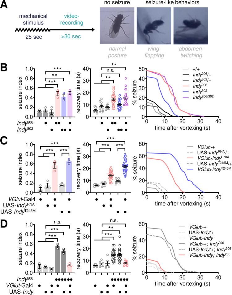
The early-onset seizure is one of the most prominent neurological symptoms observed in KTS patients. We hypothesized that Drosophila mutants of KTS-associated genes might recapitulate genetic conditions in KTS patients and display seizure-like behaviors. After a mechanical stimulus (vortexing for 25 s), wild-type flies immediately recovered a normal posture and resumed their locomotion (Fig 1A). By contrast, Indy mutants homozygous or trans-heterozygous for loss-of-function alleles exhibited “bang-induced” seizure-like behaviors (e.g., severe wing-flapping, abdominal contractions, leg-twitching, or failure to stand upright), as reflected in a high seizure index and prolonged recovery time (Fig 1B and S1 Movie). The bang-sensitive seizure (BSS) phenotypes in Indy mutants were comparable to those observed in other seizure mutants, such as easily shocked or slamdance but weaker than bang senseless mutants [35–37] (S1A Fig). Moreover, Indy mutant seizure displayed transient resistance to the second mechanical stimulus after the first BSS (S1B Fig), indicating the seizure threshold shifts during the refractory period as observed in other bang-sensitive mutants [38]. To map a neural locus responsible for Indy mutant BSS, we silenced Indy expression by overexpressing an RNA interference (RNAi) transgene in select groups of cells and examined subsequent effects on BSS (S2A Fig). INDY depletion in vesicular glutamate transporter (VGlut)-expressing neurons phenocopied BSS in Indy mutants (Fig 1C), whereas the Indy RNAi in other groups of neurons defined by their specific neurotransmitters (e.g., GABAergic, cholinergic, or dopaminergic neurons) did not induce BSS (S2B Fig). The INDY-depletion phenotypes were confirmed by independent Indy RNAi transgenes (i.e., IndyRNAi #2, #3, and #4), possibly excluding off-target effects (S2C Fig). We generated a mutant INDY transgene that harbored a KTS-associated allele (S3 Fig, INDYT245M) and expressed mutant INDY proteins with no citrate transporter activity [7–9,39]. Overexpression of INDYT245M in VGlut-expressing neurons similarly induced BSS in a wild-type Indy background (Fig 1C). Moreover, transgenic expression of wild-type Indy cDNA in VGlut-expressing neurons was sufficient to rescue BSS in Indy mutants (Fig 1D). These results suggest that Indy function in glutamatergic neurons is necessary and sufficient for seizure suppression. We further found that rogdi mutants displayed BSS comparably to Indy mutants (S4A Fig), and its seizure-suppressor function was similarly mapped to glutamatergic neurons (S4B–S4D Fig). Nevertheless, our subsequent analyses focused on elucidating Indy-dependent mechanisms of seizure control since the molecular function of ROGDI has been poorly defined.
Fig 1. Loss of Indy function in glutamatergic neurons induces BSS.
(A) Experimental design for the quantitative analysis of bang-induced seizure-like behaviors in Drosophila. A group of flies (n = 5) was vortexed for 25 s, after which post-stimulus responses were video recorded. Seizure-like behaviors, including wing-flapping and abdomen-twitching, were scored in individual flies. (B) Quantitative analyses of BSS in Indy mutants homozygous or trans-heterozygous for loss-of-function alleles. A seizure index was calculated as the ratio of the number of BSS-positive flies to the total number of flies tested in each experiment (n = 20; 5 flies per group × 4 groups per experiment) and averaged from three independent experiments. Recovery time was calculated individually for BSS-positive flies as the latency to normal posture after vortexing and was averaged for each genotype (n = 7–29 flies). Percent seizure was calculated as the percentage of BSS-positive flies per genotype at each second after vortexing (n = 60 flies; 20 flies per experiment × 3 experiments). Data represent means ± SEM. n.s., not significant; **P < 0.01, ***P < 0.001, as determined by one-way ANOVA with Holm-Sidak’s multiple comparisons test (seizure index) or by Kruskal Wallis test with Dunn’s multiple comparisons test (recovery time). (C) Silencing of Indy function in glutamatergic neurons by transgenic overexpression of IndyRNAi or KTS-associated IndyT245M is sufficient to induce BSS. Quantitative analyses of BSS in individual flies were performed as described above. Data represent means ± SEM (seizure index, n = 60 flies in 3 independent experiments; recovery time, n = 5–39 flies). ***P < 0.001, as determined by one-way ANOVA with Holm-Sidak’s multiple comparisons test. (D) Transgenic overexpression of wild-type INDY in glutamatergic neurons rescues BSS in Indy mutants. Data represent means ± SEM (seizure index, n = 60 flies in 3 independent experiments; recovery time, n = 5–33 flies). n.s., not significant; **P < 0.01, ***P < 0.001, as determined by two-way ANOVA with Holm-Sidak’s multiple comparisons test.
Down-regulation of glutamate transmission induces BSS in Indy mutants
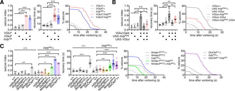
To determine if glutamate transmission actually contributes to BSS phenotypes in Indy mutants, we genetically manipulated the expression of VGLUT, a vesicular transporter that incorporates glutamate into synaptic vesicles [40], and examined subsequent effects on Indy-dependent BSS. The heterozygosity of VGlut did not induce seizure-like behaviors in wild-type flies (Fig 2A); however, it substantially increased seizure susceptibility in Indy heterozygous mutants. VGLUT overexpression in glutamatergic neurons partially but significantly rescued BSS phenotypes in INDY-depleted flies (Fig 2B). It has been shown that VGLUT overexpression increases synaptic vesicle size in larval motor neurons and their spontaneous release of glutamate at the larval neuromuscular junction [40–42], whereas it could lead to neurodegeneration in a cell type-specific manner [42,43]. Nonetheless, no gross effects of VGLUT overexpression were detected on the seizure susceptibility in wild-type flies (Fig 2B). We further mapped two glutamate receptors, N-methyl-D-aspartic acid receptors (Nmdar1 and Nmdar2) and glutamate-gated chloride channel (GluClα), that mediate Indy-dependent BSS in trans-heterozygous mutants (Fig 2C). However, hemizygous Nmdar2 mutants exhibited significant BSS even in a wild-type Indy background. These genetic interactions suggest that down-scaling of glutamate transmission via the ionotropic glutamate receptors may underlie BSS phenotypes in Indy mutants.
Fig 2. Down-regulation of glutamatergic transmission is responsible for Indy-dependent BSS.
(A) Heterozygosity of VGlut induces BSS in heterozygous Indy mutants. Quantitative analyses of BSS in individual flies were performed as described in Fig 1. Data represent means ± SEM. ***P < 0.001, as determined by one-way ANOVA with Holm-Sidak’s multiple comparisons test (seizure index, n = 55–60 flies in 3 independent experiments) or by Welch’s ANOVA with Dunnett’s multiple comparisons test (recovery time, n = 7–32 flies). (B) Overexpression of wild-type VGLUT suppresses BSS induced by transgenic depletion of INDY in glutamatergic neurons. Data represent means ± SEM (seizure index, n = 55–60 flies in 3 independent experiments; recovery time, n = 5–34 flies). n.s., not significant; *P < 0.05, **P < 0.01, ***P < 0.001, as determined by one-way ANOVA with Holm-Sidak’s multiple comparisons test. (C) Heterozygosity of ionotropic glutamate receptor Nmdar or GluClα induces BSS in heterozygous Indy mutants. Significant Indy x glutamate receptor interaction effects on seizure index (P = 0.0047 for Nmdar1; P = 0.0023 for Nmdar2; P = 0.0009 for GluClα) and recovery time (P = 0.0136 for Nmdar2; P = 0.0049 for GluClα) were detected by two-way ANOVA. Data represent means ± SEM (seizure index, n = 60 flies in 3 independent experiments; recovery time, n = 2–37). *P < 0.05, **P < 0.01, ***P < 0.001, as determined by Holm-Sidak’s multiple comparisons test.
A metabolic link between the TCA cycle and glutamate transmission underlies Indy-dependent seizure suppression
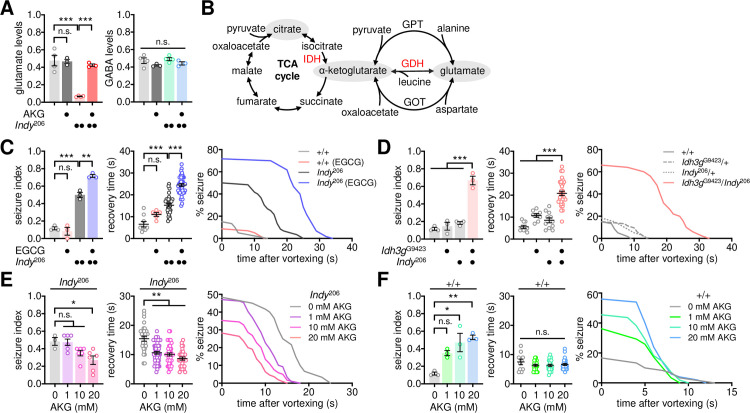
Two classes of presynaptic neurons—glutamatergic and GABAergic—are intrinsically coupled to astrocytes via the glutamate/GABA-glutamine cycle, mediating excitatory and inhibitory transmissions, respectively. An imbalance in their activity is associated with neurological disorders, including a seizure [44–46]. In contrast to the mammalian central nervous system, acetylcholine is the primary excitatory neurotransmitter in the fly brain whereas glutamate plays this role at the neuromuscular junction [47–49]. Nonetheless, our genetic evidence indicated that low glutamate transmission likely induced seizure-like behaviors in Indy mutants. We thus examined if the loss of Indy function impaired the biosynthesis of glutamate or GABA. Our quantitative assessment of free amino acids revealed that glutamate levels were substantially reduced in Indy mutants (Fig 3A). In contrast, no significant differences in GABA levels were detected between wild-type and Indy mutant flies. Considering that glutamate dehydrogenase (GDH) mediates the interconversion of glutamate and α-ketoglutarate—one of the TCA cycle intermediates (Fig 3B)—we reasoned that loss of INDY-dependent import of extracellular citrate might limit glutamate biosynthesis via the TCA cycle.
Fig 3. Indy-dependent BSS involves a metabolic link between the TCA cycle and glutamate transmission.
(A) Indy mutants display low levels of free glutamate that are rescued by oral administration of α-ketoglutarate (AKG). Flies were fed control or AKG-containing food (20 mM) for 3 d before harvesting. Free amino acids in whole-body extracts were quantified using ion-exchange chromatography. Relative levels of glutamate and GABA were calculated by normalizing to glycine levels. A significant Indy x AKG interaction effect on glutamate levels (P = 0.0006) was detected by two-way ANOVA. Data represent means ± SEM (n = 3–4). n.s., not significant; ***P < 0.001, as determined by Holm-Sidak’s multiple comparisons test. (B) Glutamate biosynthesis via the TCA cycle. IDH, isocitrate dehydrogenase; GDH, glutamate dehydrogenase; GPT, glutamate-pyruvate transaminase; GOT, glutamate-oxaloacetate transaminase. (C) Oral administration of the GDH inhibitor epigallocatechin gallate (EGCG) exaggerates BSS phenotypes in Indy mutants. Flies were fed control or EGCG-containing food (5 mg/ml) for 3 d before the BSS assessment. Quantitative analyses of BSS in individual flies were performed as described in Fig 1. Two-way ANOVA detected a significant Indy x EGCG interaction effect on seizure index (P = 0.0025). Data represent means ± SEM (seizure index, n = 58–60 flies in 3 independent experiments; recovery time, n = 5–43 flies). n.s., not significant; **P < 0.01, ***P < 0.001, as determined by Holm-Sidak’s multiple comparisons test. (D) Idh3g heterozygosity induces BSS phenotypes in heterozygous Indy mutants. Significant Indy x Idh3g interaction effects on seizure index (P = 0.0002 by two-way ANOVA) and recovery time (P = 0.0086 by Aligned ranks transformation ANOVA) were detected. Data represent means ± SEM (seizure index, n = 50–60 flies in 3 independent experiments; recovery time, n = 8–33 flies). ***P < 0.001, as determined by Holm-Sidak’s multiple comparisons test (seizure index) or by Wilcoxon rank sum test (recovery time). (E and F) Oral administration of AKG rescues BSS phenotypes in Indy mutants. Flies were fed control or AKG-containing food (20 mM) for 3 d before the BSS assessment. Data represent means ± SEM (seizure index, n = 50–119 flies in 3 independent experiments; recovery time, n = 10–56 flies). n.s., not significant; *P < 0.05, **P < 0.01, as determined by one-way ANOVA with Holm-Sidak’s multiple comparisons test (seizure index), by Welch’s ANOVA with Dunnett’s T3 multiple comparisons test (E, recovery time), or by Kruskal-Wallis test with Dunn’s multiple comparisons (F, recovery time).
To validate this hypothesis, we fed the GDH inhibitor, epigallocatechin gallate [50], to adult flies and tested its effects on BSS phenotypes. Pharmacological inhibition of GDH robustly increased both seizure index and recovery time after BSS in Indy mutants, but it did not induce BSS in wild-type flies (Fig 3C). Similar results were obtained using another GDH inhibitor (i.e., diethylstilbestrol), excluding possible off-target effects (S5 Fig). Accordingly, these data suggest that GDH-mediated metabolic flux is limiting for seizure suppression, particularly in the context of Indy deficiency. To further examine the involvement of the TCA cycle in seizure suppression, we genetically manipulated isocitrate dehydrogenase 3 (IDH3), a hetero-tetrameric enzyme that converts isocitrate into α-ketoglutarate in the rate-limiting step in the TCA cycle (Fig 3B). Heterozygosity of the Idh3g mutant allele induced BSS in Indy heterozygous mutants, but not in wild-type flies (Fig 3D). In addition, RNAi-mediated depletion of individual IDH3 subunit proteins in wild-type glutamatergic neurons alone was sufficient to induce BSS (S6 Fig). These lines of pharmacological and genetic evidence indicate that reducing the metabolic flux from the TCA cycle to glutamate biosynthesis may down-scale seizure-suppressing glutamate transmission, thereby causing BSS phenotypes in Indy mutants.
We further hypothesized that dietary supplements of TCA cycle intermediates should compensate for the genetic deficits in flies with loss of Indy function and rescue their BSS phenotypes. Oral administration of α-ketoglutarate indeed ameliorated seizure phenotypes in Indy mutants in a dose-dependent manner (Fig 3E). Moreover, α-ketoglutarate supplementation restored glutamate levels in Indy mutants to wild-type levels (Fig 3A). Unexpectedly, we found that α-ketoglutarate supplementation induced a dose-dependent increase in seizure index, but not recovery time, in wild-type flies (Fig 3F). A possible explanation for this observation is that an excess of α-ketoglutarate may lead to an imbalance in the metabolic flux between the TCA cycle and glutamate in wild-type flies, thereby lowing the threshold for seizure initiation. Alternatively, surplus α-ketoglutarate may deplete synaptic vesicles containing glutamate by promoting their fusion with the synaptic membrane [51] (see Discussion).
A pair of glutamatergic neurons mediates Indy-dependent seizure suppression
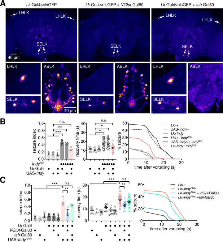
Genetic and biochemical analyses in Indy mutants revealed the contribution of low glutamate transmission to their seizure phenotypes. We found that daily locomotor activity was reduced in Indy mutants, but their waking activity (i.e., activity count per minute awake) was indistinguishable from wild-type control (S7A Fig). Moreover, wild-type and Indy mutants displayed similar climbing activities in a negative geotaxis assay (S7B Fig). We thus reasoned that the glutamate transmission in Indy mutants might not be limiting for general motor function as reported previously [13,16,52–54], but a subset of the glutamatergic neurons would be somehow sensitized to loss of Indy function for seizure control. Our transgenic mapping of the Indy RNAi phenotypes actually identified a very small group of neurons expressing the neuropeptide leucokinin [55] (hereafter, LK neurons) as a neural locus important for Indy-dependent seizure suppression (Figs 4A and S2B). Introduction of a temperature-sensitive Gal80 transgene allowed us to turn off the Gal4-driven expression of the Indy RNAi transgene at low temperature (i.e., 21°C) but silence endogenous Indy expression in LK neurons at high temperature (i.e., 29°C). The conditional RNAi confirmed that Indy was necessary in adult LK neurons for seizure suppression (S8 Fig). Conversely, transgenic expression of the wild-type Indy cDNA in LK neurons was sufficient to rescue BSS in Indy mutants (Fig 4B).
Fig 4. A pair of LK neurons mediates Indy-dependent BSS.
(A) A glutamatergic transgene specifically targets a pair of LHLK neurons among LK neurons. LK neurons in the adult brain (LHLK and SELK) and ventral nerve cord (ABLK) were visualized by transgene expression of nuclear GFP (nlsGFP). The VGlut-Gal80 transgene suppressed GFP expression only in LHLK neurons. Representative confocal images of each genotype were shown. (B) INDY overexpression in LK neurons is sufficient to rescue BSS phenotypes in Indy mutants. Quantitative analyses of BSS in individual flies were performed as described in Fig 1. Data represent means ± SEM. n.s., not significant; *P < 0.05, **P < 0.01, ***P < 0.001, as determined by two-way ANOVA with Holm-Sidak’s multiple comparisons test (seizure index, n = 60 flies in 3 independent experiments) or by Aligned ranks transformation ANOVA with Wilcoxon rank sum test (recovery time, n = 9–29). (C) INDY depletion in a pair of LHLK neurons is necessary for BSS phenotypes in Indy RNAi flies. LK neuron-specific expression of the Indy RNAi transgene was inhibited specifically in LHLK neurons by the VGlut-Gal80 transgene. Data represent means ± SEM (seizure index, n = 55–60 flies in 3 independent experiments; recovery time, n = 3–28 flies). n.s., not significant; **P < 0.01, ***P < 0.001, as determined by one-way ANOVA with Holm-Sidak’s multiple comparisons test.
LK neurons can be divided into three groups based on their neuroanatomical positions in the adult brain and ventral nerve cord [56]. These include lateral horn LK (LHLK), subesophageal ganglion LK (SELK), and abdominal LK (ABLK) neurons (Fig 4A). To determine which subset of LK neurons contributed to Indy-dependent seizure suppression, we employed additional Gal80 transgenes that were constitutively expressed in specific subgroups of LK neurons and inhibited Gal4 activity to restrict their expression of the Indy RNAi transgene. This intersectional strategy revealed that ABLK neurons in the ventral nerve cord, targeted by tsh-Gal80 [57], were dispensable for BSS control (Fig 4A and 4C). In contrast, INDY depletion in a single pair of LHLK neurons, specifically targeted by a glutamatergic VGlut-Gal80 transgene, was necessary to induce BSS (Fig 4A and 4C).
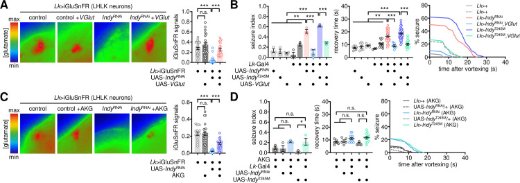
A previous study suggested that LK neurons are unlikely glutamatergic [56]. However, we found additional evidence supporting that LHLK neurons indeed express the glutamatergic marker VGlut. LK neuron-specific expression of B3 recombinase led to the genomic excision of VGlut coding sequence flanked by the B3 recombination target sites from the genome-edited VGlut allele (B3RT-VGlut-B3RT-LexA) [58], thereby driving the downstream expression of a transgenic LexA driver only in VGlut-expressing LK neurons (S9A Fig). Assessment of the transgene expression in the adult fly brain revealed that LHLK neurons, but not SELK neurons, expressed the VGlut-derived LexA (S9B Fig), indicating VGlut expression only in LHLK neurons. Given these observations, we asked whether Indy controls the glutamate transmission from LHLK neurons for seizure suppression. A transgenic fluorescence sensor for detecting synaptic release of glutamate [59] validated that INDY depletion in LHLK neurons lowered the levels of glutamate release (Fig 5A). Transgenic overexpression of wild-type VGLUT not only rescued the glutamate transmission in INDY-depleted LHLK neurons (Fig 5A) but also suppressed their BSS phenotypes (Fig 5B). These observations indicate that the glutamate transmission from INDY-depleted LHLK neurons is likely limiting for seizure suppression.
Fig 5. LK neurons suppress seizures via metabolic control of glutamate transmission.
(A) Overexpression of wild-type VGLUT rescues low glutamate levels in INDY-depleted LHLK neurons. The fluorescent glutamate sensor, iGluSnFR, was co-expressed with IndyRNAi and wild-type VGLUT transgenes in LK neurons. Fluorescence images of LHLK neurons in dissected brains were recorded using photoactivated localization microscopy and analyzed using ZEN software. Relative fluorescence was calculated by normalizing to backgrounds and was averaged for each genotype (n = 19–38). A significant Indy x VGlut interaction effect on the iGluSnFR signal (P < 0.0001) was detected by Aligned ranks transformation ANOVA. Error bars indicate SEM. n.s., not significant; ***P < 0.001, as determined by Wilcoxon rank sum test. (B) Overexpression of wild-type VGLUT suppresses BSS caused by the loss of Indy function in LK neurons. Quantitative analyses of BSS in individual flies were performed as described in Fig 1. Data represent means ± SEM (seizure index, n = 56–60 flies in 3 independent experiments; recovery time, n = 3–37 flies). **P < 0.01, ***P < 0.001, as determined by one-way ANOVA with Holm-Sidak’s multiple comparisons test. (C) Oral administration of AKG rescues low glutamate levels in INDY-depleted LHLK neurons. Transgenic flies were fed control or AKG-containing food (20 mM) for 3 d before live-brain imaging. A significant Indy x AKG interaction effect on the iGluSnFR signal (P = 0.0014) was detected by Aligned ranks transformation ANOVA. Data represent means ± SEM (n = 16–24). n.s., not significant; ***P < 0.001, as determined by Wilcoxon rank sum test. (D) Oral administration of AKG suppresses BSS caused by the loss of Indy function in LK neurons. Data represent means ± SEM (seizure index, n = 60 flies in 3 independent experiments; recovery time, n = 3–13 flies). n.s., not significant; *P < 0.05, as determined by one-way ANOVA with Holm-Sidak’s multiple comparisons test.
To validate the Indy-relevant link between the TCA cycle and glutamate transmission in seizure-suppressing LHLK neurons, we performed two additional experiments. First, oral administration of α-ketoglutarate partially but significantly restored the glutamate transmission in INDY-depleted LHLK neurons (Fig 5C) and suppressed the BSS phenotypes induced by loss of Indy function in LK neurons (Fig 5D). Second, IDH3 depletion in LK neurons was sufficient to induce BSS (S10 Fig), phenocopying the seizure induction by the pan-glutamatergic IDH3 depletion (S6 Fig). Taken together, these results demonstrate that a pair of glutamatergic LK neurons mediates Indy-dependent seizure suppression.
LK neuron activity gates the behavioral output from seizure initiation
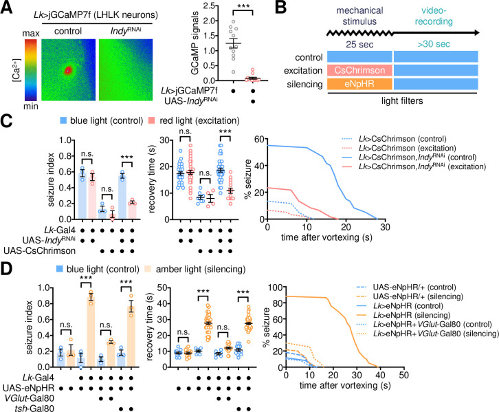
LK signaling has been implicated in neural physiology and behaviors relevant to metabolism in Drosophila [55,57,60–64]. It has also been shown that Indy mutant flies physiologically mimic calorie-restricted animals and exhibit low fat levels [16], raising the possibility that energy deficiency might underlie Indy mutant seizure. Nonetheless, we found some evidence against this idea. First, a lipid-rich diet did not rescue Indy mutant seizures while significantly suppressing BSS phenotypes in other seizure mutants [37] (S11A Fig). We note, however, the lipid-rich diet did not rescue low glucose levels in Indy mutants (S11B Fig). Second, LK neuron-specific loss of Indy function did not affect the baseline levels of triglyceride and glucose (S11C Fig) and the corresponding seizure was insensitive to the lipid-rich supplement (S11D Fig). Finally, hypomorphic mutants of Lk and Lk receptor (Lkr) genes did not exhibit any detectable phenotypes in seizure index under our experimental conditions (S12 Fig). We thus reasoned that LK neuron activity, but not the LK signaling or metabolism per se, might be critical for Indy-dependent seizure suppression. Live-brain imaging revealed that INDY depletion reduced the baseline levels of intracellular Ca2+ in LHLK neurons (Fig 6A), possibly reflecting their low activity. We asked whether transgenic manipulations of LK neuron activity could either suppress or mimic INDY-depletion phenotypes in BSS.
Fig 6. LK neuron activity gates the behavioral output from seizure initiation.
(A) INDY depletion reduces intracellular Ca2+ levels in LHLK neurons. The codon-optimized calcium sensor, jGCaMP7f, was co-expressed with IndyRNAi in LK neurons. The fluorescence signal in LHLK neurons was analyzed as described in Fig 5A. Data represent means ± SEM (n = 11–12). ***P < 0.001, as determined by Mann-Whitney test. (B) Experimental design for optogenetic manipulations of neural activity during the assessment of BSS. (C) Optogenetic excitation of LK neurons substantially suppresses BSS in INDY-depleted flies. Transgenic flies were crossed and kept in constant darkness. The mechanical stimulus was given under blue light (no excitation) or red light (excitation by CsChrimson), and BSS phenotypes were then assessed under blue light. Quantitative analyses of BSS in individual flies were performed as described in Fig 1. Data represent means ± SEM (seizure index, n = 56–60 flies in 3 independent experiments; recovery time, n = 4–33 flies). n.s., not significant; ***P < 0.001, as determined by two-way ANOVA with Holm-Sidak’s multiple comparisons test. (D) Optogenetic silencing of glutamatergic LK neurons induces BSS in a wild-type background. The mechanical stimulus was given under blue light (no silencing) or amber light (silencing by eNpHR). Data represent means ± SEM (seizure index, n = 59–60 flies in 3 independent experiments; recovery time, n = 6–52 flies). n.s., not significant; ***P < 0.001, as determined by two-way ANOVA with Holm-Sidak’s multiple comparisons test.
To this end, optogenetic transgenes were combined with different genetic tools to excite or silence LK neuron activity only during seizure induction for the assessment of their effects on BSS (Fig 6B). Transgenic flies expressing CsChrimson [65] were exposed to red light to transiently excite LK neurons during a mechanical stimulus. This manipulation significantly suppressed BSS phenotypes in Indy RNAi flies (Fig 6C), whereas the light transition per se negligibly affected Indy-dependent seizure. These observations validate that Indy RNAi phenotypes are readily reversible in adult LK neurons within a range of seconds, excluding the possibility that any long-term effects of INDY depletion on neuronal development or metabolic stress contribute to the seizure control. We consistently found that amber light-dependent silencing of LHLK neurons by the eNpHR transgene [66] was sufficient to induce BSS, even in a wild-type background (Fig 6D). These results demonstrate that LK neuron activity inversely correlates with seizure susceptibility. Nonetheless, the optogenetic inhibition of LK neuron activity alone did not trigger seizure-like behaviors in the absence of a mechanical stimulus (S13 Fig). We reason that this neural locus may not initiate seizure-like behaviors but inhibit the behavioral output from an as-yet-unmapped seizure-initiating locus in the adult Drosophila brain.
NMDA receptors in dorsal fan-shaped body neurons act downstream of LHLK neurons to suppress BSS
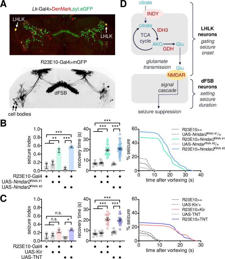
Although the implication of LK-LKR signaling in Indy-dependent seizure was not evident, we reasoned that LKR-expressing neurons could still act downstream of LHLK neurons via the seizure-suppressing glutamate transmission. It has been shown that LKR neurons are present in distinct parts of the adult fly brain, including the pars intercerebralis, ellipsoid body, and fan-shaped body (FSB) [55,57,60]. Also, our transgenic expression of the GFP-fused synaptotagmin 1 revealed LHLK projections in the LH and FSB (Fig 7A). We thus employed a dorsal FSB (dFSB)-specific Gal4 driver (i.e., R23E10-Gal4) to examine whether glutamate receptors expressed in dFSB neurons contribute to the susceptibility of BSS. We indeed found that dFSB-specific depletion of NMDAR2 was sufficient to cause BSS phenotypes, comparable to the high seizure index and long recovery time observed in Nmdar2 mutants (Fig 7B). Neither electrical silencing of dFSB neurons by the inwardly rectifying Kir2.1 channel [67] nor blocking their synaptic transmission by tetanus toxin light chain (TNT) [68] significantly affected seizure index; however, both the transgenic manipulations lengthened the recovery time in BSS-positive animals (Fig 7C). These data together map the metabolic control of seizure onset to the glutamate transmission between LHLK and dFSB neurons and suggest that the subsequent output of dFSB neurons may govern the duration of seizure-like behaviors.
Fig 7. NMDA receptors in dorsal fan-shaped body neurons relay seizure-suppressing glutamate transmission.
(A) Representative confocal images of dendrites (DenMark, red) and axonal projections (syt.eGFP, green) from LHLK neurons (top) and mGFP expression from dFSB-specific R23E10-Gal4 driver (bottom) in the adult fly brain. (B) Depletion of NMDA receptors in dFSB neurons induces BSS. Quantitative analyses of BSS in individual flies were performed as described in Fig 1. Data represent means ± SEM. **P < 0.01, ***P < 0.001, as determined by one-way ANOVA with Holm-Sidak’s multiple comparisons test (seizure index, n = 60 in 3 independent experiments) or by Welch’s ANOVA with Dunnett’s multiple comparisons test (recovery time, n = 3–34). (C) Genetic silencing of dFSB neurons lengthens recovery time after BSS. Data represent means ± SEM. n.s., not significant; *P < 0.05, ***P < 0.001, as determined by one-way ANOVA with Holm-Sidak’s multiple comparisons test (seizure index, n = 60 in 3 independent experiments; recovery time, n = 5–16 for TNT) or by Welch’s ANOVA with Dunnett’s multiple comparisons test (recovery time, n = 7–14 for Kir). (D) A working model for the neurometabolic pathway of Indy-dependent seizure suppression.
Discussion
Human genetic studies have previously identified several loss-of-function alleles of ROGDI or SLC13A5 among KTS-associated loci. In the current study, we behaviorally assessed the susceptibility to a specific form of seizure-like activity in Drosophila mutants of the KTS-associated genes. We defined genetic, biochemical, and neural pathways of SLC13A5/Indy-relevant seizure in the context of the citric acid cycle, glutamate metabolism, and glutamatergic transmission (Fig 7D). We further mapped the reversible and scalable functions of the seizure suppressor gene to a surprisingly small group of glutamatergic neurons (i.e., a pair of LHLK neurons) in the adult fly brain. In a broad sense, these findings demonstrate a specific micro-neural circuit that gates both initiation and duration of seizure-like behaviors.
Given the citrate transporter activity of SLC13A5/INDY, it has been suggested that neuronal energy failure may cause SLC13A5-associated epilepsy [7,8]. Citrate acts as a precursor of fatty acid biosynthesis while also serving as a key intermediate in the TCA cycle. Accordingly, an Indy mutation mimics calorie-restricted conditions and thereby causes metabolic and longevity phenotypes in Drosophila and mouse models, including alterations in lipid metabolism (i.e., low-fat storage), insulin signaling, and mitochondrial biogenesis [16,21]. Perturbations of the TCA cycle and altered levels of extracellular TCA metabolites are consistently observed in SLC13A5-associated KTS patients [69]. In fact, genetic losses of other metabolic enzymes involved in the TCA cycle (e.g., IDH3, malate dehydrogenase, fumarase hydratase) have been implicated in several neurological disorders, including early-onset seizure and developmental delay [70–74]. Nonetheless, there are conflicting reports on the effects of a ketogenic diet on KTS-associated epilepsy [8].
Genetic or pharmacological manipulations of metabolic enzymes and relevant genes likely impact on general cell physiology. The long-term metabolic stress may thus lead to pleiotropic effects and poor phenotypic outcomes, possibly explaining the seizure phenotypes observed in our genetic models. Nonetheless, several lines of our evidence support a more specific mechanism for Indy-dependent seizure suppression. First, Indy mutants did not display any gross motor defects, while their seizure phenotypes were mapped very specifically to a small group of the glutamatergic LK neurons among other broadly defined groups of neurons (e.g., GABAergic or cholinergic neurons). Second, Indy displayed specific genetic interactions with Idh3, VGlut, and select glutamate receptors on BSS in trans-heterozygous conditions, whereas BSS were not detected in individual heterozygous mutants. Third, genetic enhancement of the glutamate transmission by VGLUT overexpression in LK neurons was sufficient to suppress BSS in INDY-depleted flies. Finally, the INDY-depletion phenotypes were readily reversible in the adult LK neurons (i.e., AKG administration, conditional RNAi) and the optogenetic excitation of INDY-depleted LK neurons only during a mechanical stimulus was sufficient to suppress BSS although the possible off-target effects of the Indy RNAi transgene were not completely excluded. These observations together support our model that impairment of the metabolic flux between the TCA cycle and glutamate biosynthesis—specifically, the conversion of α-ketoglutarate to glutamate—down-scales the seizure-suppressing transmission from a specific subset of glutamatergic neurons, resulting in BSS phenotypes.
Previous studies in animal models have actually implicated low levels of TCA metabolites and glutamate in epilepsy [73,75–79]. Moreover, co-injection of α-ketoglutarate suppresses chemically induced epilepsy in mice [80,81], consistent with our results. Intriguingly, the recent observation that α-ketoglutarate promotes the interaction between synaptotagmin 1 and phospholipids demonstrates an unexpected role of α-ketoglutarate in synaptic vesicle fusion [51]. Thus, we reasoned that an excess of α-ketoglutarate might deplete synaptic vesicle pools and thereby interfere with their transmission. This explains why oral administration of α-ketoglutarate to wild-type flies induces rather than suppresses BSS under our experimental conditions.
Although Drosophila genetic studies have led to the isolation and characterization of a number of seizure-related mutants, much less is known about the neural loci responsible for inducing seizure and sustaining seizure-like activity [82]. For instance, the seizure-suppressing function of the potassium-chloride symporter kazachoc and seizure-inducing neural locus in a BSS mutant of the voltage-gated sodium channel paralytic have been mapped to the mushroom body in the adult brain [83,84]. Seizure suppression by the product of the transmembrane domain gene, julius seizure, was more broadly mapped to cholinergic or GABAergic neurons but not to glutamatergic neurons [85]. The emergence of a seizure has been thought to involve three distinct components of neural circuits that mediate seizure initiation, seizure buildup, and seizure spread, respectively [86]. Modulatory neurons likely regulate the excitability of these neural components and thus shape seizure intensity or duration. Based on this model, we propose that glutamatergic LK neurons mediate a seizure-inhibitory pathway such that their conditional silencing de-represses both seizure onset and duration. Accordingly, down-regulation of the glutamatergic transmission from LK neurons induces BSS, as opposed to the general involvement of excitatory glutamate in seizure induction [44–46].
Our data suggest that LK signaling is not directly involved in controlling seizure susceptibility, which makes sense considering the temporal scale of neuropeptide signaling in general. Nonetheless, we found that the metabolic flux of the TCA cycle in LK neurons and their glutamate transmission onto dFSB neurons might be closely linked to the control of BSS. Evidence for the involvement of LK neurons in feeding, metabolism, and associated physiology (e.g., circadian behaviors, sleep, memory) is abundant [55,57,60–64]. The activity of LHLK neurons is also sensitive to the metabolic state and the time of day [57,60,63]. Thus, we hypothesize that LK neurons act as a neural sensor that integrates internal cues (e.g., circadian clocks, sleep state, and metabolic state) while engaging different postsynaptic partners and divergent pathways for relevant physiological outputs through distinct signaling molecules. In particular, dFSB neurons are a key sleep-promoting locus in the adult fly brain, analogous to the sleep-promoting ventrolateral preoptic nucleus (VLPO) in mammals [87,88]. Given the implication of VLPO in consciousness loss during epileptogenesis [89], the LK-dFSB pathway may reveal the conserved neural principles underlying intimate interactions among metabolism, epilepsy, and sleep [90–92].
Drosophila models of human genetic disorders have proven to be valuable tools for elucidating underlying pathogenic mechanisms. Our findings enrich this body of knowledge by providing a genetic, biochemical, and neural map of the seizure suppressor pathway related to KTS. The complexity of the mammalian brain and species-specific organization of excitatory neurotransmitters in the nervous systems may limit the direct relevance of our working model to KTS pathogenesis at neural-circuit levels. Nonetheless, given the strong conservation of SLC13A5/Indy homologs and their physiological function among different species, we propose that similar cellular mechanisms may underlie early-onset seizures in KTS patients and explain their relative resistance to generic antiepileptic drugs.
Materials and methods
Drosophila stocks
All flies were maintained in standard cornmeal–yeast–agar medium at room temperature. w1118 (BL5905; a wild-type control), Indy206 (BL27901), Indy302 (BL27902), Idh3gG9423 (BL30194; CG5028), LkC275 (BL16324), LkrMI06336 (BL41520), LkrMI08640 (BL51094), Nmdar1EP331 (BL17331), Nmdar2attP (BL84548), GluRIAattP (BL84506), GluRIBattP (BL84507), mGluRMI02169(BL32830), GluClαglc1(BL6353), VGlutOK371-Gal4 (BL26160), R23E10-Gal4 (BL49032), VGlut-Gal80 (BL58448), tub-Gal80ts (BL7018), 20XUAS-eNpHR3.0.YFP (BL36350), 20XUAS-IVS-CsChrimson.mVenus (BL55135), 20XUAS-iGluSnFR.A184A (BL59609), UAS-Denmark, UAS-syt.eGFP (BL33065), UAS-Nmdar2RNAi (#1, BL26019), UAS-Nmdar2RNAi (#2, BL40846), 20XUAS-IVS-jGCaMP7f (BL80906), Tdc2[B3RT]-LexA; UAS-B3R, LexAop2-His2B-mCherry, UAS-His2A-GFP (BL91248), and VGlut[B3RT]-LexA (BL91249) were obtained from Bloomington Drosophila Stock Center. UAS-IndyRNAi #1 (3979R-1), UAS-IndyRNAi #2 (3979R-3), UAS-Idh3aRNAi (12233R-3), UAS-Idh3bRNAi (6439R-2), and UAS-Idh3gRNAi (5028R-2) were obtained from National Institute of Genetics. UAS-IndyRNAi #3 (v9981), UAS-IndyRNAi #4 (v9982), UAS-rogdiRNAi (v107310) was obtained from Vienna Drosophila Resource Center. parabss, eas2, and sdaiso7.8 were gifts from Dr. Kitamoto [36] and Dr. Kuebler [37]. VGlut1, VGlut2, and UAS-VGlut were gifts from Dr. DiAntonio [40]. rogdidel, rogdiP1, UAS-rogdi-3xFLAG, UAS-Kir2.1, UAS-TNT, Lk-Gal4, and tsh-Gal80 have been described previously [32,56,67,68,93]. Indy-PB cDNA was PCR-amplified from a cDNA clone (RH67364, Drosophila Genomics Resource Center) and inserted into pUAST-attB-V5 [94]. Subsequently, a T[ACT]>M[ATG] mutation was introduced to the wild-type cDNA to mimic the KTS-associated 680C>T allele. UAS-Indy and UAS-IndyT245M transgenic lines were generated by site-specific transformation on the attP40 landing site (BestGene Inc.)
Bang-sensitive seizure analysis
To avoid any genetic-background effects on bang-sensitive seizure, Indy mutant stocks were outcrossed six times to the wild-type control (w1118) for isogenization. All the behavioral tests for heterozygous, homozygous, and trans-heterozygous mutants were conducted with the isogenized Indy lines and w1118 control. For transgenic lines, seizure behaviors in trans-heterozygous animals were compared to those in all appropriate heterozygous controls. Where applicable, additional transgenic combinations or independent transgenes were tested for validation. Unless otherwise indicated, BSS was assessed in male flies. Three to seven-day-old flies were fed cornmeal–yeast–agar medium in LD cycles at 25°C for 3 d. They were CO2-anesthetized and harvested into fresh vials containing the same food >3 h before a group of flies (n = 5 for non-optogenetic experiments; n = 5–10 for optogenetic experiments) was transferred to an empty vial using an aspirator. Each vial was vortexed at a maximum speed for 25 s using Vortex-Genie 2 (Scientific Industries, Inc.) and then video-recorded for >30 s using a cellular phone (LG G5 Pro or Samsung Galaxy Note 8). BSS-positive flies were defined if they displayed initial paralysis upon mechanical stimulus, followed by uncoordinated movements such as wing-flapping, abdominal contractions, or leg-twitching before their recovery of a normal posture. BSS was quantitatively assessed by three parameters. A seizure index was calculated as the ratio of the number of BSS-positive flies to the total number of flies tested in each experiment (n = ~20 flies per genotype or condition) and averaged from three independent experiments. Recovery time was calculated individually for BSS-positive flies as the latency to normal posture after vortexing and was averaged for each genotype or condition. Percent seizure was calculated as the percentage of BSS-positive flies at each second after vortexing (n = ~60 flies per genotype or condition from all the three experiments). To determine the length of a refractory period in Indy mutants, the first mechanical stimulus was given to each fly and then the second mechanical stimulus was given only to BSS-positive animals at the indicated time after recovery from their first BSS. The seizure index was calculated in each experiment (n = 10 flies per time point) and averaged from three independent experiments.
Locomotor behavior analysis
Locomotor activities in individual male flies were measured in 1-min bins using the Drosophila Activity Monitor system (TriKinetics). Daily locomotor activity (total activity count per 12-hour light: 12-hour dark cycle) and waking activity (averaged activity count per minute awake) were analyzed as described previously [95,96]. For the climbing activity measurement, a group of 10 male flies was kept in the climbing chamber. Flies were allowed to climb for 5 seconds after gentle tapping-down. Climbing distance in each fly was measured as the highest position during the recording.
Oral administration of chemicals
Each chemical was dissolved in 5% sucrose and 2% agar food (behavior food) at the indicated concentration and fed to flies for 3 d before the BSS assessment. The chemicals tested included epigallocatechin gallate (EGCG, Sigma), diethylstilbestrol (DES, Sigma), α-ketoglutarate (Sigma), and KetoCal 4: 1 (fat: carbohydrate plus protein, Nutricia).
Optogenetics
Transgenic flies were grown in light-tight vials and kept in constant dark. They were loaded on to the behavior food containing 0.1 mM all-trans retinal (Sigma) and entrained at 25°C for 3 d. A light source with the indicated wavelength was turned on during the mechanical stimulation. A white LED was covered with a 585-nm or 470-nm filter to generate amber or blue light, respectively, while the 630-nm LED provided red light.
Immunofluorescence imaging
Immunostaining of whole-mount adult brains was performed as described previously [97]. Dissected brains were fixed with 3.7% formaldehyde in PBS, blocked with PBS containing 0.3% Triton X-100 and 5% normal goat serum, and incubated with anti-GFP antibody (Invitrogen, A-11122; diluted at 1:1000) or anti-mCherry antibody (Developmental Studies Hybridoma Bank, DSHB-mCherry-3A11; diluted at 1:20) at 4°C overnight. After washing in PBS containing Triton X-100, immunostained samples were further incubated with species-specific Alexa Fluor secondary antibodies (Jackson ImmunoResearch) at 4°C overnight, washed with PBS containing Triton X-100, and then mounted using VECTASHIELD mounting medium (Vector Laboratories). Confocal images were obtained using an FV1000 microscope (Olympus) and analyzed using ImageJ software.
Live-brain imaging
Transgenic flies were loaded on to the behavior food containing 0 mM (control) or 20 mM α-ketoglutarate (Sigma), entrained at 25°C for 3 d, and then anesthetized in ice. Whole brains were dissected out in hemolymph-like HL3 solution (5 mM HEPES pH 7.2, 70 mM NaCl, 5 mM KCl, 1.5 mM CaCl2, 20 mM MgCl2, 10 mM NaHCO3, 5 mM trehalose, 115 mM sucrose) and then transferred on cover glass in a magnetic imaging chamber (Chamlide CMB, Live Cell Instrument) filled with HL3 buffer. Fluorescence images from live brains were recorded at room temperature using photoactivated localization microscopy (ELYRA P.1, Carl Zeiss) with a C-Apochromat 40x/1.20 W Korr M27 at a pixel resolution of 512 x 512. Fluorescence signals were quantified by background subtraction and analyzed using ZEN software (Carl Zeiss).
Quantitative transcript analysis
Total RNA was purified from thirty fly heads using TRIzol reagent (Thermo Fisher Scientific). After DNase I digestion, cDNA was synthesized using M-MLV Reverse Transcriptase (Promega) and random hexamers. Quantitative real-time PCR was performed using Prime Q-Mastermix (GeNet Bio) and LightCycler 480 Instrument II (Roche). The qPCR primers used in this study were as follows: 5’-TTC ATC GCT TCA CGT CAC TC-3’ (forward) and 5’-TGC TGA CTT GGT GGA TTT TG-3’ (reverse) for rogdi; 5’-ATC TCC CAC AGG ACG TCA AC-3’ (forward) and 5’-GCG ACG AAG AGA AGG ATC AC-3’ (reverse) for pabp (internal control). Indy primers have been described previously [16].
Amino acid profiling
Male flies were loaded on to behavior food containing either 0 mM (control) or 20 mM α-ketoglutarate and then entrained in LD cycles at 25°C for 3 d before harvest. Whole-body extracts were prepared from 50 flies per condition, and the levels of free amino acids were quantitatively measured using ion-exchange chromatography as described previously [95,96].
Metabolite measurements
Whole-body extracts were prepared by homogenizing 8 male flies per sample in 200 μl of PBS containing 0.05% Tween-20. Triglyceride and glucose measurements were performed using Triglyceride Reagent (Sigma-Aldrich, T2449), Free Glycerol Reagent (Sigma-Aldrich, F6428), and Glucose Assay Reagent (Sigma-Aldrich, G3293), respectively, according to the manufacturers’ instructions.
Statistical analysis
Statistical analyses were performed using GraphPad Prism, R (version 3.5.3), or Microsoft Excel. Shapiro-Wilk and Brown-Forsythe tests were conducted to check normality (P < 0.05) and equality of variances (P < 0.05), respectively. For comparisons among multiple samples: 1) Parametric datasets with equal variance were further analyzed by ordinary ANOVA. 2) Parametric datasets with unequal variance were further analyzed by Welch’s ANOVA (one-way) or Aligned ranks transformation ANOVA (two-way). 3) Nonparametric datasets with equal variance were analyzed by Kruskal-Wallis ANOVA (one-way) or Aligned ranks transformation ANOVA (two-way). 4) Nonparametric datasets with unequal variance were analyzed by Aligned ranks transformation ANOVA. Post hoc multiple comparisons were performed by Holm-Sidak’s (ordinary ANOVA), Dunnett’s T3 (Welch’s ANOVA), Wilcoxon rank sum (Aligned ranks transformation ANOVA), or Dunn’s test (Kruskal-Wallis ANOVA). For comparisons between two samples, nonparametric datasets were analyzed by Mann-Whitney U test. Datasets including any sample size less than 7 were analyzed by Student’s t-test or ordinary ANOVA. Sample sizes and P values obtained from individual statistical analyses were indicated in the figure legends.
Supporting information
(A) The seizure phenotypes in Indy mutants were comparable to those observed in easily shocked (eas2) and slamdance (sdaiso7.8) but weaker than bang senseless (parabss) mutants. Quantitative analyses of BSS in individual flies were performed as described in Fig 1. Data represent means ± SEM (seizure index, n = 60 flies in 3 independent experiments; recovery time, n = 2–59 flies). n.s., not significant; *P < 0.05, ***P < 0.001, as determined by one-way ANOVA with Holm-Sidak’s multiple comparisons test. (B) Indy mutant seizure displays a refractory period after seizure recovery. The first mechanical stimulus was given to each fly and then the second mechanical stimulus was given only to BSS-positive animals at the indicated time after recovery from their first BSS. The seizure index was calculated in each experiment (n = 10 flies per time point) and averaged from three independent experiments. Error bars indicate SEM.
(TIF)
(A) Pan-neuronal overexpression of IndyRNAi transgene reduces endogenous Indy expression. Total RNA was purified from fly heads. Quantitative analyses of Indy and poly(A)-binding protein (normalizing control) mRNAs were performed using real-time PCR with gene-specific primer sets. The relative levels of Indy mRNA in each genetic background were calculated by normalizing to Elav>Dcr2 control (set as 1). Data represent means ± SEM (n = 3). ***P < 0.001 as determined by one-way ANOVA with Holm-Sidak’s multiple comparisons test. (B) A genetic screen identifies VGlut- and Lk-expressing neurons as neural loci important for Indy-dependent control of the seizure susceptibility. Quantitative analyses of BSS in individual flies were performed as described in Fig 1. Data represent means ± SEM (n = 60 flies in 3 independent experiments). **P < 0.01, ***P < 0.001, as determined by one-way ANOVA with Holm-Sidak’s multiple comparisons test. (C) The BSS induction by glutamatergic INDY depletion is consistently observed by independent Indy RNAi transgenes. Data represent means ± SEM (seizure index, n = 60 flies in 3 independent experiments; recovery time, n = 1–38 flies). *P < 0.05, **P < 0.01, ***P < 0.001, as determined by one-way ANOVA with Holm-Sidak’s multiple comparisons test.
(TIF)
Drosophila IndyT245M mutant mimics the gene product of the KTS-associated 680C>T allele. TM, a transmembrane region.
(TIF)
(A) rogdi mutants homozygous or trans-heterozygous for loss-of-function alleles display BSS. Quantitative analyses of BSS in individual flies were performed as described in Fig 1. Data represent means ± SEM. **P < 0.01, ***P < 0.001 as determined by one-way ANOVA with Holm-Sidak’s multiple comparisons test (seizure index, n = 60 flies in 3 independent experiments) or by Aligned ranks transformation ANOVA with Wilcoxon rank sum test (recovery time, n = 7–29 flies). (B) Pan-neuronal overexpression of rogdiRNAi transgene reduces endogenous rogdi expression in fly heads. Quantitative transcript analyses were performed as described in S2A Fig. Data represent means ± SEM (n = 3). ***P < 0.001 as determined by one-way ANOVA with Holm-Sidak’s multiple comparisons test. (C) ROGDI depletion in glutamatergic neurons is sufficient to induce BSS. Quantitative analyses of BSS in individual flies were performed as described in Fig 1. Data represent means ± SEM (seizure index, n = 60 flies in 3 independent experiments; recovery time, n = 5–27 flies). ***P < 0.001, as determined by one-way ANOVA with Holm-Sidak’s multiple comparisons test. (D) Transgenic overexpression of wild-type ROGDI in glutamatergic neurons rescues BSS in rogdi mutants. Data represent means ± SEM (seizure index, n = 49–60 flies in 3 independent experiments; recovery time, n = 6–26 flies). n.s., not significant; **P < 0.01, ***P < 0.001, as determined by two-way ANOVA with Holm-Sidak’s multiple comparisons test.
(TIF)
Flies were fed control or DES-containing food (10 or 20 μg/mg) for 3 d before the BSS assessment. Quantitative analyses of BSS in individual flies were performed as described in Fig 1. Two-way ANOVA detected a significant Indy x DES interaction effect on recovery time (P = 0.0421). Data represent means ± SEM (seizure index, n = 60 flies in 3 independent experiments; recovery time, n = 3–49 flies). n.s., not significant; **P < 0.01, ***P < 0.001, as determined by Holm-Sidak’s multiple comparisons test.
(TIF)
Quantitative analyses of BSS in individual flies were performed as described in Fig 1. Data represent means ± SEM. **P < 0.01, ***P < 0.001 as determined by one-way ANOVA with Holm-Sidak’s multiple comparisons test (seizure index, n = 60 flies in 3 independent experiments), by Aligned ranks transformation ANOVA with Wilcoxon rank sum test (recovery time, n = 8–43 flies for Idh3aRNAi or Idh3bRNAi), or by Welch’s ANOVA with Dunnett’s T3 multiple comparisons test (recovery time, n = 8–35 flies for Idh3gRNAi).
(TIF)
(A) w1118 control and Indy mutant flies show similar waking activities under 12-h light: 12-h dark (LD) cycles. Individual male flies were transferred to 65 × 5 mm glass tubes containing 5% sucrose and 2% agar food and entrained in LD cycles. Locomotor activities were indirectly measured by infrared beam crosses per minute using the Drosophila Activity Monitor system. Daily locomotor activity (activity/day) and waking activity (activity/min awake) were calculated in each fly on the fourth LD cycles and averaged (n = 25 and 29 flies for w1118 control and Indy206 mutants, respectively). Error bars indicate SEM. n.s., not significant; *P < 0.05 as determined by Mann-Whitney U test. (B) w1118 control and Indy mutant flies display comparable climbing activities. A group of 10 male flies was kept in the climbing chamber and then allowed to climb for 5 seconds after gentle tapping-down. Climbing distance in each fly was measured as the highest position during the recording and averaged (n = 30). Error bars indicate SEM. n.s., not significant as determined by Student’s t-test.
(TIF)
Adult-specific INDY depletion in LK neurons is sufficient to induce BSS. Transgenic flies were crossed and kept at 21°C to block the expression of IndyRNAi transgene using tub-Gal80ts. Adult flies were then incubated at 21°C (no depletion) or 29°C (RNAi-mediated depletion) for >24 hours prior to the assessment of BSS at the same temperature. Quantitative analyses of BSS in individual flies were performed as described in Fig 1. Data represent means ± SEM (seizure index, n = 60 flies in 3 independent experiments; recovery time, n = 5–28). n.s., not significant; **P < 0.01, ***P < 0.001, as determined by one-way ANOVA with Holm-Sidak’s multiple comparisons test.
(TIF)
(A) A transgenic strategy for visualizing VGlut-expressing LK neurons by the fluorescent reporter proteins. The CRISPR-edited VGlut locus includes two B3 recombination target (B3RT) sites upstream of the LexA-coding sequence. LK neuron-specific overexpression of the B3 recombinase leads to the genomic excision, thereby allowing LexA expression only in VGlut-expressing LK neurons. LexA expression could be indirectly visualized by the transgenic mCherry reporter. (B) LHLK neurons, but not SELK neurons, express B3RT-LexA transgene from the VGlut-deleted locus. Representative confocal images of LHLK and SELK neurons in the adult fly brain were shown along with the full genotype.
(TIF)
Quantitative analyses of BSS in individual flies were performed as described in Fig 1. Data represent means ± SEM (seizure index, n = 44–60 in 3 independent experiments; recovery time, n = 8–27). *P < 0.05, **P < 0.01, ***P < 0.001, as determined by one-way ANOVA with Holm-Sidak’s multiple comparisons test (seizure index), by Aligned ranks transformation ANOVA with Wilcoxon rank sum test (recovery time, Idh3a and Idh3b RNAi), or by Welch’s ANOVA with Dunnett’s multiple comparisons test (recovery time, Idh3g RNAi).
(TIF)
(A) A lipid-rich diet rescues BSS in eas2 mutants but not in Indy206 mutants. Flies were fed control or 5% KetoCal food for 3 d before the BSS assessment. Quantitative analyses of BSS in individual flies were performed as described in Fig 1. Two-way ANOVA detected a significant eas x KetoCal interaction effect on seizure index (P = 0.0006). Data represent means ± SEM (seizure index, n = 60 flies in 3 independent experiments; recovery time, n = 4–34). n.s., not significant; *P < 0.05, **P < 0.01, ***P < 0.001, as determined by Holm-Sidak’s multiple comparisons test. (B) The lipid-rich diet does not rescue low glucose levels in Indy mutants. Flies were fed control or 5% KetoCal (4: 1 = fat: carbohydrate plus protein) food for 3 d before harvesting. Triglyceride and glucose levels in whole-body extracts were quantified using standard curves. Two-way ANOVA detected significant Indy x KetoCal interaction effects on triglyceride (P = 0.0019) and glucose levels (P = 0.0228). Data represent means ± SEM (n = 5–8). n.s., not significant; *P < 0.05, ***P < 0.001, as determined by Holm-Sidak’s multiple comparisons test. (C) Loss of Indy function in LK neurons does not cause a change in metabolite levels. Data represent means ± SEM (n = 4–5). n.s., not significant as determined by two-way ANOVA with Holm-Sidak’s multiple comparisons test. (D) The lipid-rich diet does not rescue BSS induced by LK neuron-specific loss of Indy function. Two-way ANOVA detected no significant Indy x KetoCal interaction effect on seizure index (P = 0.7802 for IndyRNAi; P = 0.1725 for IndyT245M) and recovery time (P = 0.9534 for IndyRNAi; P = 0.7273 for IndyT245M). n.s., not significant; *P < 0.05, **P < 0.01, ***P < 0.001, as determined by Holm-Sidak’s multiple comparisons test.
(TIF)
Quantitative analyses of BSS in individual flies were performed as described in Fig 1. Data represent means ± SEM. n.s., not significant; *P < 0.05 as determined by Mann-Whitney U test (seizure index, n = 60 in 3 independent experiments for Lk; recovery time, n = 8–9 flies for Lk) or by one-way ANOVA with Holm-Sidak’s multiple comparisons test (seizure index, n = 54–58 in 3 independent experiments for Lkr; recovery time, n = 4–10 flies for Lkr).
(TIF)
Transgenic flies were crossed and kept in constant dark. Any behavioral changes upon exposure to blue (no silencing) or amber light (silencing by eNpHR) condition were examined accordingly. Data represent means ± SEM (seizure index, n = 30 flies in 3 independent experiments; recovery time, n = 2–6 flies). No significant differences in seizure index and recovery time were detected by two-way ANOVA with Holm-Sidak’s multiple comparisons test.
(TIF)
(MP4)
(XLSX)
Acknowledgments
We thank A. DiAntonio, S.J. Certel, D. Dickman, T. Kitamoto, D. Kuebler, R.S. Stowers, Bloomington Drosophila Stock Center, Korea Drosophila Resource Center, National Institute of Genetics, Vienna Drosophila Resource Center, Drosophila Genomics Resource Center, and Developmental Studies Hybridoma Bank for reagents; E.Y. Suh at Chungnam National University for amino acid analyses.
Data Availability
All relevant data are within the manuscript and its Supporting Information files.
Funding Statement
This work was supported by grants from the Suh Kyungbae Foundation (SUHF-17020101[CL]); from the National Research Foundation funded by the Ministry of Science and Information & Communication Technology (MSIT), Republic of Korea (NRF-2018R1A5A1024261[CL]; NRF-2021M3A9G8022960[CL]), and by the Ministry of Education, Republic of Korea (NRF-2019R1I1A1A01063087[JL]). The funders had no role in study design, data collection and analysis, decision to publish, or preparation of the manuscript.
References
- 1.Schossig A, Wolf NI, Kapferer I, Kohlschutter A, Zschocke J. Epileptic encephalopathy and amelogenesis imperfecta: Kohlschutter-Tonz syndrome. Eur J Med Genet. 2012;55(5):319–22. doi: 10.1016/j.ejmg.2012.02.008 . [DOI] [PubMed] [Google Scholar]
- 2.Huckert M, Mecili H, Laugel-Haushalter V, Stoetzel C, Muller J, Flori E, et al. A Novel Mutation in the ROGDI Gene in a Patient with Kohlschutter-Tonz Syndrome. Mol Syndromol. 2014;5(6):293–8. Epub 2015/01/08. doi: 10.1159/000366252 ; PubMed Central PMCID: PMC4281576. [DOI] [PMC free article] [PubMed] [Google Scholar]
- 3.Mory A, Dagan E, Illi B, Duquesnoy P, Mordechai S, Shahor I, et al. A nonsense mutation in the human homolog of Drosophila rogdi causes Kohlschutter-Tonz syndrome. Am J Hum Genet. 2012;90(4):708–14. Epub 2012/04/10. doi: 10.1016/j.ajhg.2012.03.005 ; PubMed Central PMCID: PMC3322231. [DOI] [PMC free article] [PubMed] [Google Scholar]
- 4.Mory A, Dagan E, Shahor I, Mandel H, Illi B, Zolotushko J, et al. Kohlschutter-Tonz syndrome: clinical and genetic insights gained from 16 cases deriving from a close-knit village in Northern Israel. Pediatr Neurol. 2014;50(4):421–6. Epub 2014/03/19. doi: 10.1016/j.pediatrneurol.2014.01.006 . [DOI] [PubMed] [Google Scholar]
- 5.Schossig A, Wolf NI, Fischer C, Fischer M, Stocker G, Pabinger S, et al. Mutations in ROGDI Cause Kohlschutter-Tonz Syndrome. Am J Hum Genet. 2012;90(4):701–7. Epub 2012/03/20. doi: 10.1016/j.ajhg.2012.02.012 ; PubMed Central PMCID: PMC3322220. [DOI] [PMC free article] [PubMed] [Google Scholar]
- 6.Tucci A, Kara E, Schossig A, Wolf NI, Plagnol V, Fawcett K, et al. Kohlschutter-Tonz syndrome: mutations in ROGDI and evidence of genetic heterogeneity. Hum Mutat. 2013;34(2):296–300. doi: 10.1002/humu.22241 ; PubMed Central PMCID: PMC3902979. [DOI] [PMC free article] [PubMed] [Google Scholar]
- 7.Hardies K, de Kovel CG, Weckhuysen S, Asselbergh B, Geuens T, Deconinck T, et al. Recessive mutations in SLC13A5 result in a loss of citrate transport and cause neonatal epilepsy, developmental delay and teeth hypoplasia. Brain. 2015;138(Pt 11):3238–50. Epub 2015/09/20. doi: 10.1093/brain/awv263 . [DOI] [PubMed] [Google Scholar]
- 8.Klotz J, Porter BE, Colas C, Schlessinger A, Pajor AM. Mutations in the Na(+)/citrate cotransporter NaCT (SLC13A5) in pediatric patients with epilepsy and developmental delay. Mol Med. 2016;22. Epub 2016/06/05. doi: 10.2119/molmed.2016.00077 ; PubMed Central PMCID: PMC5023510. [DOI] [PMC free article] [PubMed] [Google Scholar]
- 9.Thevenon J, Milh M, Feillet F, St-Onge J, Duffourd Y, Juge C, et al. Mutations in SLC13A5 cause autosomal-recessive epileptic encephalopathy with seizure onset in the first days of life. Am J Hum Genet. 2014;95(1):113–20. Epub 2014/07/06. doi: 10.1016/j.ajhg.2014.06.006 ; PubMed Central PMCID: PMC4085634. [DOI] [PMC free article] [PubMed] [Google Scholar]
- 10.Schossig A, Bloch-Zupan A, Lussi A, Wolf NI, Raskin S, Cohen M, et al. SLC13A5 is the second gene associated with Kohlschutter-Tonz syndrome. J Med Genet. 2016. doi: 10.1136/jmedgenet-2016-103988 . [DOI] [PubMed] [Google Scholar]
- 11.Inoue K, Zhuang L, Maddox DM, Smith SB, Ganapathy V. Structure, function, and expression pattern of a novel sodium-coupled citrate transporter (NaCT) cloned from mammalian brain. J Biol Chem. 2002;277(42):39469–76. Epub 2002/08/15. doi: 10.1074/jbc.M207072200 . [DOI] [PubMed] [Google Scholar]
- 12.Knauf F, Rogina B, Jiang Z, Aronson PS, Helfand SL. Functional characterization and immunolocalization of the transporter encoded by the life-extending gene Indy. Proc Natl Acad Sci U S A. 2002;99(22):14315–9. Epub 2002/10/23. doi: 10.1073/pnas.222531899 ; PubMed Central PMCID: PMC137881. [DOI] [PMC free article] [PubMed] [Google Scholar]
- 13.Rogina B, Reenan RA, Nilsen SP, Helfand SL. Extended life-span conferred by cotransporter gene mutations in Drosophila. Science. 2000;290(5499):2137–40. Epub 2000/12/16. doi: 10.1126/science.290.5499.2137 . [DOI] [PubMed] [Google Scholar]
- 14.Toivonen JM, Gems D, Partridge L. Longevity of Indy mutant Drosophila not attributable to Indy mutation. Proc Natl Acad Sci U S A. 2009;106(21):E53; author reply E4. Epub 2009/05/14. doi: 10.1073/pnas.0902462106 ; PubMed Central PMCID: PMC2688976. [DOI] [PMC free article] [PubMed] [Google Scholar]
- 15.Toivonen JM, Walker GA, Martinez-Diaz P, Bjedov I, Driege Y, Jacobs HT, et al. No influence of Indy on lifespan in Drosophila after correction for genetic and cytoplasmic background effects. PLoS Genet. 2007;3(6):e95. Epub 2007/06/19. doi: 10.1371/journal.pgen.0030095 ; PubMed Central PMCID: PMC1892600. [DOI] [PMC free article] [PubMed] [Google Scholar]
- 16.Wang PY, Neretti N, Whitaker R, Hosier S, Chang C, Lu D, et al. Long-lived Indy and calorie restriction interact to extend life span. Proc Natl Acad Sci U S A. 2009;106(23):9262–7. Epub 2009/05/28. doi: 10.1073/pnas.0904115106 ; PubMed Central PMCID: PMC2685744. [DOI] [PMC free article] [PubMed] [Google Scholar]
- 17.Neretti N, Wang PY, Brodsky AS, Nyguyen HH, White KP, Rogina B, et al. Long-lived Indy induces reduced mitochondrial reactive oxygen species production and oxidative damage. Proc Natl Acad Sci U S A. 2009;106(7):2277–82. Epub 2009/01/24. doi: 10.1073/pnas.0812484106 ; PubMed Central PMCID: PMC2629441. [DOI] [PMC free article] [PubMed] [Google Scholar]
- 18.Rogers RP, Rogina B. Increased mitochondrial biogenesis preserves intestinal stem cell homeostasis and contributes to longevity in Indy mutant flies. Aging (Albany NY). 2014;6(4):335–50. Epub 2014/05/16. doi: 10.18632/aging.100658 ; PubMed Central PMCID: PMC4032799. [DOI] [PMC free article] [PubMed] [Google Scholar]
- 19.Fei YJ, Liu JC, Inoue K, Zhuang L, Miyake K, Miyauchi S, et al. Relevance of NAC-2, an Na+-coupled citrate transporter, to life span, body size and fat content in Caenorhabditis elegans. Biochem J. 2004;379(Pt 1):191–8. Epub 2003/12/18. doi: 10.1042/BJ20031807 ; PubMed Central PMCID: PMC1224044. [DOI] [PMC free article] [PubMed] [Google Scholar]
- 20.Schwarz F, Karadeniz Z, Fischer-Rosinsky A, Willmes DM, Spranger J, Birkenfeld AL. Knockdown of Indy/CeNac2 extends Caenorhabditis elegans life span by inducing AMPK/aak-2. Aging (Albany NY). 2015;7(8):553–67. Epub 2015/09/01. doi: 10.18632/aging.100791 ; PubMed Central PMCID: PMC4586101. [DOI] [PMC free article] [PubMed] [Google Scholar]
- 21.Birkenfeld AL, Lee HY, Guebre-Egziabher F, Alves TC, Jurczak MJ, Jornayvaz FR, et al. Deletion of the mammalian INDY homolog mimics aspects of dietary restriction and protects against adiposity and insulin resistance in mice. Cell Metab. 2011;14(2):184–95. Epub 2011/08/02. doi: 10.1016/j.cmet.2011.06.009 ; PubMed Central PMCID: PMC3163140. [DOI] [PMC free article] [PubMed] [Google Scholar]
- 22.Brachs S, Winkel AF, Tang H, Birkenfeld AL, Brunner B, Jahn-Hofmann K, et al. Inhibition of citrate cotransporter Slc13a5/mINDY by RNAi improves hepatic insulin sensitivity and prevents diet-induced non-alcoholic fatty liver disease in mice. Mol Metab. 2016;5(11):1072–82. Epub 2016/11/08. doi: 10.1016/j.molmet.2016.08.004 ; PubMed Central PMCID: PMC5081411. [DOI] [PMC free article] [PubMed] [Google Scholar]
- 23.Pesta DH, Perry RJ, Guebre-Egziabher F, Zhang D, Jurczak M, Fischer-Rosinsky A, et al. Prevention of diet-induced hepatic steatosis and hepatic insulin resistance by second generation antisense oligonucleotides targeted to the longevity gene mIndy (Slc13a5). Aging (Albany NY). 2015;7(12):1086–93. Epub 2015/12/10. doi: 10.18632/aging.100854 ; PubMed Central PMCID: PMC4712334. [DOI] [PMC free article] [PubMed] [Google Scholar]
- 24.Li L, Li H, Garzel B, Yang H, Sueyoshi T, Li Q, et al. SLC13A5 is a novel transcriptional target of the pregnane X receptor and sensitizes drug-induced steatosis in human liver. Mol Pharmacol. 2015;87(4):674–82. Epub 2015/01/30. doi: 10.1124/mol.114.097287 ; PubMed Central PMCID: PMC4366797. [DOI] [PMC free article] [PubMed] [Google Scholar]
- 25.Neuschafer-Rube F, Lieske S, Kuna M, Henkel J, Perry RJ, Erion DM, et al. The mammalian INDY homolog is induced by CREB in a rat model of type 2 diabetes. Diabetes. 2014;63(3):1048–57. Epub 2013/11/14. doi: 10.2337/db13-0749 ; PubMed Central PMCID: PMC3968437. [DOI] [PMC free article] [PubMed] [Google Scholar]
- 26.Neuschafer-Rube F, Schraplau A, Schewe B, Lieske S, Krutzfeldt JM, Ringel S, et al. Arylhydrocarbon receptor-dependent mIndy (Slc13a5) induction as possible contributor to benzo[a]pyrene-induced lipid accumulation in hepatocytes. Toxicology. 2015;337:1–9. Epub 2015/08/26. doi: 10.1016/j.tox.2015.08.007 . [DOI] [PubMed] [Google Scholar]
- 27.von Loeffelholz C, Lieske S, Neuschafer-Rube F, Willmes DM, Raschzok N, Sauer IM, et al. The human longevity gene homolog INDY and interleukin-6 interact in hepatic lipid metabolism. Hepatology. 2017;66(2):616–30. Epub 2017/01/31. doi: 10.1002/hep.29089 ; PubMed Central PMCID: PMC5519435. [DOI] [PMC free article] [PubMed] [Google Scholar]
- 28.Rogers RP, Rogina B. The role of INDY in metabolism, health and longevity. Front Genet. 2015;6:204. Epub 2015/06/25. doi: 10.3389/fgene.2015.00204 ; PubMed Central PMCID: PMC4460575. [DOI] [PMC free article] [PubMed] [Google Scholar]
- 29.Rogina B. INDY-A New Link to Metabolic Regulation in Animals and Humans. Front Genet. 2017;8:66. Epub 2017/06/10. doi: 10.3389/fgene.2017.00066 ; PubMed Central PMCID: PMC5442177. [DOI] [PMC free article] [PubMed] [Google Scholar]
- 30.Willmes DM, Kurzbach A, Henke C, Schumann T, Zahn G, Heifetz A, et al. The longevity gene INDY (I’m Not Dead Yet) in metabolic control: Potential as pharmacological target. Pharmacol Ther. 2018;185:1–11. Epub 2017/10/11. doi: 10.1016/j.pharmthera.2017.10.003 . [DOI] [PubMed] [Google Scholar]
- 31.Dubnau J, Chiang AS, Grady L, Barditch J, Gossweiler S, McNeil J, et al. The staufen/pumilio pathway is involved in Drosophila long-term memory. Curr Biol. 2003;13(4):286–96. Epub 2003/02/21. doi: 10.1016/s0960-9822(03)00064-2 . [DOI] [PubMed] [Google Scholar]
- 32.Kim M, Jang D, Yoo E, Oh Y, Sonn JY, Lee J, et al. Rogdi Defines GABAergic Control of a Wake-promoting Dopaminergic Pathway to Sustain Sleep in Drosophila. Sci Rep. 2017;7(1):11368. Epub 2017/09/14. doi: 10.1038/s41598-017-11941-3 ; PubMed Central PMCID: PMC5595912. [DOI] [PMC free article] [PubMed] [Google Scholar]
- 33.Riemann D, Wallrafen R, Dresbach T. The Kohlschutter-Tonz syndrome associated gene Rogdi encodes a novel presynaptic protein. Sci Rep. 2017;7(1):15791. Epub 2017/11/19. doi: 10.1038/s41598-017-16004-1 ; PubMed Central PMCID: PMC5693994. [DOI] [PMC free article] [PubMed] [Google Scholar]
- 34.Lee H, Jeong H, Choe J, Jun Y, Lim C, Lee C. The crystal structure of human Rogdi provides insight into the causes of Kohlschutter-Tonz Syndrome. Sci Rep. 2017;7(1):3972. Epub 2017/06/24. doi: 10.1038/s41598-017-04120-x ; PubMed Central PMCID: PMC5479863. [DOI] [PMC free article] [PubMed] [Google Scholar]
- 35.Parker L, Howlett IC, Rusan ZM, Tanouye MA. Seizure and epilepsy: studies of seizure disorders in Drosophila. Int Rev Neurobiol. 2011;99:1–21. Epub 2011/09/13. doi: 10.1016/B978-0-12-387003-2.00001-X ; PubMed Central PMCID: PMC3532860. [DOI] [PMC free article] [PubMed] [Google Scholar]
- 36.Kasuya J, Iyengar A, Chen HL, Lansdon P, Wu CF, Kitamoto T. Milk-whey diet substantially suppresses seizure-like phenotypes of para(Shu), a Drosophila voltage-gated sodium channel mutant. J Neurogenet. 2019;33(3):164–78. Epub 2019/05/18. doi: 10.1080/01677063.2019.1597082 ; PubMed Central PMCID: PMC6641994. [DOI] [PMC free article] [PubMed] [Google Scholar]
- 37.Radlicz C, Chambers A, Olis E, Kuebler D. The addition of a lipid-rich dietary supplement eliminates seizure-like activity and paralysis in the drosophila bang sensitive mutants. Epilepsy Res. 2019;155:106153. Epub 2019/07/02. doi: 10.1016/j.eplepsyres.2019.106153 . [DOI] [PubMed] [Google Scholar]
- 38.Kuebler D, Tanouye MA. Modifications of seizure susceptibility in Drosophila. J Neurophysiol. 2000;83(2):998–1009. Epub 2000/02/11. doi: 10.1152/jn.2000.83.2.998 . [DOI] [PubMed] [Google Scholar]
- 39.Selch S, Chafai A, Sticht H, Birkenfeld AL, Fromm MF, Konig J. Analysis of naturally occurring mutations in the human uptake transporter NaCT important for bone and brain development and energy metabolism. Sci Rep. 2018;8(1):11330. Epub 2018/07/29. doi: 10.1038/s41598-018-29547-8 ; PubMed Central PMCID: PMC6063891. [DOI] [PMC free article] [PubMed] [Google Scholar]
- 40.Daniels RW, Collins CA, Chen K, Gelfand MV, Featherstone DE, DiAntonio A. A single vesicular glutamate transporter is sufficient to fill a synaptic vesicle. Neuron. 2006;49(1):11–6. Epub 2006/01/03. doi: 10.1016/j.neuron.2005.11.032 ; PubMed Central PMCID: PMC2248602. [DOI] [PMC free article] [PubMed] [Google Scholar]
- 41.Daniels RW, Collins CA, Gelfand MV, Dant J, Brooks ES, Krantz DE, et al. Increased expression of the Drosophila vesicular glutamate transporter leads to excess glutamate release and a compensatory decrease in quantal content. J Neurosci. 2004;24(46):10466–74. Epub 2004/11/19. doi: 10.1523/JNEUROSCI.3001-04.2004 ; PubMed Central PMCID: PMC6730318. [DOI] [PMC free article] [PubMed] [Google Scholar]
- 42.Daniels RW, Miller BR, DiAntonio A. Increased vesicular glutamate transporter expression causes excitotoxic neurodegeneration. Neurobiol Dis. 2011;41(2):415–20. Epub 2010/10/19. doi: 10.1016/j.nbd.2010.10.009 ; PubMed Central PMCID: PMC3014407. [DOI] [PMC free article] [PubMed] [Google Scholar]
- 43.Steinkellner T, Zell V, Farino ZJ, Sonders MS, Villeneuve M, Freyberg RJ, et al. Role for VGLUT2 in selective vulnerability of midbrain dopamine neurons. J Clin Invest. 2018;128(2):774–88. Epub 2018/01/18. doi: 10.1172/JCI95795 ; PubMed Central PMCID: PMC5785252. [DOI] [PMC free article] [PubMed] [Google Scholar]
- 44.Bradford HF. Glutamate, GABA and epilepsy. Prog Neurobiol. 1995;47(6):477–511. Epub 1995/12/01. doi: 10.1016/0301-0082(95)00030-5 . [DOI] [PubMed] [Google Scholar]
- 45.DiNuzzo M, Mangia S, Maraviglia B, Giove F. Physiological bases of the K+ and the glutamate/GABA hypotheses of epilepsy. Epilepsy Res. 2014;108(6):995–1012. Epub 2014/05/14. doi: 10.1016/j.eplepsyres.2014.04.001 ; PubMed Central PMCID: PMC4838019. [DOI] [PMC free article] [PubMed] [Google Scholar]
- 46.Rowley NM, Madsen KK, Schousboe A, Steve White H. Glutamate and GABA synthesis, release, transport and metabolism as targets for seizure control. Neurochem Int. 2012;61(4):546–58. Epub 2012/03/01. doi: 10.1016/j.neuint.2012.02.013 . [DOI] [PubMed] [Google Scholar]
- 47.Chvilicek MM, Titos I, Rothenfluh A. The Neurotransmitters Involved in Drosophila Alcohol-Induced Behaviors. Front Behav Neurosci. 2020;14:607700. Epub 2021/01/02. doi: 10.3389/fnbeh.2020.607700 ; PubMed Central PMCID: PMC7770116. [DOI] [PMC free article] [PubMed] [Google Scholar]
- 48.Kolodziejczyk A, Sun X, Meinertzhagen IA, Nassel DR. Glutamate, GABA and acetylcholine signaling components in the lamina of the Drosophila visual system. PLoS One. 2008;3(5):e2110. Epub 2008/05/10. doi: 10.1371/journal.pone.0002110 ; PubMed Central PMCID: PMC2373871. [DOI] [PMC free article] [PubMed] [Google Scholar]
- 49.Liu WW, Wilson RI. Glutamate is an inhibitory neurotransmitter in the Drosophila olfactory system. Proc Natl Acad Sci U S A. 2013;110(25):10294–9. Epub 2013/06/05. doi: 10.1073/pnas.1220560110 ; PubMed Central PMCID: PMC3690841. [DOI] [PMC free article] [PubMed] [Google Scholar]
- 50.Li C, Allen A, Kwagh J, Doliba NM, Qin W, Najafi H, et al. Green tea polyphenols modulate insulin secretion by inhibiting glutamate dehydrogenase. J Biol Chem. 2006;281(15):10214–21. Epub 2006/02/16. doi: 10.1074/jbc.M512792200 . [DOI] [PubMed] [Google Scholar]
- 51.Ugur B, Bao H, Stawarski M, Duraine LR, Zuo Z, Lin YQ, et al. The Krebs Cycle Enzyme Isocitrate Dehydrogenase 3A Couples Mitochondrial Metabolism to Synaptic Transmission. Cell Rep. 2017;21(13):3794–806. Epub 2017/12/28. doi: 10.1016/j.celrep.2017.12.005 ; PubMed Central PMCID: PMC5747319. [DOI] [PMC free article] [PubMed] [Google Scholar]
- 52.Gargano JW, Martin I, Bhandari P, Grotewiel MS. Rapid iterative negative geotaxis (RING): a new method for assessing age-related locomotor decline in Drosophila. Exp Gerontol. 2005;40(5):386–95. Epub 2005/05/28. doi: 10.1016/j.exger.2005.02.005 . [DOI] [PubMed] [Google Scholar]
- 53.Marden JH, Rogina B, Montooth KL, Helfand SL. Conditional tradeoffs between aging and organismal performance of Indy long-lived mutant flies. Proc Natl Acad Sci U S A. 2003;100(6):3369–73. Epub 2003/03/11. doi: 10.1073/pnas.0634985100 ; PubMed Central PMCID: PMC152299. [DOI] [PMC free article] [PubMed] [Google Scholar]
- 54.Martin I, Grotewiel MS. Distinct genetic influences on locomotor senescence in Drosophila revealed by a series of metrical analyses. Exp Gerontol. 2006;41(9):877–81. Epub 2006/08/08. doi: 10.1016/j.exger.2006.06.052 . [DOI] [PubMed] [Google Scholar]
- 55.Al-Anzi B, Armand E, Nagamei P, Olszewski M, Sapin V, Waters C, et al. The leucokinin pathway and its neurons regulate meal size in Drosophila. Curr Biol. 2010;20(11):969–78. Epub 2010/05/25. doi: 10.1016/j.cub.2010.04.039 ; PubMed Central PMCID: PMC2896026. [DOI] [PMC free article] [PubMed] [Google Scholar]
- 56.de Haro M, Al-Ramahi I, Benito-Sipos J, Lopez-Arias B, Dorado B, Veenstra JA, et al. Detailed analysis of leucokinin-expressing neurons and their candidate functions in the Drosophila nervous system. Cell Tissue Res. 2010;339(2):321–36. Epub 2009/11/27. doi: 10.1007/s00441-009-0890-y . [DOI] [PubMed] [Google Scholar]
- 57.Yurgel ME, Kakad P, Zandawala M, Nassel DR, Godenschwege TA, Keene AC. A single pair of leucokinin neurons are modulated by feeding state and regulate sleep-metabolism interactions. PLoS Biol. 2019;17(2):e2006409. Epub 2019/02/14. doi: 10.1371/journal.pbio.2006409 ; PubMed Central PMCID: PMC6391015. [DOI] [PMC free article] [PubMed] [Google Scholar]
- 58.Sherer LM, Catudio Garrett E, Morgan HR, Brewer ED, Sirrs LA, Shearin HK, et al. Octopamine neuron dependent aggression requires dVGLUT from dual-transmitting neurons. PLoS Genet. 2020;16(2):e1008609. Epub 2020/02/26. doi: 10.1371/journal.pgen.1008609 ; PubMed Central PMCID: PMC7059954. [DOI] [PMC free article] [PubMed] [Google Scholar]
- 59.Marvin JS, Borghuis BG, Tian L, Cichon J, Harnett MT, Akerboom J, et al. An optimized fluorescent probe for visualizing glutamate neurotransmission. Nat Methods. 2013;10(2):162–70. Epub 2013/01/15. doi: 10.1038/nmeth.2333 ; PubMed Central PMCID: PMC4469972. [DOI] [PMC free article] [PubMed] [Google Scholar]
- 60.Cavey M, Collins B, Bertet C, Blau J. Circadian rhythms in neuronal activity propagate through output circuits. Nat Neurosci. 2016;19(4):587–95. Epub 2016/03/02. doi: 10.1038/nn.4263 ; PubMed Central PMCID: PMC5066395. [DOI] [PMC free article] [PubMed] [Google Scholar]
- 61.Murakami K, Yurgel ME, Stahl BA, Masek P, Mehta A, Heidker R, et al. translin Is Required for Metabolic Regulation of Sleep. Curr Biol. 2016;26(7):972–80. Epub 2016/03/30. doi: 10.1016/j.cub.2016.02.013 ; PubMed Central PMCID: PMC4846466. [DOI] [PMC free article] [PubMed] [Google Scholar]
- 62.Murphy KR, Deshpande SA, Yurgel ME, Quinn JP, Weissbach JL, Keene AC, et al. Postprandial sleep mechanics in Drosophila. Elife. 2016;5. Epub 2016/11/23. doi: 10.7554/eLife.19334 ; PubMed Central PMCID: PMC5119887. [DOI] [PMC free article] [PubMed] [Google Scholar]
- 63.Senapati B, Tsao CH, Juan YA, Chiu TH, Wu CL, Waddell S, et al. A neural mechanism for deprivation state-specific expression of relevant memories in Drosophila. Nat Neurosci. 2019;22(12):2029–39. Epub 2019/10/30. doi: 10.1038/s41593-019-0515-z ; PubMed Central PMCID: PMC6885014. [DOI] [PMC free article] [PubMed] [Google Scholar]
- 64.Zandawala M, Yurgel ME, Texada MJ, Liao S, Rewitz KF, Keene AC, et al. Modulation of Drosophila post-feeding physiology and behavior by the neuropeptide leucokinin. PLoS Genet. 2018;14(11):e1007767. Epub 2018/11/21. doi: 10.1371/journal.pgen.1007767 ; PubMed Central PMCID: PMC6245514. [DOI] [PMC free article] [PubMed] [Google Scholar]
- 65.Klapoetke NC, Murata Y, Kim SS, Pulver SR, Birdsey-Benson A, Cho YK, et al. Independent optical excitation of distinct neural populations. Nat Methods. 2014;11(3):338–46. Epub 2014/02/11. doi: 10.1038/nmeth.2836 ; PubMed Central PMCID: PMC3943671. [DOI] [PMC free article] [PubMed] [Google Scholar]
- 66.Petersen LK, Stowers RS. A Gateway MultiSite recombination cloning toolkit. PLoS One. 2011;6(9):e24531. Epub 2011/09/21. doi: 10.1371/journal.pone.0024531 ; PubMed Central PMCID: PMC3170369. [DOI] [PMC free article] [PubMed] [Google Scholar]
- 67.Baines RA, Uhler JP, Thompson A, Sweeney ST, Bate M. Altered electrical properties in Drosophila neurons developing without synaptic transmission. J Neurosci. 2001;21(5):1523–31. Epub 2001/02/27. doi: 10.1523/JNEUROSCI.21-05-01523.2001 ; PubMed Central PMCID: PMC6762927. [DOI] [PMC free article] [PubMed] [Google Scholar]
- 68.Sweeney ST, Broadie K, Keane J, Niemann H, O’Kane CJ. Targeted expression of tetanus toxin light chain in Drosophila specifically eliminates synaptic transmission and causes behavioral defects. Neuron. 1995;14(2):341–51. Epub 1995/02/01. doi: 10.1016/0896-6273(95)90290-2 . [DOI] [PubMed] [Google Scholar]
- 69.Bainbridge MN, Cooney E, Miller M, Kennedy AD, Wulff JE, Donti T, et al. Analyses of SLC13A5-epilepsy patients reveal perturbations of TCA cycle. Mol Genet Metab. 2017;121(4):314–9. Epub 2017/07/05. doi: 10.1016/j.ymgme.2017.06.009 . [DOI] [PMC free article] [PubMed] [Google Scholar]
- 70.Ait-El-Mkadem S, Dayem-Quere M, Gusic M, Chaussenot A, Bannwarth S, Francois B, et al. Mutations in MDH2, Encoding a Krebs Cycle Enzyme, Cause Early-Onset Severe Encephalopathy. Am J Hum Genet. 2017;100(1):151–9. Epub 2016/12/19. doi: 10.1016/j.ajhg.2016.11.014 ; PubMed Central PMCID: PMC5223029. [DOI] [PMC free article] [PubMed] [Google Scholar]
- 71.Fattal-Valevski A, Eliyahu H, Fraenkel ND, Elmaliach G, Hausman-Kedem M, Shaag A, et al. Homozygous mutation, p.Pro304His, in IDH3A, encoding isocitrate dehydrogenase subunit is associated with severe encephalopathy in infancy. Neurogenetics. 2017;18(1):57–61. Epub 2017/01/07. doi: 10.1007/s10048-016-0507-z . [DOI] [PubMed] [Google Scholar]
- 72.Kovac S, Abramov AY, Walker MC. Energy depletion in seizures: anaplerosis as a strategy for future therapies. Neuropharmacology. 2013;69:96–104. Epub 2012/06/05. doi: 10.1016/j.neuropharm.2012.05.012 . [DOI] [PubMed] [Google Scholar]
- 73.McDonald T, Puchowicz M, Borges K. Impairments in Oxidative Glucose Metabolism in Epilepsy and Metabolic Treatments Thereof. Front Cell Neurosci. 2018;12:274. Epub 2018/09/21. doi: 10.3389/fncel.2018.00274 ; PubMed Central PMCID: PMC6127311. [DOI] [PMC free article] [PubMed] [Google Scholar]
- 74.Ottolenghi C, Hubert L, Allanore Y, Brassier A, Altuzarra C, Mellot-Draznieks C, et al. Clinical and biochemical heterogeneity associated with fumarase deficiency. Hum Mutat. 2011;32(9):1046–52. Epub 2011/05/12. doi: 10.1002/humu.21534 . [DOI] [PubMed] [Google Scholar]
- 75.Alvestad S, Hammer J, Eyjolfsson E, Qu H, Ottersen OP, Sonnewald U. Limbic structures show altered glial-neuronal metabolism in the chronic phase of kainate induced epilepsy. Neurochem Res. 2008;33(2):257–66. Epub 2007/08/22. doi: 10.1007/s11064-007-9435-5 . [DOI] [PubMed] [Google Scholar]
- 76.Melo TM, Nehlig A, Sonnewald U. Metabolism is normal in astrocytes in chronically epileptic rats: a (13)C NMR study of neuronal-glial interactions in a model of temporal lobe epilepsy. J Cereb Blood Flow Metab. 2005;25(10):1254–64. Epub 2005/05/20. doi: 10.1038/sj.jcbfm.9600128 . [DOI] [PubMed] [Google Scholar]
- 77.Smeland OB, Hadera MG, McDonald TS, Sonnewald U, Borges K. Brain mitochondrial metabolic dysfunction and glutamate level reduction in the pilocarpine model of temporal lobe epilepsy in mice. J Cereb Blood Flow Metab. 2013;33(7):1090–7. Epub 2013/04/25. doi: 10.1038/jcbfm.2013.54 ; PubMed Central PMCID: PMC3705438. [DOI] [PMC free article] [PubMed] [Google Scholar]
- 78.Fergestad T, Bostwick B, Ganetzky B. Metabolic disruption in Drosophila bang-sensitive seizure mutants. Genetics. 2006;173(3):1357–64. Epub 2006/05/02. doi: 10.1534/genetics.106.057463 ; PubMed Central PMCID: PMC1526683. [DOI] [PMC free article] [PubMed] [Google Scholar]
- 79.Jakkamsetti V, Marin-Valencia I, Ma Q, Good LB, Terrill T, Rajasekaran K, et al. Brain metabolism modulates neuronal excitability in a mouse model of pyruvate dehydrogenase deficiency. Sci Transl Med. 2019;11(480). Epub 2019/02/23. doi: 10.1126/scitranslmed.aan0457 ; PubMed Central PMCID: PMC6637765. [DOI] [PMC free article] [PubMed] [Google Scholar]
- 80.Yamamoto H. Protection against cyanide-induced convulsions with alpha-ketoglutarate. Toxicology. 1990;61(3):221–8. Epub 1990/04/30. doi: 10.1016/0300-483x(90)90172-d . [DOI] [PubMed] [Google Scholar]
- 81.Yamamoto HA, Mohanan PV. Effect of alpha-ketoglutarate and oxaloacetate on brain mitochondrial DNA damage and seizures induced by kainic acid in mice. Toxicol Lett. 2003;143(2):115–22. Epub 2003/05/17. doi: 10.1016/s0378-4274(03)00114-0 . [DOI] [PubMed] [Google Scholar]
- 82.Saras A, Simon LE, Brawer HJ, Price RE, Tanouye MA. Drosophila seizure disorders: genetic suppression of seizure susceptibility. Frontiers in Biology. 2016;11(2):96–108. doi: 10.1007/s11515-016-1395-1 [DOI] [Google Scholar]
- 83.Hekmat-Scafe DS, Mercado A, Fajilan AA, Lee AW, Hsu R, Mount DB, et al. Seizure sensitivity is ameliorated by targeted expression of K+-Cl- cotransporter function in the mushroom body of the Drosophila brain. Genetics. 2010;184(1):171–83. Epub 2009/11/04. doi: 10.1534/genetics.109.109074 ; PubMed Central PMCID: PMC2815914. [DOI] [PMC free article] [PubMed] [Google Scholar]
- 84.Saras A, Wu VV, Brawer HJ, Tanouye MA. Investigation of Seizure-Susceptibility in a Drosophila melanogaster Model of Human Epilepsy with Optogenetic Stimulation. Genetics. 2017;206(4):1739–46. Epub 2017/06/21. doi: 10.1534/genetics.116.194779 ; PubMed Central PMCID: PMC5560784. [DOI] [PMC free article] [PubMed] [Google Scholar]
- 85.Horne M, Krebushevski K, Wells A, Tunio N, Jarvis C, Francisco G, et al. julius seizure, a Drosophila Mutant, Defines a Neuronal Population Underlying Epileptogenesis. Genetics. 2017;205(3):1261–9. Epub 2017/01/14. doi: 10.1534/genetics.116.199083 ; PubMed Central PMCID: PMC5340337. [DOI] [PMC free article] [PubMed] [Google Scholar]
- 86.Bertram EH. Neuronal circuits in epilepsy: do they matter? Exp Neurol. 2013;244:67–74. Epub 2012/02/22. doi: 10.1016/j.expneurol.2012.01.028 ; PubMed Central PMCID: PMC4266595. [DOI] [PMC free article] [PubMed] [Google Scholar]
- 87.Liu Q, Liu S, Kodama L, Driscoll MR, Wu MN. Two dopaminergic neurons signal to the dorsal fan-shaped body to promote wakefulness in Drosophila. Curr Biol. 2012;22(22):2114–23. Epub 2012/10/02. doi: 10.1016/j.cub.2012.09.008 ; PubMed Central PMCID: PMC3505250. [DOI] [PMC free article] [PubMed] [Google Scholar]
- 88.Donlea JM, Pimentel D, Talbot CB, Kempf A, Omoto JJ, Hartenstein V, et al. Recurrent Circuitry for Balancing Sleep Need and Sleep. Neuron. 2018;97(2):378–89 e4. Epub 2018/01/09. doi: 10.1016/j.neuron.2017.12.016 ; PubMed Central PMCID: PMC5779612. [DOI] [PMC free article] [PubMed] [Google Scholar]
- 89.Feng M, He Z, Liu B, Li Z, Tao G, Wu D, et al. Consciousness loss during epileptogenesis: implication for VLPO-PnO circuits. Int J Physiol Pathophysiol Pharmacol. 2017;9(1):1–7. Epub 2017/03/25. ; PubMed Central PMCID: PMC5344992. [PMC free article] [PubMed] [Google Scholar]
- 90.Jin B, Aung T, Geng Y, Wang S. Epilepsy and Its Interaction With Sleep and Circadian Rhythm. Front Neurol. 2020;11:327. Epub 2020/05/28. doi: 10.3389/fneur.2020.00327 ; PubMed Central PMCID: PMC7225332. [DOI] [PMC free article] [PubMed] [Google Scholar]
- 91.Lucey BP, Leahy A, Rosas R, Shaw PJ. A new model to study sleep deprivation-induced seizure. Sleep. 2015;38(5):777–85. Epub 2014/12/18. doi: 10.5665/sleep.4674 ; PubMed Central PMCID: PMC4402675. [DOI] [PMC free article] [PubMed] [Google Scholar]
- 92.Petruccelli E, Lansdon P, Kitamoto T. Exaggerated Nighttime Sleep and Defective Sleep Homeostasis in a Drosophila Knock-In Model of Human Epilepsy. PLoS One. 2015;10(9):e0137758. Epub 2015/09/12. doi: 10.1371/journal.pone.0137758 ; PubMed Central PMCID: PMC4567262. [DOI] [PMC free article] [PubMed] [Google Scholar]
- 93.Clyne JD, Miesenbock G. Sex-specific control and tuning of the pattern generator for courtship song in Drosophila. Cell. 2008;133(2):354–63. Epub 2008/04/22. doi: 10.1016/j.cell.2008.01.050 . [DOI] [PubMed] [Google Scholar]
- 94.Lim C, Allada R. ATAXIN-2 activates PERIOD translation to sustain circadian rhythms in Drosophila. Science. 2013;340(6134):875–9. Epub 2013/05/21. doi: 10.1126/science.1234785 . [DOI] [PubMed] [Google Scholar]
- 95.Ki Y, Lim C. Sleep-promoting effects of threonine link amino acid metabolism in Drosophila neuron to GABAergic control of sleep drive. Elife. 2019;8. Epub 2019/07/18. doi: 10.7554/eLife.40593 ; PubMed Central PMCID: PMC6636906. [DOI] [PMC free article] [PubMed] [Google Scholar]
- 96.Sonn JY, Lee J, Sung MK, Ri H, Choi JK, Lim C, et al. Serine metabolism in the brain regulates starvation-induced sleep suppression in Drosophila melanogaster. Proc Natl Acad Sci U S A. 2018;115(27):7129–34. Epub 2018/06/20. doi: 10.1073/pnas.1719033115 ; PubMed Central PMCID: PMC6142195. [DOI] [PMC free article] [PubMed] [Google Scholar]
- 97.Lee J, Yoo E, Lee H, Park K, Hur JH, Lim C. LSM12 and ME31B/DDX6 Define Distinct Modes of Posttranscriptional Regulation by ATAXIN-2 Protein Complex in Drosophila Circadian Pacemaker Neurons. Mol Cell. 2017;66(1):129–40 e7. Epub 2017/04/08. doi: 10.1016/j.molcel.2017.03.004 . [DOI] [PubMed] [Google Scholar]


