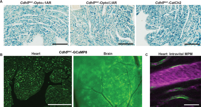
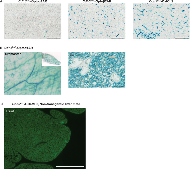
Figure 6. Endothelial cell optoeffector and optosensor expression.
(A) X-gal staining of heart cryosections from Cdh5BAC-Optoα1AR_IRES_lacZ, Cdh5BAC-Optoß2AR_IRES_lacZ, and Cdh5BAC-CatCh2_IRES_lacZ mice. Scale bars: 200 µm. (B) Native fluorescence of GCaMP8 protein in heart cryosection (left panel) and brain dorsal surface (right panel). Scale bar: 200 µm. (C) Intravital two-photon imaging of Cdh5 BAC-GCaMP8 mice shows endothelial GCaMP8 (green) labeling of coronary microvasculature. Intravenous Texas Red-conjugated 70 kDa dextran identifies the vessel lumen (magenta). All images shown are representative images from three animals unless otherwise specified.


