

Abstract
Extensive glenoid labral tears, whether the result of repetitive instability or first-time dislocation, compromise the mechanical stability of the glenohumeral joint due to disruption of the anterior, inferior, posterior, and/or superior portions of the labrum. These lesions often result in recurrent multiplanar instability and pain that is nonresponsive to conservative management and difficult to diagnose due to variability in clinical presentation and advanced imaging findings. Arthroscopic repair techniques to address symptomatic shoulder instability have showed positive patient-reported outcomes, low failure rates, and high return-to-sport rates. The evolution of knotless suture anchors offers a fixation method that has proven to be functionally equivalent to knotted suture anchors while avoiding the risks of knotted anchors (knot loosening, knot migration, articular abrasion) and allowing easier placement and decreased operative time. The purpose of this technique is to describe our preferred method to treat a 270° labral tear through arthroscopic knotless anchor repair and demonstrate the expanded application of this technique for extensive glenoid labral pathology.
Technique Video
For the treatment of a 270° labral tear in this left shoulder, the patient is placed in the lateral decubitus position and the standard diagnostic arthroscopy is performed using a posterior portal, anterior superior portal, and mid glenoid portal. Initial evaluation includes confirmation of the labral tear extending from 2 o’clock anteriorly to 11 o’clock posteriorly. Then attention is turned to the glenoid rim and torn labrum in preparation for repair. An arthroscopic shaver is used to debride the damaged labral tissue and create a bleeding surface on the glenoid rim to allow for better suture fixation and facilitate healing of the repaired labrum. A curved elevator is used to lyse capsulolabral adhesions and mobilize the labrum to allow for it to fall at the level of the glenoid articular surface. Suture repair is then performed using a ReelPass SutureLasso (Arthrex) to pass the repair suture through the capsule and grasp a several-millimeter (approximately 2-3 mm) portion of the labrum to integrate it into the repair. To address the anterior portion of the 270° labral tear, three 2.9 mm knotless PEEK PushLock Anchors (Arthrex) are fixed 5-7 mm apart from the 6 o’clock to 4 o’clock position. An accessory 7 o’clock portal is established for better accessibility to the posterior portion of the labral tear. A 3 mm knotless SutureTak Anchor (Arthrex) is placed at the 7 o’clock position followed by three additional 2.9 mm knotless PushLock Anchors 2-3 mm apart to the 11 o’clock position. A total of seven anchors are required to fully complete the capsulolabral repair and provide restoration of the labrum contour anterior, inferiorly, and posteriorly. Inspection with an arthroscopic probe is performed to check the integrity of the repair. The portal sites are then closed with no. 3-0 Monocryl suture, Dermabond, and Steri-Strips. A sterile dressing is placed, and then the shoulder is placed into a padded abduction sling.
Introduction
Reinforcing the stability of the glenohumeral joint is the glenoid labrum, a fibrocartilaginous structure that encircles the glenoid to supplement concavity of the shoulder.1,2 Despite its extensive network of dynamic and static stabilizers, the delicate architectural anatomy of the glenoid labrum is inherently predisposed to injury.3,4 Disruption to the glenohumeral balance can result in glenoid labral tears and sequential glenohumeral instability. In the setting of 270° labral tears, the anterior, inferior and posterior aspects of the glenoid labrum are detached from the glenoid, rendering the antero- and posteroinferior regions of the glenohumeral joint unstable.5,6
Open and arthroscopic techniques have been described to address glenohumeral shoulder instability, both producing positive patient-reported outcomes, low risk of recurrence, and high return-to-activity rates.6, 7, 8, 9 The modern mainstay of anterior instability treatment involves capsulolabral plication using knotted and/or knotless suture anchors. While knotted all-suture anchors remain widely utilized, recent research has supported the functional equivalence of knotless all-suture anchors which offer the advantage of potentially quicker application, tensionability, with a lower risk of subsequent soft-tissue and cartilage abrasions.10, 11, 12, 13
Previously, the senior author has described a technique with knotless suture anchor fixation for addressing recurrent posterior shoulder instability.14 The purpose of this Technical Note is to outline the expanded application of the knotless suture anchor technique to treat a 270° labral tear in order to address multiplanar instability.
Surgical Technique
A narrated video with demonstration of the surgical technique described in the following may be reviewed (Video 1).
Patient Positioning and Anesthesia
Prior to transfer to the operating room, an interscalene nerve block is placed by the regional anesthesia team using ultrasound guidance and a catheter. Following administration of anesthesia, the patient is brought into the operating room and positioned in the lateral decubitus position with balanced suspension (10-15 pounds depending on patient size) utilized for the duration of the case. The lateral decubitus position is preferred to allow for optimal visualization and access of the glenohumeral joint and reduction of the torn labrum onto the glenoid face during repair.
Diagnostic Arthroscopy and Debridement
Following a surgical safety time out, a standard diagnostic arthroscopy is performed utilizing three working portals: a standard posterior portal, anterior superior portal and mid-glenoid portal. Any concomitant glenohumeral pathology including loose bodies or synovitis is addressed and a complete diagnostic examination is performed. Attention is then turned toward the labral repair. The preoperative imaging showed an anteroinferior labral tear (Figure 1). The labrum is first mobilized using a series of specially designed elevators (Arthrex, Naples, FL). Depending on the size and extent of the tear, the arthroscope and elevators are switched amongst the various portals to gain the appropriate trajectory (Figure 2). Complete mobilization of the tear is achieved once the labral tissue falls at a resting position at the level of the glenoid articular surface (Figure 3). Next, a Torpedo (Arthrex, Naples, FL) and other zone-specific curved rasping instruments are used to create a bleeding bony surface for labral reattachment.
Fig 1.
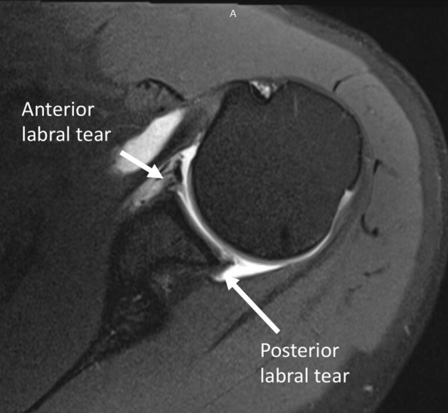
Sagittal fluid sensitive MRI of a left shoulder demonstrating an anteroinferior and posteroinferior labral tear with intact glenohumeral articular cartilage.
Fig 2.

Arthroscopic view from the posterior portal using a 30-degree arthroscope in a left shoulder in the lateral position demonstrating an anterior and posterior labral tear.
Fig 3.

Arthroscopic view from the posterior portal using a 30-degree arthroscope in a left shoulder in the lateral position demonstrating appropriate mobilization of the tear: the resting position of the labrum should fall at the level of the glenoid articular surface.
Posterior Capsulolabral Repair
As many Bankart lesions tend to extend past the 6 o’clock position, in order to restore anatomy, the repair begins at the posterior extent. Under most circumstances an accessory 7 o’clock portal is established using a percutaneous kit (Arthrex, Naples, FL) which allows for initial spinal needle localization in order to determine the desired trajectory. While viewing the posterior labrum from one of the anterior portals, the first anchor, a 3 mm knotless SutureTak (Arthrex, Naples, FL), is placed at the 7 o’clock position. Then using a crescent shaped REEL PASS device (Arthrex, Naples, FL) through an 8 mm canula, the suture hook is passed inferiorly through the capsule, then directly under and hugging the labrum to avoid the axilliary nerve and is used to shuttle the repair suture. Next, both the repair suture and the shuttling sutures are retrieved through the same cannula and the shuttling suture is used to pass the repair suture. The repair suture is pulled in order to tension the repair to the desired degree. Three additional 2.9 mm knotless PushLock Anchors 2-3 mm apart to the 11 o’clock position (Figure 4A and B).
Fig 4.

Arthroscopic view from the posterior portal using a 30-degree arthroscope in a left shoulder in the lateral position demonstrating posterior labral repair with placement of 2.9 mm knotless PushLock Anchors 2-3 mm apart at the 8 o’clock (A) and 9 o’clock (B) positions.
Anterior Capsulolabral Repair
Once fixed at 7 o’clock, the remainder of the anchors utilized are 2.9 mm knotless PEEK PushLocks (Arthrex, Naples, FL), which are placed while viewing from posteriorly (Fig 5). For these anchors, a curved REEL PASS device is used to first pass a #1 monofilament suture through both capsule and labrum at the desired location, near 5:30 o’clock, spacing 2-3mm from the 7 o’clock anchor. The monofilament is then used to shuttle a 1.3mm FiberLink SutureTape with care to keep the looped end of the SutureTape away from the articular side of the repair in order to provide a broader surface area of compression. This is then fixed onto the articular margin with 0.5 mm of superior advancement from the SutureTape entry using a 2.9 mm PushLock device (Fig 6). These steps are repeated for completion of the repair with two additional anchors spaced every 2-3mm (Fig 7A, B).
Fig 5.

Arthroscopic view from the posterior portal using a 30-degree arthroscope in a left shoulder in the lateral position demonstrating the ideal position for the low anterior portal (dashed circle) for anterior bankart repairs.
Fig 6.

Arthroscopic view from the posterior portal using a 30-degree arthroscope in a left shoulder in the lateral position demonstrating the appropriate location and trajectory for placement of anchors, on the glenoid rim.
Fig 7.

Arthroscopic view from the posterior portal using a 30-degree arthroscope in a left shoulder in the lateral position demonstrating anterior labral repair with synching of the repair suture (A) and placement of a 2.9 mm knotless PEEK PushLock Anchor (B) at the 4 o’clock position.
Final Inspection and Closure
The final construct results in a 270-degree repair with a total of seven knotless anchors to provide excellent restoration of the labrum, contour anteriorly, inferiorly, and posteriorly. Final images of the resulting stabilization construct are taken, and range of motion is assessed to present excellent abduction and external rotation with stability to load and shift testing (Fig 8). The wounds were closed with 3-0 Monocryl suture, Dermabond, and Steri-strips. Sterile dressing is placed and the shoulder is placed in a padded abduction sling. The pearls and pitfalls of the described technique are show in Table 1.
Fig 8.

Arthroscopic view from the anterior portal using a 30-degree arthroscope in a left shoulder in the lateral position demonstrating the final repair construct with 270-degree restoration of the capsulolabral bumper.
Table 1.
Pearls and Pitfalls of 270-Degree Labral Repair with Knotless Sutures
| Pearls | Pitfalls |
|---|---|
| The lateral decubitus position is utilized for shoulder instability to ensure circumferential access to the shoulder joint and to allow for capsular shift. | Safe positioning in the lateral decubitus position requires protection of neurovascular structures: an axillary roll and padding of the peroneal nerves should be utilized in all cases to prevent postoperative neuropraxias. |
| A well placed 7 o’clock portal is essential for safe proper trajectory. The starting point is approximately 5-6 cm lateral from the posterolateral corner of the acromion. | A spinal needle should be placed first through the expected 7 o’clock portal before making an incision to ensure appropriate trajectory for both capsulolabral suture passage and anchor placement. |
| For most anterior instability cases, the capsulolabral repair should start posteriorly and progress anteriorly with progressive anterior and superior shifting with each anchor. | Care should be taken at the 5:30 position when using a curved suture passing device to only grab capsulolabral tissue as the axillary nerve is close and can be entrapped. |
| Anchors should be placed on the rim of the glenoid and care should be taken to ensure both the suture material utilized and the anchor are seated beneath the cartilaginous surface. | Proud sutures and anchors can lead to mechanical abrasion, pain, and early failure known as anchor arthropathy. |
| When using the FiberLink SutureTape for labral repair, the looped end should always exit the capsular, not the glenoid, end of the repair. | If the looped end FiberLink SutureTape exits on the glenoid end, the sutures will be prominent, produce less compression across the repair, and can potentially lead to mechical abrasion of the humeral head cartilage and should be avoided. |
Postoperative Rehabilitation
The patient remains in the sling with full elbow support for 6 weeks to decrease the tension on the reconstruction while supporting the arm as the regional anesthesia persists. During this period passive range of motion at the elbow and wrist is initiated. Passive shoulder motion is limited to less than 90 degrees of abduction and forward flexion. Physical therapy begins immediately for guidance on passive range of motion and restrictions. Active assist range of motion begins at 6 weeks post-operatively and strengthening begins at 3 months post-operatively. The patient resumes full activities at 6 months or earlier if cleared by the physician.
Discussion
This Technical Note highlights our preferred arthroscopic repair of a 270° labral tear with knotless suture anchor fixation. Following traumatic dislocations, should conservative treatment fail to resolve symptomatic instability, especially in high-demand patients, we recommend our technique to restore function without the requirement of capsular plication, rotator interval closure, or volume-reduction procedures.
Large lesions of the glenoid labrum, whether the result of repetitive instability or first-time dislocation, represent a unique subpopulation of shoulder instability due to the variability in clinical presentation and advanced imaging findings, often challenging to diagnose until direct visualization with arthroscopy.9 These complex labral tears compromise the mechanical stability of the shoulder leading to multiplanar instability of the humeral head that is commonly nonresponsive to conservative management. A systematic review by Ernat et al., including 128 patients from 6 level IV studies, found improved outcomes and return to work/sport was achieved with arthroscopic repair of 270° and 360° glenoid labrum tears.6 Recently, Pounder et al. published an outcomes study on 25 patients following arthroscopic repair of 270° labral tears and found a high return to sport rate (76%), only one case of recurrent subluxation, no recurrent dislocation, and no revision surgeries required at mean follow-up of 42.2 months.15 Therefore, arthroscopic management of large labral tears proves to be the favorable treatment option to restore stability of the glenohumeral joint for patients with high functional demand and recurrent symptoms.
While the use of suture anchors for glenoid labral repair has become the gold standard, demonstrating superior outcomes compared to other arthroscopic fixation techniques, the evolution of knotless anchors has unveiled a more essential debate whether any significant difference exists between knotted and knotless suture anchor methods. Advantages to knotless anchors include minimized risk and avoidance of knot loosening and migration while also preventing articular abrasion from the knot prominence.11,14 Furthermore, use of knotless sutures can decrease operative time as advanced arthroscopic knot tying skills are not required. Newer knotless anchor designs feature curved drill guides and smaller profile for easier anchor placement and preservation of glenoid bone stock. When used with the patient in lateral decubitus position, as described by the current technique, this offers optimal visualization and access to all four-quadrants of the glenoid, which is important for 270° and other extensive labral tears that often involve the inferior and posterior portions.
A disadvantage of knotless sutures is that some designs prevent further tensioning after initial fixation. However, a recent systematic review by Matache et al. found conflicting evidence supporting knotless and knotted anchors but no major differences in any biomechanical outcomes across the majority of studies reviewed, including load to failure and stiffness.11 This study reported no significant difference in clinical outcomes between use of knotless or knotted anchors for Bankart, SLAP, or posterior labral repairs, but did find operative time was reduced with knotless anchors.11 Similarly, Wu et al. published a matched cohort study that found knotless anchor Bankart repairs have comparable patient-reported outcomes and revision rates to knotted repairs at mean 4.8 year follow-up, but demonstrated lower rates of recurrent subluxation.13 A systematic review by Knapik et al. examined a total of 234 patients who underwent arthroscopic repair of isolated type II SLAP lesions and showed patients treated with knotted anchors were significantly more likely to experience postoperative complications than patients treated with knotless anchors.12
The current literature supports the use of knotless suture anchors as an effective technique to repair glenoid labral tears. However, there is conflicting evidence on the difference in complications and failure rates between knotless and knotted anchor fixation methods along with a paucity in clinical outcomes published on the use of these techniques to repair extensive labral tears. Future studies are needed with longer follow-up time and evaluation of more complex glenoid labral pathology in order to determine the viability of these techniques in addressing multiplanar shoulder instability and better guide clinical application.
In conclusion, we recommend arthroscopic repair with knotless suture anchor fixation for patients with 270° labral tears who present with recurrent shoulder instability nonresponsive to conservative management. Further research is required to validate the safety and long-term efficacy of this procedure for treatment of extensive glenoid labral tears.
Footnotes
The authors report the following potential conflicts of interest or sources of funding: M.T.P. has royalties or licenses with Arthrex and Elsevier; receives consulting fees from Arthrex, SLACK, and Joint Research Foundation; receives payment or honoraria for lectures, presentations, speakers bureaus, manuscript writing or educational events from ArthroSurface; receives support for attending meetings and/or travel from Arthrex; has the following patents planned, issued, or pending: 9226743, 20150164498, 20150150594, and 20110040339; and has leadership or fiduciary role in the following board, society, committee or advocacy group, paid or unpaid: AAOS: Board or Committee member; AANA: Board or Committee member; AOSSM: Board or Committee member; ASES: Board or Committee member; Arthroscopy: Editorial or governing board; ISAKOS: Board or Committee member; Knee: Editorial or governing board; Orthopedics: Editorial or governing board; San Diego Shoulder Institute: Board or Committee member; SLACK Inc: Editorial or governing board; Society of Military Orthopaedic Surgeons: Board or Committee member. Full ICMJE author disclosure forms are available for this article online, as supplementary material.
Supplementary Data
For the treatment of a 270° labral tear in this left shoulder, the patient is placed in the lateral decubitus position and the standard diagnostic arthroscopy is performed using a posterior portal, anterior superior portal, and mid glenoid portal. Initial evaluation includes confirmation of the labral tear extending from 2 o’clock anteriorly to 11 o’clock posteriorly. Then attention is turned to the glenoid rim and torn labrum in preparation for repair. An arthroscopic shaver is used to debride the damaged labral tissue and create a bleeding surface on the glenoid rim to allow for better suture fixation and facilitate healing of the repaired labrum. A curved elevator is used to lyse capsulolabral adhesions and mobilize the labrum to allow for it to fall at the level of the glenoid articular surface. Suture repair is then performed using a ReelPass SutureLasso (Arthrex) to pass the repair suture through the capsule and grasp a several-millimeter (approximately 2-3 mm) portion of the labrum to integrate it into the repair. To address the anterior portion of the 270° labral tear, three 2.9 mm knotless PEEK PushLock Anchors (Arthrex) are fixed 5-7 mm apart from the 6 o’clock to 4 o’clock position. An accessory 7 o’clock portal is established for better accessibility to the posterior portion of the labral tear. A 3 mm knotless SutureTak Anchor (Arthrex) is placed at the 7 o’clock position followed by three additional 2.9 mm knotless PushLock Anchors 2-3 mm apart to the 11 o’clock position. A total of seven anchors are required to fully complete the capsulolabral repair and provide restoration of the labrum contour anterior, inferiorly, and posteriorly. Inspection with an arthroscopic probe is performed to check the integrity of the repair. The portal sites are then closed with no. 3-0 Monocryl suture, Dermabond, and Steri-Strips. A sterile dressing is placed, and then the shoulder is placed into a padded abduction sling.
References
- 1.Yoshida M., Goto H., Nozaki M. Quantitative analysis of attachment of the labrum to the glenoid fossa: a cadaveric study. J Orthop Sci. 2015;20:823–829. doi: 10.1007/s00776-015-0742-4. [DOI] [PubMed] [Google Scholar]
- 2.Itoigawa Y., Itoi E. Anatomy of the capsulolabral complex and rotator interval related to glenohumeral instability. Knee Surg Sports Traumatol Arthrosc. 2016;24:343–349. doi: 10.1007/s00167-015-3892-1. [DOI] [PubMed] [Google Scholar]
- 3.De Coninck T., Ngai S.S., Tafur M., Chung C.B. Imaging the glenoid labrum and labral tears. Radiographics. 2016;36:1628–1647. doi: 10.1148/rg.2016160020. [DOI] [PubMed] [Google Scholar]
- 4.Mlynarek R.A., Lee S., Bedi A. Shoulder injuries in the overhead throwing athlete. Hand Clin. 2017;33:19–34. doi: 10.1016/j.hcl.2016.08.014. [DOI] [PubMed] [Google Scholar]
- 5.Alpert J.M., Verma N., Wysocki R., Yanke A.B., Romeo A.A. Arthroscopic treatment of multidirectional shoulder instability with minimum 270 degrees labral repair: minimum 2-year follow-up. Arthroscopy. 2008;24:704–711. doi: 10.1016/j.arthro.2008.01.008. [DOI] [PubMed] [Google Scholar]
- 6.Ernat J.J., Yheulon C.G., Shaha J.S. Arthroscopic repair of 270- and 360-degree glenoid labrum tears: a systematic review. Arthroscopy. 2020;36:307–317. doi: 10.1016/j.arthro.2019.07.027. [DOI] [PubMed] [Google Scholar]
- 7.DeFroda S., Bokshan S., Stern E., Sullivan K., Owens B.D. Arthroscopic bankart repair for the management of anterior shoulder instability: indications and outcomes. Curr Rev Musculoskelet Med. 2017;10:442–451. doi: 10.1007/s12178-017-9435-2. [DOI] [PMC free article] [PubMed] [Google Scholar]
- 8.Feng S., Song Y., Li H., Chen J., Chen J., Chen S. Outcomes for arthroscopic repair of combined bankart/SLAP lesions in the treatment of anterior shoulder instability: a systematic review and meta-analysis. Orthop J Sports Med. 2019;7 doi: 10.1177/2325967119877804. :2325967119877804. [DOI] [PMC free article] [PubMed] [Google Scholar]
- 9.Mazzocca A.D., Cote M.P., Solovyova O., Rizvi S.H., Mostofi A., Arciero R.A. Traumatic shoulder instability involving anterior, inferior, and posterior labral injury: a prospective clinical evaluation of arthroscopic repair of 270° labral tears. Am J Sports Med. 2011;39:1687–1696. doi: 10.1177/0363546511405449. [DOI] [PubMed] [Google Scholar]
- 10.Nolte P.C., Midtgaard K.S., Ciccotti M. Biomechanical comparison of knotless all-suture anchors and knotted all-suture anchors in Type II SLAP lesions: A cadaveric study. Arthroscopy. 2020;36:2094–2102. doi: 10.1016/j.arthro.2020.04.026. [DOI] [PubMed] [Google Scholar]
- 11.Matache B.A., Hurley E.T., Kanakamedala A.C. Knotted versus knotless anchors for labral repair in the shoulder - a systematic review. Arthroscopy. 2021;37:1314–1321. doi: 10.1016/j.arthro.2020.11.056. [DOI] [PubMed] [Google Scholar]
- 12.Knapik D.M., Kolaczko J.G., Gillespie R.J., Salata M.J., Voos J.E. Complications and return to activity after arthroscopic repair of isolated Type II SLAP lesions: A Systematic Review comparing knotted versus knotless suture anchors. Orthop J Sports Med. 2020;8 doi: 10.1177/2325967120911361. :2325967120911361. [DOI] [PMC free article] [PubMed] [Google Scholar]
- 13.Wu I.T., Desai V.S., Mangold D.R. Comparable clinical outcomes using knotless and knot-tying anchors for arthroscopic capsulolabral repair in recurrent anterior glenohumeral instability at mean 5-year follow-up. Knee Surg Sports Traumatol Arthrosc. 2021;29:2077–2084. doi: 10.1007/s00167-020-06057-7. [DOI] [PubMed] [Google Scholar]
- 14.Sanchez G., Kennedy N.I., Ferrari M.B., Mannava S., Frangiamore S.J., Provencher M.T. Arthroscopic labral repair in the setting of recurrent posterior shoulder instability. Arthrosc Tech. 2017;6:e1789–e1794. doi: 10.1016/j.eats.2017.06.055. [DOI] [PMC free article] [PubMed] [Google Scholar]
- 15.Pounder E.J., Hurley E.T., Ali Z.S., Pauzenberger L., Mullett H. Return to sport following arthroscopic repair of 270° labral tears. Arthrosc Sports Med Rehabil. 2020;2:e237–e240. doi: 10.1016/j.asmr.2020.02.009. [DOI] [PMC free article] [PubMed] [Google Scholar]
Associated Data
This section collects any data citations, data availability statements, or supplementary materials included in this article.
Supplementary Materials
For the treatment of a 270° labral tear in this left shoulder, the patient is placed in the lateral decubitus position and the standard diagnostic arthroscopy is performed using a posterior portal, anterior superior portal, and mid glenoid portal. Initial evaluation includes confirmation of the labral tear extending from 2 o’clock anteriorly to 11 o’clock posteriorly. Then attention is turned to the glenoid rim and torn labrum in preparation for repair. An arthroscopic shaver is used to debride the damaged labral tissue and create a bleeding surface on the glenoid rim to allow for better suture fixation and facilitate healing of the repaired labrum. A curved elevator is used to lyse capsulolabral adhesions and mobilize the labrum to allow for it to fall at the level of the glenoid articular surface. Suture repair is then performed using a ReelPass SutureLasso (Arthrex) to pass the repair suture through the capsule and grasp a several-millimeter (approximately 2-3 mm) portion of the labrum to integrate it into the repair. To address the anterior portion of the 270° labral tear, three 2.9 mm knotless PEEK PushLock Anchors (Arthrex) are fixed 5-7 mm apart from the 6 o’clock to 4 o’clock position. An accessory 7 o’clock portal is established for better accessibility to the posterior portion of the labral tear. A 3 mm knotless SutureTak Anchor (Arthrex) is placed at the 7 o’clock position followed by three additional 2.9 mm knotless PushLock Anchors 2-3 mm apart to the 11 o’clock position. A total of seven anchors are required to fully complete the capsulolabral repair and provide restoration of the labrum contour anterior, inferiorly, and posteriorly. Inspection with an arthroscopic probe is performed to check the integrity of the repair. The portal sites are then closed with no. 3-0 Monocryl suture, Dermabond, and Steri-Strips. A sterile dressing is placed, and then the shoulder is placed into a padded abduction sling.
For the treatment of a 270° labral tear in this left shoulder, the patient is placed in the lateral decubitus position and the standard diagnostic arthroscopy is performed using a posterior portal, anterior superior portal, and mid glenoid portal. Initial evaluation includes confirmation of the labral tear extending from 2 o’clock anteriorly to 11 o’clock posteriorly. Then attention is turned to the glenoid rim and torn labrum in preparation for repair. An arthroscopic shaver is used to debride the damaged labral tissue and create a bleeding surface on the glenoid rim to allow for better suture fixation and facilitate healing of the repaired labrum. A curved elevator is used to lyse capsulolabral adhesions and mobilize the labrum to allow for it to fall at the level of the glenoid articular surface. Suture repair is then performed using a ReelPass SutureLasso (Arthrex) to pass the repair suture through the capsule and grasp a several-millimeter (approximately 2-3 mm) portion of the labrum to integrate it into the repair. To address the anterior portion of the 270° labral tear, three 2.9 mm knotless PEEK PushLock Anchors (Arthrex) are fixed 5-7 mm apart from the 6 o’clock to 4 o’clock position. An accessory 7 o’clock portal is established for better accessibility to the posterior portion of the labral tear. A 3 mm knotless SutureTak Anchor (Arthrex) is placed at the 7 o’clock position followed by three additional 2.9 mm knotless PushLock Anchors 2-3 mm apart to the 11 o’clock position. A total of seven anchors are required to fully complete the capsulolabral repair and provide restoration of the labrum contour anterior, inferiorly, and posteriorly. Inspection with an arthroscopic probe is performed to check the integrity of the repair. The portal sites are then closed with no. 3-0 Monocryl suture, Dermabond, and Steri-Strips. A sterile dressing is placed, and then the shoulder is placed into a padded abduction sling.